. 2010 Jun 23:9:161.
doi: 10.1186/1476-4598-9-161.

Tumor promoting effects of CD95 signaling in chemoresistant cells

Affiliations

Tumor promoting effects of CD95 signaling in chemoresistant cells

Elisabet Ametller et al. Mol Cancer. .

Abstract

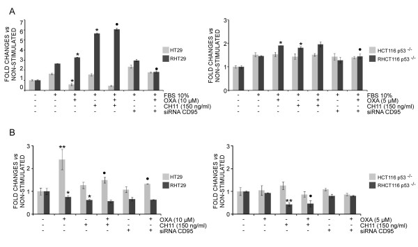
Background: CD95 is a death receptor controlling not only apoptotic pathways but also activating mechanisms promoting tumor growth. During the acquisition of chemoresistance to oxaliplatin there is a progressive loss of CD95 expression in colon cancer cells and a decreased ability of this receptor to induce cell death. The aim of this study was to characterize some key cellular responses controlled by CD95 signaling in oxaliplatin-resistant colon cancer cells.

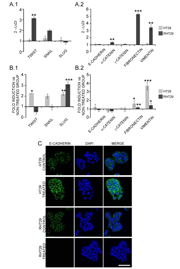
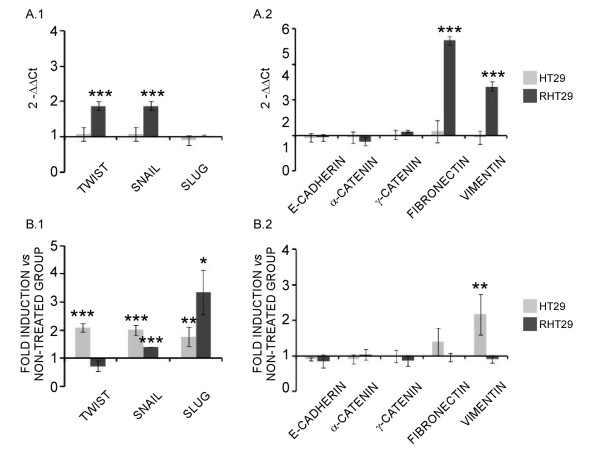
Results: We show that CD95 triggering results in an increased metastatic ability in resistant cells. Moreover, oxaliplatin treatment itself stimulates cell migration and decreases cell adhesion through CD95 activation, since CD95 expression inhibition by siRNA blocks the promigratory effects of oxaliplatin. These promigratory effects are related to the epithelia-to-mesenchymal transition (EMT) phenomenon, as evidenced by the up-regulation of some transcription factors and mesenchymal markers both in vitro and in vivo.

Conclusions: We conclude that oxaliplatin treatment in cells that have acquired resistance to oxaliplatin-induced apoptosis results in tumor-promoting effects through the activation of CD95 signaling and by inducing EMT, all these events jointly contributing to a metastatic phenotype.


Figures

Figure 1
Figure 1
Effects of oxaliplatin and CD95 activation on cell migration and adhesion in oxaliplatin-resistant cells. A) To determine the effects of oxaliplatin or CD95 triggering on cell migration different sensitive (HT29 and HCT116 p53-/-) and resistant (RHT29 and RHCT116 p53-/-) cell lines were treated with oxaliplatin (10 μM for the HT29 and RHT29 and 5 μM for the HCT116 p53-/- and RHTC116 p53-/-) or/and the CD95 agonistic antibody CH11 (150 ng/ml) for 24 hours, and their migratory ability was assessed in transwell assays. The contribution of CD95 activation to oxaliplatin-induced cell migration was determined by inhibiting CD95 expression with siRNA. B) The effects of oxaliplatin or CD95 triggering on the adhesion ability of the different cell lines or the contribution of CD95 activation to oxaliplatin-induced cell adhesion were determined under the same experimental conditions on fibronectin coated plates. The experiments were performed in triplicate and results represent the mean ± SEM. Values that are significantly different from control group by ANOVA's analysis are indicated by *p < 0.05, **p < 0.01, and those different from the oxaliplatin-treated group are indicated by •p < 0.05. OXA: oxaliplatin, FBS: Fetal bovine serum.
Figure 2
Figure 2
Differential activation of MAPK pathways by CD95 in cells resistant to oxaliplatin-induced cell death. To determine the contribution of oxaliplatin-induced CD95 to the activation of A) p42/44 MAPK, B) p38 MAPK signaling pathways and to the activation of C) NF-κB (p65 NLS), cells were treated with oxaliplatin (10 μM), the CD95 agonistic antibody CH11 (150 ng/ml) or the combination of both for 48 hours, and the protein levels determined by Western blot.
Figure 3
Figure 3
Oxaliplatin regulation of cell cycle-related proteins in the HT29 and RHT29 cell lines. A) HT29 and RHT29 cell lines were treated with oxaliplatin 10 μM for 24 and 48 hours and the expression of the cell-cycle related proteins cyclin D1, p27, CDK2 and CDK4 determined by Western blot. Results shown are the mean ± SEM of the densitometric quantification three independent experiments. Values that are significantly different between groups by ANOVA's analysis are indicated by *p < 0.05, and those different between non-treated and treated groups are indicated by •p < 0.05, ••p < 0.01, •••p < 0.001. B) Cell cycle studies: the distribution of the different phases of the cell cycle was determined in HT29 and RHT29 cells by flow cytometry. CDK2: cyclin-dependent kinase 2, CDK4: cyclin-dependent kinase 4.
Figure 4
Figure 4
Oxaliplatin regulation of apoptosis-related proteins in the HT29 and RHT29 cell lines. HT29 and RHT29 cell lines were treated with oxaliplatin 10 μM for 24 and 48 hours and the expression of the A) active caspase 8 and C) Bcl-2 and Bax were determined by Western blot. B) Densitometry of active caspase 8. D-E) Ratio between Bcl-2 and Bax for the HT29 and RHT29 cell lines based on the densitometry quantification.
Figure 5
Figure 5
Analysis of EMT markers and E-cadherin expression on HT29 and RHT29 cells. (A1) Differential basal expression of some transcription factors related to the EMT process and (A2) markers of epithelial and mesenchymal phenotype were determined in the HT29 and RHT29 cell lines by qPCR. (B1) The effects of oxaliplatin in the gene expression of those transcription factors and (B2) EMT markers were also analyzed in cells treated with oxaliplatin 10 μM for 48 hours and in this case the results are shown as fold induction versus non-treated cells. Results represent the mean ± SEM of triplicates. Values that are significantly different between (A) groups or between (B) non-treated and treated groups by ANOVA's analysis are indicated by *p < 0.05, **p < 0.01, ***p < 0.001. C) HT29 and RHT29 cells were treated with oxaliplatin 10 μM for 48 hours and E-cadherin expression was detected by immunofluorescence and confocal microscope analysis. Scale bar is 50 μm.
Figure 6
Figure 6
Analysis of EMT markers on HT29 and RHT29 xenografts. (A1) Differential basal expression of some transcription factors related to the EMT process and (A2) markers of epithelial and mesenchymal phenotype were determined by qPCR in tumor xenografts derived from the HT29 and RHT29 cell lines. (B1) The effects of oxaliplatin in the gene expression of those transcription factors and (B2) EMT markers were also analyzed in xenografted tumors treated with oxaliplatin (i.p. 10 mg/kg) once per week during 27 days and in this case the results are shown as fold induction versus non-treated tumors. Results represent the mean ± SEM for 6 xenografts per group. Values that are significantly different between (A) groups or between (B) non-treated and treated groups by ANOVA's analysis are indicated by *p < 0.05, **p < 0.01, ***p < 0.001.

Similar articles

Cited by

References

    1. Fulda S, Meyer E, Friesen C, Susin SA, Kroemer G, Debatin KM. Cell type specific involvement of death receptor and mitochondrial pathways in drug-induced apoptosis. Oncogene. 2001;20:1063–1075. doi: 10.1038/sj.onc.1204141. - DOI - PubMed
    1. Scaffidi C, Fulda S, Srinivasan A, Friesen C, Li F, Tomaselli KJ, Debatin KM, Krammer PH, Peter ME. Two CD95 (APO-1/Fas) signaling pathways. EMBO J. 1998;17:1675–1687. doi: 10.1093/emboj/17.6.1675. - DOI - PMC - PubMed
    1. Algeciras-Schimnich A, Pietras EM, Barnhart BC, Legembre P, Vijayan S, Holbeck SL, Peter ME. Two CD95 tumor classes with different sensitivities to antitumor drugs. Proc Natl Acad Sci USA. 2003;100:11445–11450. doi: 10.1073/pnas.2034995100. - DOI - PMC - PubMed
    1. Khong HT, Restifo NP. Natural selection of tumor variants in the generation of "tumor escape" phenotypes. Nat Immunol. 2002;3:999–1005. doi: 10.1038/ni1102-999. - DOI - PMC - PubMed
    1. Peter ME, Legembre P, Barnhart BC. Does CD95 have tumor promoting activities? Biochim Biophys Acta. 2005;1755:25–36. - PubMed

Publication types