. 2010 Jun 24:11:45.
doi: 10.1186/1471-2121-11-45.

An autocrine sphingosine-1-phosphate signaling loop enhances NF-kappaB-activation and survival

Affiliations

An autocrine sphingosine-1-phosphate signaling loop enhances NF-kappaB-activation and survival

Tomas Blom et al. BMC Cell Biol. .

Abstract

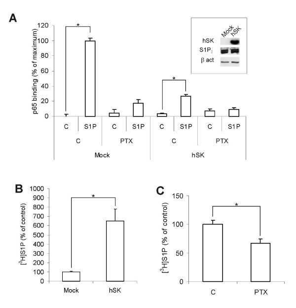
Background: Sphingosine-1-phosphate (S1P) is a bioactive lipid that regulates a multitude of cellular functions, including cell proliferation, survival, migration and angiogenesis. S1P mediates its effects either by signaling through G protein-coupled receptors (GPCRs) or through an intracellular mode of action. In this study, we have investigated the mechanism behind S1P-induced survival signalling.

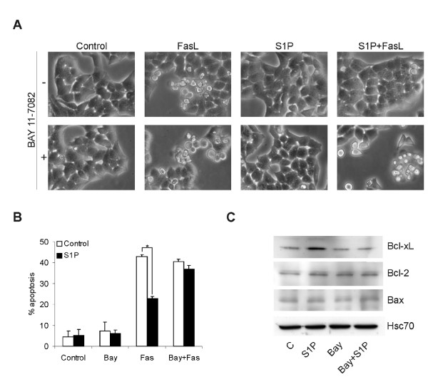
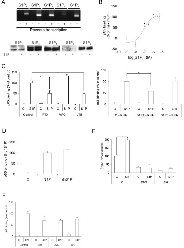
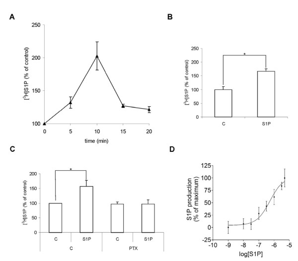
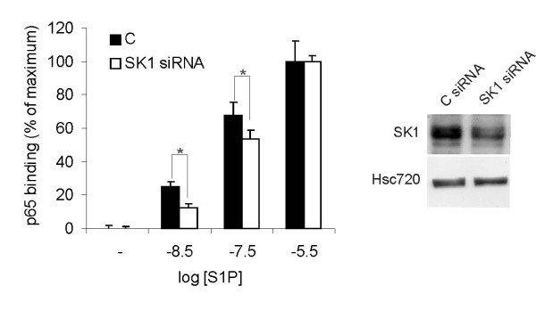
Results: We found that S1P protected cells from FasL-induced cell death in an NF-kappaB dependent manner. NF-kappaB was activated by extracellular S1P via S1P2 receptors and Gi protein signaling. Our study also demonstrates that extracellular S1P stimulates cells to rapidly produce and secrete additional S1P, which can further amplify the NF-kappaB activation.

Conclusions: We propose a self-amplifying loop of autocrine S1P with capacity to enhance cell survival. The mechanism provides increased understanding of the multifaceted roles of S1P in regulating cell fate during normal development and carcinogenesis.


Figures

Figure 1
Figure 1
S1P confers NF-κB dependent protection from death receptor induced apoptosis. Figure A shows representative phase contrast pictures of HeLa cells treated ±5 μM Bay 11-7082 (NF-κB inhibitor) for 6 h, followed by addition of vehicle or 3 μM S1P for 8 hours. Apoptosis was then induced by addition of 50 ng/ml superFas ligand (Fas) for 16 hours. B. HeLa cells were treated as in A, and were fixed, stained for active caspase-3, and were analysed by FACS. The bars denote the mean ± SEM of at least three independent experiments (*, p < 0.05). C. Western blot showing Bcl-xL, Bcl-2 and Bax expression in cells pre-treated with 5 μM Bay 11-7082 or vehicle for 6 h, followed by stimulation with vehicle or 3 μM S1P for 12 hours. Hsc70 was used as a loading control. The results are representative of three independent experiments.
Figure 2
Figure 2
S1P activates NF-κB through G protein coupled receptors. A. The upper panel shows the presence of mRNAs encoding G protein coupled S1P receptors in HeLa cells. + and - denote whether the extracted RNA was reverse transcribed or not. The lower panel shows Western blots of the S1P receptors in HeLa cells stimulated ± 3 μM S1P for 30 minutes. B. Concentration-response curve for S1P-induced activation of NF-κB (p65). HeLa cells were stimulated with varying concentrations of S1P for 30 minutes. Protein extracts were analyzed by NF-κB (p65) ELISA. 5 μg of protein was used from each extract. C. HeLa cells were preincubated with 50 ng/ml PTX for 16 h, 10 μM VPC23019 for 30 minutes or 10 μM JTE013 for 30 minutes prior to stimulation with 3 μM S1P for 30 minutes (left panel), or treated with S1P2 or S1P5 siRNA for 48 h prior to S1P stimulation. Proteins were then extracted and NF-κB (p65)-activation was assayed by ELISA. D. HeLa cells were stimulated with either vehicle, 3 μM S1P or 3 μM dihydro-S1P for 30 minutes. 5 μg of protein from each extract was used for the NF-κB (p65) ELISA. E. HeLa cells were pre-incubated with either vehicle, 10 μM DMS or 10 μM SKi for 5 minutes. The cells were then stimulated with 3 μM S1P for 10 minutes in the presence of [3H]sphingosine. The bars show synthesized cellular [3H]S1P as percent of the unstimulated control. F. The cells were pre-incubated either with vehicle, 10 μM sphingosine, 10 μM DMS or 10 μM SKi for 5 minutes. Following a 30-minute stimulation with 3 μM S1P, the cells were harvested, lyzed and the DNA-binding activity of p65 was measured from 5 μg of protein extract. The data points and bars in panels B-F denote the mean ± SEM of at least three independent experiments (*, p < 0.05).
Figure 3
Figure 3
S1P induces cellular S1P-production and -secretion through Gi protein coupled receptors. A. HeLa cells were exposed to either vehicle or 3 μM S1P for the indicated period of times in the presence of [3H]sphingosine. The amount of produced [3H]S1P is expressed as percent of that in vehicle-treated cells. B. Secreted [3H]S1P was extracted from the cell culture medium following a 10 minute stimulation with vehicle or 3 μM S1P, and was analyzed using thin layer chromatography and scintillation counting. C. The cells were treated with 50 ng/ml pertussis toxin or vehicle for 16 h. Cellular lipids were extracted following a 10 minute stimulation with 3 μM S1P or vehicle in the presence of [3H]sphingosine. The formed [3H]S1P was assayed using thin layer chromatography and scintillation counting. D. Concentration-response curve for S1P-induced S1P production. HeLa cells were stimulated with varying concentrations of S1P or vehicle together with [3H]sphingosine for 10 minutes. Cellular lipids were extracted and separated by HPTLC. The results are expressed as percent increased [3H]S1P production in S1P stimulated cells compared with control. The data points and bars in panels A-D denote the mean ± SEM of at least three independent experiments (*, p < 0.05).
Figure 4
Figure 4
NF-κB activation induced by exogenously added S1P is enhanced by S1P produced by the cells. Cells transfected with control siRNA or SK1 siRNA were treated with the indicated concentrations of S1P for 30 minutes and were analyzed for active NF-κB (p65) using ELISA. The panel to the right shows representative western blots of control and SphK1 siRNA treated cells. The bars denote the mean ± SEM of at least three independent experiments (*, p < 0.05).
Figure 5
Figure 5
Effect of prolonged overexpression of SphK on NF-κB activation and S1P synthesis. A. Control cells and cells overexpressing SphK (hSK) were pretreated ± 50 ng/ml pertussis toxin for 16 h before stimulation with vehicle or 3 μM S1P for 30 minutes. Proteins were extracted and NF-κB (p65)-activation was assayed by ELISA. The inset shows Western blots of 10 μg of protein from mock transduced or SphK1 overexpressing HeLa cells. The cell extracts were probed for sphingosine kinase and S1P2. B. [3H]S1P was extracted from the mock- or SphK transduced cells following a 10-minute incubation with [3H]sphingosine, and was analyzed using thin layer chromatography and scintillation counting. C. The SphK overexpressing cells were treated with 50 ng/ml pertussis toxin or vehicle for 16 h. Secreted [3H]S1P was extracted from the medium following a 10-minute incubation with [3H]sphingosine. The formed [3H]S1P was assayed using thin layer chromatography and scintillation counting. The bars in panels A-C denote the mean ± SEM of at least three independent experiments (*, p < 0.05).
Figure 6
Figure 6
Signaling cascades regulating S1P-induced S1P-production and S1P-induced activation of NF-κB. A. NF-κB-activation induced after stimulating HeLa cells for 30 minutes with 3 μM S1P. The cells were pre-treated with 10 μM BAPTA-AM for 30 minutes, 50 μM PD98059 for 30 min, 1 μM GF109203× for 5 min or 30 nM Wortmannin for 5 minutes. B. The cells were treated with inhibitors the same way as in A. Following a 10-minute stimulation with 3 μM S1P or vehicle together with [3H]sphingosine, lipids were extracted and the produced [3H]S1P was measured by thin layer chromatography and scintillation counting. The bars in A and B denote the mean ± SEM of at least three independent experiments (*, p < 0.05).
Figure 7
Figure 7
S1P-induced secretion of S1P in the malignant tumor cell lines MEL-7 and WM35. WM35 (A.) or MEL-7 (B.) cells were exposed to either vehicle, 3 μM S1P or 10 μM SKi for 10 minutes in the presence of [3H]sphingosine. Secreted [3H]S1P was then extracted from the cell culture medium and analysed by HPTLC. C. MEL-7 cells were treated with 10 μM SKi or vehicle for 4 h. Apoptosis was then induced by addition of 100 ng/ml Tumor necrosis factor-related apoptosis-inducing ligand (TRAIL) for 4 h. D. MEL-7 cells were treated with vehicle or 10 μM JTE013 and VPC23019 for 4 h. Apoptosis was induced by addition of TRAIL for 4 h, and cells positive for cleaved caspase-3 was measured using FACS. The bars in panels A-D denote the mean ± SEM of three independent experiments (*, p < 0.05).

References

    1. Taha TA, Argraves KM, Obeid LM. Sphingosine phosphate receptors: receptor specificity versus functional redundancy. Biochim Biophys Acta. 2004;1682:48–55. - PubMed
    1. Futerman AH. The complex life of simple sphingolipids. EMBO Rep. 2004;5:777–782. doi: 10.1038/sj.embor.7400208. - DOI - PMC - PubMed
    1. Sanchez T, Hla T. Structural and functional characteristics of S1P receptors. J Cell Biochem. 2004;92:913–922. doi: 10.1002/jcb.20127. - DOI - PubMed
    1. Balthasar S, Samulin J, Ahlgren H, Bergelin N, Lundquist M, Toescu EC, Eggo MC, Törnquist K. Sphingosine 1-phosphate receptor expression profile and regulation of migration in human thyroid cancer cells. Biochem J. 2006;398:547–556. doi: 10.1042/BJ20060299. - DOI - PMC - PubMed
    1. Waters CM, Long J, Gorshkova I, Fujiwara Y, Connell M, Belmonte KE, Tigyi G, Natarajan V, Pyne S, Pyne NJ. Cell migration activated by platelet-derived growth factor receptor is blocked by an inverse agonist of the Sphingosine 1-phosphate receptor-1. FASEB J. 2006;20:509–511. - PubMed

Publication types

MeSH terms

LinkOut - more resources