

Case Reports
doi: 10.1308/147870810X12699662980952.
Epub 2010 Jun 23.
Neuroendocrine carcinoma arising within a retroperitoneal mature teratoma
Affiliations
- PMID: 20573313
- PMCID: PMC5696860
- DOI: 10.1308/147870810X12699662980952
Item in Clipboard
Case Reports
Neuroendocrine carcinoma arising within a retroperitoneal mature teratoma
Ann R Coll Surg Engl.
2010 Sep.
Abstract
We discuss an unusual case of a large cystic mass arising in the left upper quadrant of a 48-year-old woman. Radiological investigations could not confirm the origin or the nature of the mass. A laparatomy revealed a large retroperitoneal cystic mass sandwiched between the left adrenal, spleen and the gastro-oesophageal junction. Histological analysis confirmed a mature teratoma of the retroperitoneum with neuroendocrine carcinoma arising within it. To our knowledge this is only the second reported case of its kind.
Figures
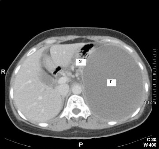
Transverse CT image of abdomen at level of T12 vertebrae. Note the large tumour mass (T) filling the left hemi-abdomen and displacing the stomach (S) superiorly.

Transverse CT image of mature teratoma (T) at level of L2 vertebrae. Note the spleen (Sp) and left kidney (LK) which have been displaced inferiorly.

Laparotomy with retroperitoneal teratoma (T) partly mobilised and adherent to the gastro-oesophageal junction (GOJ).
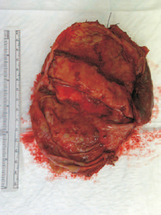
Mature teratoma – cyst wall incised and internal surface displayed.
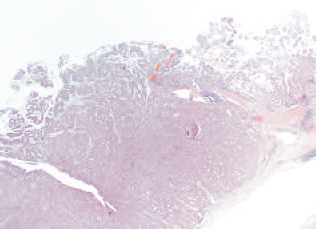
Solid nodule component of cyst wall which displays neuroendocrine morphology (×5, H&E).

Solid neuroendocrine carcinoma with atypical mitotic figures (arrows; x20, H&E).
Similar articles
-
Primary carcinoid tumor arising in a retroperitoneal mature teratoma in an adult.Int J Urol. 2004 Oct;11(10):912-5. doi: 10.1111/j.1442-2042.2004.00918.x. Int J Urol. 2004. PMID: 15479301
-
[A case of retroperitoneal teratoma in an adult resembling adrenal tumors].Hinyokika Kiyo. 1999 Jan;45(1):37-9. Hinyokika Kiyo. 1999. PMID: 10086264 Japanese.
-
Primary retroperitoneal teratoma in an adult.J Chin Med Assoc. 2003 Aug;66(8):497-500. J Chin Med Assoc. 2003. PMID: 14604315
-
Large cell neuroendocrine carcinoma arising in mature cystic teratoma: a case report and review of the literature.Eur J Gynaecol Oncol. 2012;33(4):414-8. Eur J Gynaecol Oncol. 2012. PMID: 23091901 Review.
-
Pelvic mature cystic teratoma with neuroendocrine carcinoma: report of a rare association and review of literature.Indian J Pathol Microbiol. 2014 Jan-Mar;57(1):113-5. doi: 10.4103/0377-4929.130916. Indian J Pathol Microbiol. 2014. PMID: 24739847 Review.
Cited by
-
Ganglioneuroblastoma arising within a retroperitoneal mature cystic teratoma.World J Clin Oncol. 2012 Dec 10;3(12):155-8. doi: 10.5306/wjco.v3.i12.155. World J Clin Oncol. 2012. PMID: 23293755 Free PMC article.
-
Case - Giant primary retroperitoneal teratoma with neuroendocrine components.Can Urol Assoc J. 2023 Nov;17(11):E405-E407. doi: 10.5489/cuaj.8401. Can Urol Assoc J. 2023. PMID: 37549343 Free PMC article. No abstract available.
-
Primary mature cystıc teratoma mimickıng an adrenal mass in an adult male patient.Korean J Urol. 2014 Feb;55(2):148-51. doi: 10.4111/kju.2014.55.2.148. Epub 2014 Feb 14. Korean J Urol. 2014. PMID: 24578814 Free PMC article.
-
Mature cystic retroperitoneal teratoma with well differentiated renal elements: relation to spinal dysraphism.European J Pediatr Surg Rep. 2014 Jun;2(1):46-9. doi: 10.1055/s-0033-1351393. Epub 2013 Aug 5. European J Pediatr Surg Rep. 2014. PMID: 25755970 Free PMC article.
-
Primary adrenal teratoma: A case series and review of the literature.Mol Clin Oncol. 2018 Oct;9(4):437-442. doi: 10.3892/mco.2018.1687. Epub 2018 Aug 1. Mol Clin Oncol. 2018. PMID: 30214733 Free PMC article.
References
-
- McKenny JK, Heerema-McKenney A, Rouse RV. Extragonadal germ cell tumors: a review with emphasis on pathologic features, clinical prognostic variables, and differential diagnostic considerations. Adv Anat Pathol 2007; : 69–92. - PubMed
-
- Robboy SJ, Norris HJ, Scully RE. Insular carcinoid primary in the ovary. A clini-copathologic analysis of 48 cases. Cancer 1975; : 404–18. - PubMed
-
- Arazi M, Toy H, Tavli L. Primary neuroendocrine carcinoma arising within a mature sacrococcygeal teratoma. Orthopaedics 2007; : 878. - PubMed
-
- Wang L, Chu S, Ng K, Wong Y. Adenocarcinomas arising from primary retroperitoneal mature teratomas: CT and MR imaging. Eur Radiol 2002; : 1546–9. - PubMed
-
- Gatcombe HG, Assikis V, Kooby D, Johnstone P. Primary retroperitoneal teratomas: a review of the literature. J Surg Oncol 2004; : 107–13. - PubMed
Publication types
MeSH terms
LinkOut - more resources
Full Text Sources