. 2010 Aug 1;137(15):2471-81.
doi: 10.1242/dev.049718. Epub 2010 Jun 23.

Proliferative and transcriptional identity of distinct classes of neural precursors in the mammalian olfactory epithelium

Affiliations

Proliferative and transcriptional identity of distinct classes of neural precursors in the mammalian olfactory epithelium

Eric S Tucker et al. Development. .

Abstract

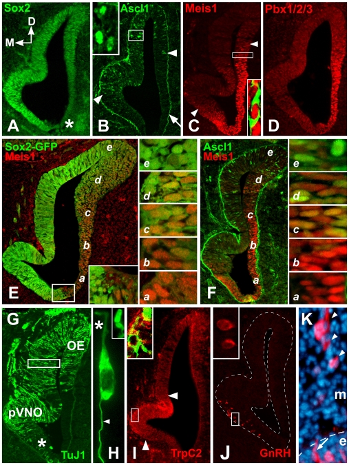
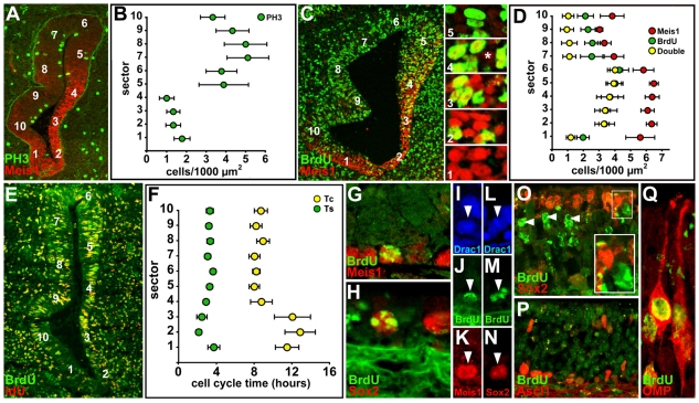
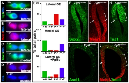
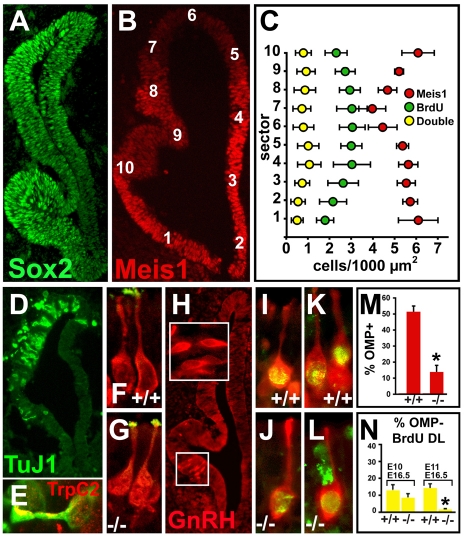
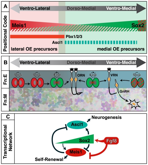
Neural precursors in the developing olfactory epithelium (OE) give rise to three major neuronal classes - olfactory receptor (ORNs), vomeronasal (VRNs) and gonadotropin releasing hormone (GnRH) neurons. Nevertheless, the molecular and proliferative identities of these precursors are largely unknown. We characterized two precursor classes in the olfactory epithelium (OE) shortly after it becomes a distinct tissue at midgestation in the mouse: slowly dividing self-renewing precursors that express Meis1/2 at high levels, and rapidly dividing neurogenic precursors that express high levels of Sox2 and Ascl1. Precursors expressing high levels of Meis genes primarily reside in the lateral OE, whereas precursors expressing high levels of Sox2 and Ascl1 primarily reside in the medial OE. Fgf8 maintains these expression signatures and proliferative identities. Using electroporation in the wild-type embryonic OE in vitro as well as Fgf8, Sox2 and Ascl1 mutant mice in vivo, we found that Sox2 dose and Meis1 - independent of Pbx co-factors - regulate Ascl1 expression and the transition from lateral to medial precursor state. Thus, we have identified proliferative characteristics and a dose-dependent transcriptional network that define distinct OE precursors: medial precursors that are most probably transit amplifying neurogenic progenitors for ORNs, VRNs and GnRH neurons, and lateral precursors that include multi-potent self-renewing OE neural stem cells.


Figures

Fig. 1.
Fig. 1.
Molecularly distinct domains in the E11.5 mouse OE. (A) Graded Sox2 expression is highest in the medial presumptive vomeronasal organ (pVNO), and lowest ventrolaterally (asterisk). (B) Ascl1 is expressed from the dorsolateral OE through medial pVNO region (between arrowheads), mostly overlapping high Sox2 expression; arrow indicates non-specific basal lamina labeling. Inset: higher magnification of the boxed region shows Ascl1-positive nuclei. (C) Meis1 expression begins ventromedially, peaks in the lateral OE and declines dorsolaterally. Inset: higher magnification of region boxed shows nuclear Meis1 in nascent TuJ1-labeled OE neurons. (D) Pbx1/2/3 is expressed in the lateral OE and, like Meis1 expression, declines dorsally. (E) Graded expression of Sox2 (Sox2-eGFP reporter) and Meis1 is complementary. There is a sharp ventral boundary (box, inset). a-e indicate the levels of sections shown in the panels on the right. Varying levels of complementary Sox2 and Meis1 expression are found in cells at distinct ventrolateral (a) to dorsomedial (e) locations. Little if any Meis1 is seen dorsally (e) where Sox2 predominates. (F) Ascl1 and Meis1 are also complementary; however, robust Ascl1 labeling begins only in dorsomedial locations (d, e, right panels), where Meis1 is nearly undetectable. (G) TuJ1-labeled OE neurons are seen at low frequency dorsolaterally, become concentrated dorsomedially through the pVNO and decline to a few scattered cells in the extreme ventromedial OE (asterisk). Box indicates region shown in H. (H) TuJ1-labeled OE neurons have cytological hallmarks of ORNs and VRNs: a single apical process (asterisk), apparent `dendritic knob' (inset) and a single axon (arrowhead). (I) VRNs are distinguished by enhanced expression of TrpC2 (red) within the pVNO (between arrowheads), apparently on the cell surface of TuJ1-labeled neurons (green, inset). (J) GnRH neurons are seen within, or ventral to, the pVNO. Inset: higher magnification of boxed region, showing two GnRH neurons within the ventromedial OE. (K) GnRH neurons delaminate from the OE (arrow, e) and migrate through the frontonasal mesenchyme (arrowheads, m).
Fig. 2.
Fig. 2.
OE precursors have distinct proliferative characteristics. (A) PH3 labeled cells (M-phase) are less frequent in lateral versus medial OE. (B) Counts of PH3 cells in 10 geometrically defined OE sectors (positions are indicated in A) confirm enhanced M-phase cell frequency in dorsomedial versus ventrolateral regions. (C) Acute BrdU labeling increases ventro- to dorsolaterally, and declines in register with diminishing levels of Meis1. (Insets) There are scattered cells lightly labeled with Meis1 (e.g. asterisk in sector 4 image) in medial regions (sectors 6-10). (D) Counts of BrdU, Meis1 and double-labeled cells in 10 equivalent OE sectors (positions are indicated in A) confirm inverse Meis1 and S-phase frequency. We counted lightly Meis1 labeled cells (* in panel 4), yielding a lower frequency of Meis1 cells especially in sectors 7-10. (E) Cell cycle times estimated using dual S-phase labeling. IdU/BrdU-labeled cells (yellow), BrdU-alone (green) and all cells (bisbenzimide; not shown) are counted to calculate cell cycle time (Tc) and S-phase duration (Ts). (F) Tc and Ts in OE sectors 1-10 (positions are indicated in E). (G) Precursors that retain BrdU for long periods (5 days, `label-retaining') are found in lateral E16.5 OE and express Meis1. (H) Similar label-retaining lateral precursors express Sox2. (I-K) A single cell in separate color channels shows nuclear, BrdU, and Meis1 labeling. (L-N) A similar color separation of a label-retaining cell confirms nuclear Sox2 expression. (O) Heavily BrdU-labeled medial cells (arrowheads) do not coincide with high Sox2 cells in medial E16.5 OE. Inset: higher magnification of boxed area shows an example of a lightly BrdU-labeled high Sox2-expressing medial OE cell. (P) Label-retaining medial cells rarely if ever coincide with Ascl1 in the medial E16.5 OE. (Q) Many heavily BrdU-labeled medial cells at E16.5 are ORNs, based upon dual labeling with the ORN marker OMP.
Fig. 3.
Fig. 3.
OE precursors have distinct modes of division and Fgf8-dependent neurogenic potential. (A-D) Examples of neuron (TuJ1 N, nuclear counterstain with bis-benzamide), precursor (Meis1 or Sox2, P) or nuclear (bisbenzamide only, P* pairs cultured from microdissected lateral and medial E11.5 OE. (E) Histograms show frequency of N-N (green, TuJ1 labeled), N-P (green-red, TuJ1 and Sox2 or Meis1), P-P (red, Meis1 or Sox2 labeled) or P*-P*(bis-benzamide only) pairs from lateral (top) and medial OE cells (middle). Bottom histogram shows influence of exogenous Fgf8 added to the culture medium on frequency of pairs generated by lateral OE cells. (F-J) In vivo consequences of reduced Fgf8 levels shown for two Fgf8 hypomorphic mouse embryos. (F) Sox2 levels are diminished in the medial OE, and a distinct mediolateral gradient is difficult to discern. (G) Meis1 expression diminishes in the dorsolateral OE (bracket), but expands into the medial OE (arrow). (H) TuJ1-positive neurons are reduced medially (arrow) and shifted laterally (bracket). (I) Ascl1-positive precursors diminish in the medial OE and shift laterally, matching TuJ1 distribution in the hypomorphic OE. (J) Meis1 and Ascl1 double-labeling shows medial loss of Ascl1 and coincident expansion of Meis1 (arrow), as well as Ascl1 lateral expansion where Meis1 declines (bracket).
Fig. 4.
Fig. 4.
Sox2 dose regulates OE precursor identity. (A) Schematic of E11.5 lateral OE explant electroporation, culture and sampling strategy. Confocal images show (B) control eGFP or (C) pCIG-Sox2-electroporated lateral OE. Boxed areas in all panels indicate regions examined for illustration and quantification. Lower (D,E) and higher (F,G) magnification confocal images show Meis1 co-expression (cyan, arrows) or lack thereof (asterisks) in control (D,F) or Sox2-electroporated (E,G) GFP-positive (green) OE cells. (H) Effects of elevated Sox2 dose on Meis1 cell frequency. Lower (I,J) and higher (K,L) magnification images show lack of Ascl1 expression (asterisks) in control cells (I,K) and enhanced Ascl1 expression (red, arrows) in Sox2-electroporated (J,L) cells. (M) Effects of elevated Sox2 dose on Ascl1 cell frequency. Confocal images show TuJ1-labeled neurons in control (N) and Sox2-electroporated (O) lateral OE. Lower (P,Q) and higher (R,S) magnification images show lack of coincidence (asterisks) with TuJ1 neurons (red) in control (P,R) or an increase (arrows) in Sox2-electroporated (Q,S) cells. (T) Effects of elevated Sox2 dose on TuJ1-labeled neuron frequency. (U) Higher magnification images show a Sox2-electroporated TuJ1-labeled neuron with a single apparent axon. *P≤0.0002.
Fig. 5.
Fig. 5.
Meis1 regulates OE precursor identity. (A-C) Confocal images show E11.5 lateral OE preparations electroporated with control (A), pCIG-Meis1 (B) or pCIG-Meis1-IRES-Pbx1 (C) and labeled for Ascl1. Inset in A shows area illustrated and sampled for Meis1 overexpression experiments. Boxed areas represent regions used for illustration and quantification. Lower (D-F) and higher (G-I) magnification comparison of Ascl1 expression in control (D,G), Meis1 (E,H) or Meis1/Pbx1 (F,I) electroporated cells in the dorsolateral OE. (J,K) Images showing E11.5 control (J) and Meis1/Pbx1 (K) electroporated preparations labeled for Sox2. (L,M) Sox2 is seen in control (L) and Meis1/Pbx1 (M) electroporated cells in the dorsolateral OE. (N) Frequency of GFP-Ascl1 doubled-labeled cells in control, Meis1 and Meis1/Pbx1 electroporated preparations. (O) Frequency of GFP-Sox2 double-labeled cells in control and Meis1-Pbx1 electroporated preparations. **P≤0.002; ***P≤0.0002.
Fig. 6.
Fig. 6.
Precursor and neuron identity in the Ascl1–/– mutant OE. (A) The Sox2 medial-lateral gradient is preserved in the E11.5 OE. (B) Heavily labeled Meis1 cells remain concentrated in the lateral OE in Ascl1–/– E11.5 mouse embryos, whereas moderately labeled cells expand medially. (C) Frequency and distribution of Meis1 cells, acutely labeled BrdU cells and Meis1/BrdU double-labeled cells is altered in the Ascl1–/– OE. Positions of the cells are indicated in B. (D) Limited neuronal differentiation, recognized with TuJ1, is seen in the E11.5 Ascl1–/– OE. (E) TrpC2 (red) expression can still be detected in a small subset of Ascl1–/– OE neurons (green). (F,G) OMP and the ORN-selective adenylyl cyclase ACIII (green) are expressed and localized appropriately in Ascl1–/– ORNs at E16.5. (H) GnRH neurons are generated in or near the pVNO in Ascl1–/– embryos. (I-L) ORNs in the E16.5 Ascl1–/– OE are birthdated with BrdU at E10 (earliest ORN genesis; I, J) and E11 (K,L). (M) Frequency of OMP-positive ORNs in the E16.5 Ascl1–/– OE is substantially reduced. (N) Near-normal frequency of ORN genesis at E10 declines to near zero by E11 in the E16.5 Ascl1–/– OE. *P≤0.02.
Fig. 7.
Fig. 7.
Precursor and neuron identity in the Sox2hyp/– OE. (A-D) Meis1 cells are distributed throughout the OE over the entire anteroposterior axis in Sox2hyp/– mouse embryos. (E) Ascl1 is not detected in Sox2hyp/– OE, but is seen in MGE precursors in the forebrain (Inset). (F) Acute BrdU labeling is substantially diminished in Sox2hyp/– OE. Scattered BrdU-positive cells are nevertheless detectable (inset). (G,H) Frequency of PH3-labeled mitotic cells diminishes towards lateral values in dorsomedial sectors of the Sox2hyp/– OE. The positions of the cells are indicated in G. (I) Neuronal differentiation is attenuated in the Sox2hyp/– OE. TuJ1-labeled neurons are seen dorsally (arrowheads) and their frequency is diminished (compare with Fig. 1G). (J) TrpC2 remains expressed in the E11.5 Sox2hyp/– OE. Arrowheads indicate the dorsomedial OE where both TuJ1- and TrpC2-labeled neurons are seen in the Sox2hyp/–.(K) A small number of ORNs that express OMP (red) and ACIII (green, inset) differentiate by E16.5 in the Sox2hyp/– OE. Arrowheads indicate the dendritic knob of an OMP-labeled ORN in the Sox2hyp/– OE. Inset is at higher magnification showing that ACIII is localized to this dendritic knob, as in wild-type E16.5 ORNS (see Fig.6F). (L) GnRH neurons are still detected, although at lower frequencies, in the Sox2hyp/– OE, adjacent mesenchyme (m) and ventral forebrain (fb). The lower inset shows GnRH-labeled cells in the OE (e), as well as the adjacent mesenchyme (m) of the E11.5 Sox2hyp/–. The upper inset shows several GnRH-labeled cells in the frontonasal mesenchyme (m).
Fig. 8.
Fig. 8.
Distinct molecular and cellular identities of OE precursors. (A) Precursor identity is established by OE position and reflected in combinatorial graded transcription factor expression. (B) Meis1 defines slowly dividing self-renewing precursors, primarily in the lateral OE; enhanced Sox2, coincident with Ascl1 and increased Fgf8 signaling, identifies transit amplifying precursors, primarily in the medial OE, that are responsible for quantitative expansion, but not specification of ORNs, VRNs and GnRH neurons. Slowly dividing precursors in the ventro-medial OE express both Meis1 and Sox2 at high levels (A), and may contribute to genesis of GnRH cells and/or VRNs, which populate this region. (C) A Sox2 dose-dependent transcriptional network, modulated by Fgf8, preserves lateral slowly dividing, multipotent OE precursors by maintaining low Sox2 levels that support Meis1 expression, or promotes medial transit amplifying cells that expand OE neuron numbers by Fgf8-dependent high Sox2-mediated downregulation of Meis1 and parallel upregulation of Ascl1.

Similar articles

Cited by

References

    1. Ache B. W., Young J. M. (2005). Olfaction: diverse species, conserved principles. Neuron 48, 417-430 - PubMed
    1. Anchan R. M., Drake D. P., Haines C. F., Gerwe E. A., LaMantia A. S. (1997). Disruption of local retinoid-mediated gene expression accompanies abnormal development in the mammalian olfactory pathway. J. Comp. Neurol. 379, 171-184 - PubMed
    1. Asson-Batres M. A., Smith W. B. (2006). Localization of retinaldehyde dehydrogenases and retinoid binding proteins to sustentacular cells, glia, Bowman's gland cells, and stroma: potential sites of retinoic acid synthesis in the postnatal rat olfactory organ. J. Comp. Neurol. 496, 149-171 - PMC - PubMed
    1. Bani-Yaghoub M., Tremblay R. G., Lei J. X., Zhang D., Zurakowski B., Sandhu J. K., Smith B., Ribecco-Lutkiewicz M., Kennedy J., Walker P. R., et al. (2006). Role of Sox2 in the development of the mouse neocortex. Dev. Biol. 295, 52-66 - PubMed
    1. Beites C. L., Kawauchi S., Crocker C. E., Calof A. L. (2005). Identification and molecular regulation of neural stem cells in the olfactory epithelium. Exp. Cell Res. 306, 309-316 - PubMed

Publication types