Comparative Study
. 2010 Jul;133(Pt 7):2022-31.
doi: 10.1093/brain/awq142. Epub 2010 Jun 23.

The transcription factor orthodenticle homeobox 2 influences axonal projections and vulnerability of midbrain dopaminergic neurons

Affiliations
Comparative Study

The transcription factor orthodenticle homeobox 2 influences axonal projections and vulnerability of midbrain dopaminergic neurons

Chee Yeun Chung et al. Brain. 2010 Jul.

Abstract

Two adjacent groups of midbrain dopaminergic neurons, A9 (substantia nigra pars compacta) and A10 (ventral tegmental area), have distinct projections and exhibit differential vulnerability in Parkinson's disease. Little is known about transcription factors that influence midbrain dopaminergic subgroup phenotypes or their potential role in disease. Here, we demonstrate elevated expression of the transcription factor orthodenticle homeobox 2 in A10 dopaminergic neurons of embryonic and adult mouse, primate and human midbrain. Overexpression of orthodenticle homeobox 2 using lentivirus increased levels of known A10 elevated genes, including neuropilin 1, neuropilin 2, slit2 and adenylyl cyclase-activating peptide in both MN9D cells and ventral mesencephalic cultures, whereas knockdown of endogenous orthodenticle homeobox 2 levels via short hairpin RNA reduced expression of these genes in ventral mesencephalic cultures. Lack of orthodenticle homeobox 2 in the ventral mesencephalon of orthodenticle homeobox 2 conditional knockout mice caused a reduction of midbrain dopaminergic neurons and selective loss of A10 dopaminergic projections. Orthodenticle homeobox 2 overexpression protected dopaminergic neurons in ventral mesencephalic cultures from Parkinson's disease-relevant toxin, 1-methyl-4-phenylpyridinium, whereas downregulation of orthodenticle homeobox 2 using short hairpin RNA increased their susceptibility. These results show that orthodenticle homeobox 2 is important for establishing subgroup phenotypes of post-mitotic midbrain dopaminergic neurons and may alter neuronal vulnerability.


Figures
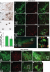
Figure 1
Figure 1
Otx2 is elevated in A10 dopaminergic neurons in adult mouse, primate and human. Otx2 mRNA levels were compared between A9 and A10 dopaminergic neurons in mouse and human using LCM and quantitative PCR. (A–C) LCM of dopaminergic neurons in the mouse midbrain. Selection of dopaminergic neurons was guided by quick tyrosine hydroxylase (TH) immunostaining. (A) Tyrosine hydroxylase-positive neurons in substantia nigra (SN) before laser capture. (B) The tyrosine hydroxylase-positive cells were targeted for laser capture with a 7.5 µm laser diameter. (C) Captured cells on the thermoplastic film were visualized before processing for RNA extraction. Quantitative PCR on the LCM samples demonstrated that Otx2 mRNA levels were elevated in A10 dopaminergic neurons compared to A9 dopaminergic neurons in mouse and human (D). Immunostaining in adult mouse SN (E–G) and lateral (H–J) and medial ventral tegmental area (VTA) (K–M and O; z-stack of the perforated square in M) regions demonstrated that Otx2 immunoreactivity was higher in A10 dopaminergic neurons in the VTA than A9 dopaminergic neurons in the mouse SN. In the primate (Macaque mulatta) midbrain (S), A9 dopaminergic neurons in the SN showed very low or undetectable Otx2 immunoreactivity (P–R), whereas A10 dopaminergic neurons in the VTA showed strong Otx2 immunoreactivity (T–V, W; z-stack image of perforated square in V). Otx2 expression was not exclusive to dopaminergic neurons.
Figure 2
Figure 2
Otx2 is elevated in A10 dopaminergic neurons in the embryonic mouse midbrain. Immunostaining of Otx2 in embroynic day (E) 17 mouse development also demonstrated that Otx2 immunoreactivity was higher in A10 dopaminergic neurons. The substantia nigra (SN) and the ventral tegmental area (VTA) are defined in a lower magnification view (A–C). Otx2 expression was absent in A9 dopaminergic neurons (D–G), whereas strong Otx2 immunoreactivity was detected in A10 dopaminergic neurons (H–K). TH = tyrosine hydroxylase.
Figure 3
Figure 3
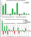
Otx2 regulates expression of known A10-elevated genes in MN9D cells and primary ventral mesencephalic (VM) cultures. (A) MN9D cells were transduced with lentivirus encoding yellow fluorescent protein (YFP)-nucleus localization signal or Otx2 and harvested 4 days after transduction for mRNA isolation and quantitative PCR analysis. Overexpression of Otx2 using lentivirus in MN9D cells induced A10 elevated guidance cue molecules including neuropilin 1 (Npn1), neuropilin 2 (Npn2) and slit2; and A10 elevated neuropeptides, vasoactive peptide (VIP) and adenylyl cylase activating peptide (Adcyap). (B) Primary ventral mesencephalic cultures were transduced with lentivirus encoding yellow fluorescent protein–nucleus localization signal, Otx2, control shRNA or shRNA against Otx2 on in vitro Day 1 and harvested at in vitro Day 8 for mRNA isolation and quantitative PCR analysis. Gene expression profiles in primary ventral mesencephalic cultures confirmed the findings in MN9D cells with the exception of VIP. Data are shown as mean ratios of lenti-Otx2 over lenti-yellow fluorescent protein–nucleus localization signal or lenti-Otx2 shRNA over lenti-control shRNA ± SEM (n = 5 for each condition; *P < 0.05 two-tailed t-test) and are representatives of two experiments with the similar trends. Plexin C1; EphnB3 = ephrin type-B receptor 3; CART = cocaine and amphetamine regulated transcript; GRP =  Gastrin releasing peptide; CCK = cholecystokinin; NT-3 = neurotrophin 3.
Figure 4
Figure 4
Adult Otx2 conditional knockout mice lack the A10 dopaminergic neuronal projection (A–F). Tyrosine hydroxylase immunostaining reveals that En1cre/+;Otx2flox/flox mice show dramatic reduction of A10 neuronal projections including prefrontal cortex (PFC; A and B), nucleus accumbens (NAc; A and C), olfactory tubercle (OT; C), septum (D) and amygdala (amg; E and F). On the other hand, A9 neuronal projection in dorsolateral striatum (Str) was not affected in these mice (C). B and F are magnified images of the perforated squares marked i and ii (from A) and iii and iv (from E), respectively.
Figure 5
Figure 5
Otx2 overexpression protects dopaminergic neurons from MPP+ toxicity in primary ventral mesencephalic cultures. Primary ventral mesencephalic cultures were transduced with lentivirus encoding yellow fluorescent protein (YFP)–nucleus localization signal, Otx2, control shRNA or shRNA against Otx2 on in vitro Day 1 and 10 µM MPP+ was applied to the culture at in vitro Day 8. Cultures were fixed at in vitro Day 10 and stained with tyrosine hydroxylase for tyrosine hydroxylase-positive cell counting. When Otx2 was overexpressed by lentiviral delivery of Otx2 with multiplicities of infection (MOI) of 5 and 10 (A–C), dopaminergic neurons were more resistant to 10 µM MPP+ compared to yellow fluorescent protein–nucleus localization signal overexpressing ventral mesencephalic cultures (D). When Otx2 levels were reduced by lentivirus encoding shRNA against Otx2 with an MOI of 10 (E–G), dopaminergic neurons become more vulnerable to 5 µM MPP+ toxicity compared to control shRNA transduced ventral mesencephalic cultures (H). Data are shown as mean ± SEM (n = 5 for each condition; *P < 0.05 two-tailed t-test) and are representatives of two experiments with the similar trends. GFP = green fluorescent protein.

References

    1. Alberi L, Sgado P, Simon HH. Engrailed genes are cell-autonomously required to prevent apoptosis in mesencephalic dopaminergic neurons. Development. 2004;131:3229–36. - PubMed
    1. Ang SL. Transcriptional control of midbrain dopaminergic neuron development. Development. 2006;133:3499–506. - PubMed
    1. Arimura A. Pituitary adenylate cyclase activating polypeptide (PACAP): discovery and current status of research. Regul Pept. 1992;37:287–303. - PubMed
    1. Arimura A. Perspectives on pituitary adenylate cyclase activating polypeptide (PACAP) in the neuroendocrine, endocrine, and nervous systems. Jpn J Physiol. 1998;48:301–31. - PubMed
    1. Borgkvist A, Puelles E, Carta M, Acampora D, Ang SL, Wurst W, et al. Altered dopaminergic innervation and amphetamine response in adult Otx2 conditional mutant mice. Mol Cell Neurosci. 2006;31:293–302. - PubMed

Publication types

MeSH terms