Review
. 2010 Sep;2(9):a001750.
doi: 10.1101/cshperspect.a001750. Epub 2010 Jun 23.

Self-avoidance and tiling: Mechanisms of dendrite and axon spacing

Affiliations
Review

Self-avoidance and tiling: Mechanisms of dendrite and axon spacing

Wesley B Grueber et al. Cold Spring Harb Perspect Biol. 2010 Sep.

Abstract

The spatial pattern of branches within axonal or dendritic arbors and the relative arrangement of neighboring arbors with respect to one another impact a neuron's potential connectivity. Although arbors can adopt diverse branching patterns to suit their functions, evenly spread branches that avoid clumping or overlap are a common feature of many axonal and dendritic arbors. The degree of overlap between neighboring arbors innervating a surface is also characteristic within particular neuron types. The arbors of some populations of neurons innervate a target with a comprehensive and nonoverlapping "tiled" arrangement, whereas those of others show substantial territory overlap. This review focuses on cellular and molecular studies that have provided insight into the regulation of spatial arrangements of neurite branches within and between arbors. These studies have revealed principles that govern arbor arrangements in dendrites and axons in both vertebrates and invertebrates. Diverse molecular mechanisms controlling the spatial patterning of sister branches and neighboring arbors have begun to be elucidated.


Figures

Figure 1.
Figure 1.
Isoneuronal neurites repel one another. (A) The branches of a mouse cerebellar Purkinje cell dendritic arbor, imaged with 2-photon microscopy in a live animal, almost never overlap with one another (image courtesy of Anna Dunaevsky, Brown University). (B) The branches of a retinal ganglion cell in culture almost never overlap with one another. (Image modified from Montague and Friedlander 1991). (C) Subfields of leech mechanosensory axons compete for territory. Simplified diagrams, based on results in (Kramer and Stent 1985). The top shows a normal leech mechanosensory neuron with three separate subfields innervating adjacent regions of the epidermis. When the growth cone of one branch is crushed (blue arrows in middle and bottom panels), either eliminating (middle) or delaying (bottom) its growth, sibling subfields grow correspondingly larger (red arrows).
Figure 2.
Figure 2.
Dscam regulates dendrite self-avoidance. (A) The Dscam locus can generate up to 152,064 distinct protein isoforms by alternative splicing, including 19,008 distinct extracellular domains (Schmucker et al. 2000; Yu et al. 2009). Exons 4 and 6 code for half Ig domains (Ig2 and Ig3 respectively) and exon 9 codes for all of Ig7. Extensive binding assays have shown isoform-specific homophilic binding (Wojtowicz et al. 2004; Wojtowicz et al. 2007). The structure of the homophilic binding region (including Ig1-Ig8) indicates that the protein folds into an S shaped molecule, and that this folding allows interactions between the three variable Ig domains. Adapted from (Sawaya et al. 2008). (B) Studies in several systems (see text) indicate that Dscam is critical for self-avoidance of dendrites and axons. Studies are consistent with a model in which sister dendrites or axons that encounter one another during development are recognized by virtue of their shared isoform repertoire. This recognition leads to repulsive signaling, the molecular basis of which is not yet understood. (C) Single Dscam isoforms are sufficient for self-recognition and avoidance but Dscam diversity is required between cells so that they can share territories (coexistence). Left: Two different neurons with coexisting arbors. When those cells are forced to express the same arbitrary isoform at high levels their arbors no longer cross each other and are thus unable to coexist (see text for details).
Figure 3.
Figure 3.
Common features of heteroneuronal tiling. (A,B) Although the cell bodies of all retinal cell subtypes are arranged in a regular mosaic, different subtypes display various degrees of overlap. Colored shapes represent the territories of individual dendrites, black circles represent cell bodies. The dendrite of one cell is diagrammed in each panel. The arbors of some retinal cell types show “perfect” tiling, with closely apposed but nonoverlapping arbors (A), whereas the arbors of other types of neurons extensively overlap (B). Dendritic fields in A are based on images from (Dacey 1993). (C,D) Dendritic arbors of at least some retinal subtypes polarize toward denervated regions. Most ganglion cell dendrites in unperturbed retinas are not polarized in a consistent direction (C), but when a region of the retina is denervated (red area in D), nearby dendritic arbors polarize toward the empty territory. Images modified from data in (Perry and Linden 1982). (E) Drosophila da neurons exemplify the principle that different neuronal subtypes innervating the same surface are spaced independently of one another. Each diagrams shows two segments of a Drosophila larva, modified from (Grueber et al. 2003). The top panel shows the dendritic fields of one subtype of da neurons that does not uniformly innervate the epidermis. The second and third show the dendritic territories of two different subtypes of da neurons that tile the epidermis almost perfectly. The bottom panel is a superimposition of the territories of the three different neuron subtypes, showing that they each independently innervate the epidermis. (F,G) Somatosensory neurons in embryonic zebrafish show virtually unlimited plasticity. Diagrams represent a dorsal view of the heads of zebrafish larvae. Based on results in (Sagasti et al. 2005). (F). In normal fish, the peripheral axon arbors of the two bilateral trigeminal ganglia are mostly confined to the ipsilateral side of the head. (G) When one trigeminal ganglion is removed early in development, arbors from the remaining ganglion freely cross the midline to innervate the opposite side of the head.
Figure 4.
Figure 4.
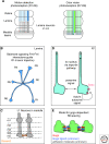
Diverse strategies for process spacing in the fly visual system. (A) Schematic of visual circuitry in Drosophila. (Left) Bundles of R1-R6 photoreceptors extend from the retina to the first order relay in the lamina. Here, the axons split and project in stereotyped directions to lamina cartridges. Each lamina cartridge is comprised of R neurons that carry visual information from a single point in space. L1-L5 neurons each project to the specific synaptic layers in the medulla neuropil. (Right) Color vision photoreceptors R7 and R8 project from the retina directly to the medulla, bypassing the lamina relay. R7 axons (light blue) project to a deeper medulla layer called m6, whereas R8 s (green) project to a more superficial medulla layer (m3). (B) R1-R6 bundles separate to take a specific trajectory to their proper lamina cartridge in a Flamingo-dependent manner (Chen and Clandinin 2008). R1-R6 neurons lacking Fmi show no obvious defects, but neurons adjacent to mutant cells do adopt the wrong trajectory. The growth cones of R1-R6 neurons may assess the relative level of Fmi signaling coming from their neighbors on either side and take a trajectory based on the opposing forces. (C) Dscam2 is essential for tiling of L1 lamina neuron axons in the medulla. L1 axons are normally restricted to a single medulla column by the action of Dscam2. Dscam2 molecules engage in homophilic interactions on the growth cones of L1 axons in m1 and m5 (Millard et al. 2007). (D) R7 axon tiling in the medulla is regulated by homotypic interactions and a TGF-β/Activin autocrine signal limits axon invasiveness for neighboring columns. The self-limiting Activin signal acts through Baboon (babo), a Type I TGF beta receptor. Babo deficient axons can respond to their neighbors, indicating that a different, as yet unknown signal mediates axon-axon interactions (Ting et al. 2007). (E) golden goal regulates R8 axon spacing. Gogo in R8 axons mediates repulsive output, probably in response to an unknown ligand also expressed on R8 neurons. R8-R8 repulsion via Gogo counteracts or silences the activity of an unknown cell adhesion molecule that would otherwise cause R8 axon clumping. Adapted from (Tomasi et al. 2008).

Similar articles

Cited by

References

    1. Amthor FR, Oyster CW 1995. Spatial organization of retinal information about the direction of image motion. Proc Natl Acad Sci 92: 4002–4005 - PMC - PubMed
    1. Andrews GL, Tanglao S, Farmer WT, Morin S, Brotman S, Berberoglu MA, Price H, Fernandez GC, Mastick GS, Charron F, et al.2008. Dscam guides embryonic axons by Netrin-dependent and -independent functions. Development 135: 3839–3848 - PMC - PubMed
    1. Ashley JA, Katz FN 1994. Competition and position-dependent targeting in the development of the Drosophila R7 visual projections. Development 120: 1537–1547 - PubMed
    1. Baker MW, Macagno ER 2000. The role of a LAR-like receptor tyrosine phosphatase in growth cone collapse and mutual-avoidance by sibling processes. J Neurobiol 44: 194–203 - PubMed
    1. Baker MW, Macagno ER 2007. In vivo imaging of growth cone and filopodial dynamics: Evidence for contact-mediated retraction of filopodia leading to the tiling of sibling processes. J Comp Neurol 500: 850–862 - PubMed

Publication types

LinkOut - more resources