Review
. 2010 Jul;2(7):a000687.
doi: 10.1101/cshperspect.a000687. Epub 2010 Jun 23.

Paraspeckles

Affiliations
Review

Paraspeckles

Archa H Fox et al. Cold Spring Harb Perspect Biol. 2010 Jul.

Abstract

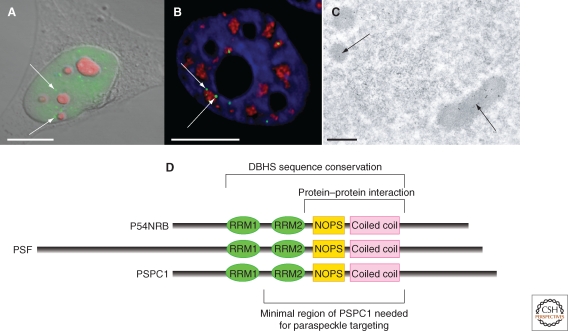
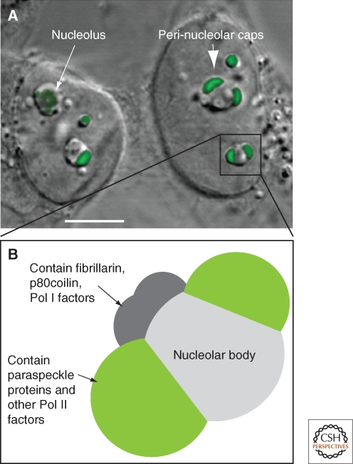
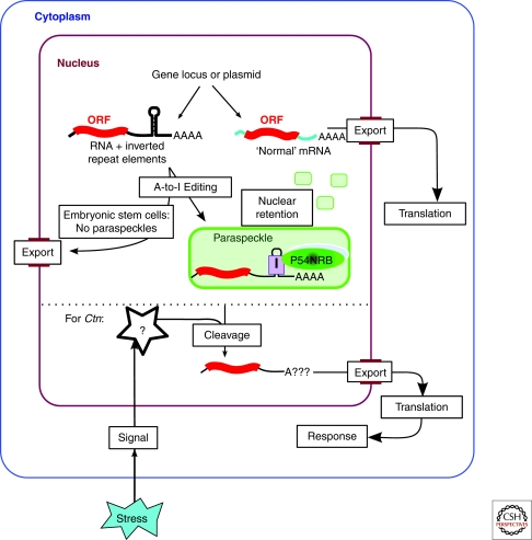
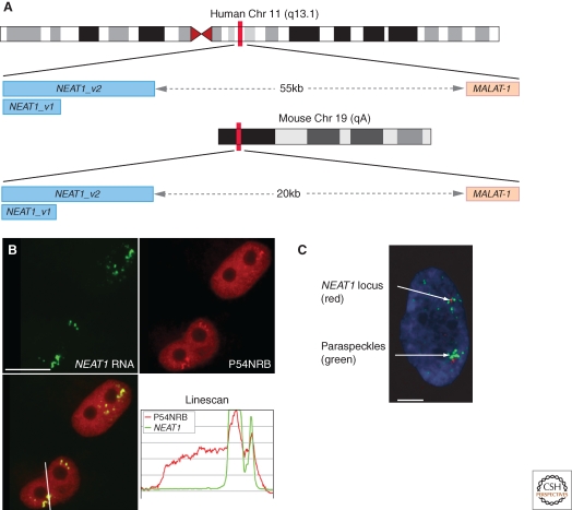
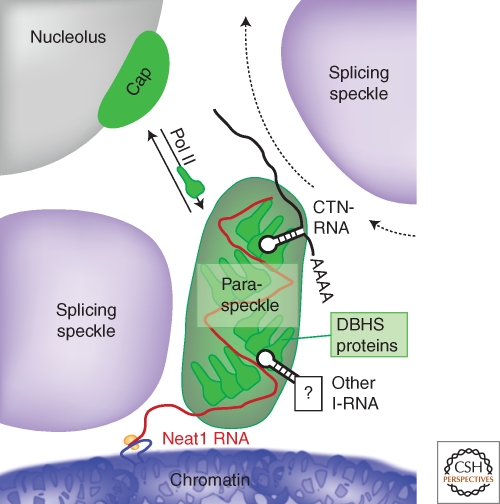
Paraspeckles are a relatively new class of subnuclear bodies found in the interchromatin space of mammalian cells. They are RNA-protein structures formed by the interaction between a long nonprotein-coding RNA species, NEAT1/Men epsilon/beta, and members of the DBHS (Drosophila Behavior Human Splicing) family of proteins: P54NRB/NONO, PSPC1, and PSF/SFPQ. Paraspeckles are critical to the control of gene expression through the nuclear retention of RNA containing double-stranded RNA regions that have been subject to adenosine-to-inosine editing. Through this mechanism paraspeckles and their components may ultimately have a role in controlling gene expression during many cellular processes including differentiation, viral infection, and stress responses.


Figures

Figure 1.
Figure 1.
Paraspeckles seen with fluorescent and electron microscopy. (A) Combined DIC and fluorescence micrograph of a HeLa cell stained with anti-PSPC1 to show paraspeckles (green) and B23 nucleolar marker (red). (B) Fluorescence micrograph of a section through a HeLa cell stained with anti-PSPC1 (green), anti-SC35 (red), and DAPI (blue) to show the relationship between paraspeckles and nuclear speckles. (C) Transmission electron micrograph of sections of HeLa cells immuno-gold labelled with anti-PSPC1: image kindly provided by Sylvie Souquere and Gerard Pierron (Villejuif, France). Scale bars in A–B, 10 µm, scale bar in C, 0.5 µm. (D) The DBHS protein family showing domain structure and indicating regions involved in paraspeckle biology.
Figure 2.
Figure 2.
Perinucleolar caps observed with RNA Pol II transcription inhibition. (A) Combined DIC and fluorescence micrograph of HeLa cells following 4 h treatment with Actinomycin D to inhibit RNA Pol II transcription. Nucleolar morphology changes under these conditions, to create a nucleolar body and a number of perinucleolar caps (arrow). Cells were transfected with a plasmid expressing YFP-PSPC1 (green), that localizes to perinucleolar caps under these conditions (large arrow); scale bar, 5 µm. (B) Perinucleolar caps form upon inhibition of RNA Pol II transcription, see text and (Shav-tal et al. 2005).
Figure 3.
Figure 3.
Gene expression by nuclear retention. RNA transcripts containing double-stranded RNA regions (formed by inverted repeat elements) are subject to A-to-I editing and retained in the nucleus and within paraspeckles. This mechanism has been shown for several endogenous genes, as well as exogenously expressed reporter genes (Prasanth et al. 2005; Chen et al. 2008; Chen and Carmichael, 2009). Nuclear retention of hyper-edited RNA is linked to the formation of paraspeckles, and does not occur efficiently in human embryonic stem cells that lack paraspeckles. In the case of Ctn, stress signals mediate cleavage of the 3′UTR and release of the RNA from the nucleus (Prasanth et al. 2005).
Figure 4.
Figure 4.
Paraspeckles contain NEAT1 ncRNA and form near to the NEAT1 gene. (A) NEAT1 and MALAT-1 gene loci on human chromosome 11 q13.1 and mouse chromosome 19qA. Two transcripts are produced from the NEAT1 gene, 3.7kb and 23kb in humans, and 3 kb and approximately 20 kb in mouse. (B) RNA-FISH with probes to NEAT1 ncRNA (green) and immunofluorescence against P54NRB (red) colocalizing in paraspeckles. The line scan is taken over the line indicated in the merged image (lower panels). (C) A HeLa cell in interphase with combined NEAT1 RNA-FISH to mark paraspeckles (green) and chromosome 11 q13.1 DNA-FISH (red). Panels B and C are reproduced from (Clemson et al. 2009) with permission. Scale bar in B, 10 µm, in panel C, 5 µm.
Figure 5.
Figure 5.
Model of paraspeckle formation. A paraspeckle forms near the NEAT1 gene locus within the interchromatin space, abutting a nuclear speckle. The paraspeckle is formed via interactions between the NEAT1 RNA and DBHS proteins. Additional RNA species regulated within paraspeckles and elsewhere in the nucleus, such as A-to-I edited mRNA, are likely recruited via interaction with DBHS proteins, and traffic through the paraspeckle. Under steady-state conditions the DBHS proteins are dynamic, and exchange between paraspeckles, the nucleoplasm and the nucleolus. When RNA Pol II transcription is inhibited, DBHS proteins accumulate at perinucleolar caps.

Similar articles

Cited by

References

    1. Andersen JS, Lyon CE, Fox AH, Leung AKL, Lam YW, Steen H, Mann M, Lamond AI 2002. Directed proteomic analysis of the human nucleolus. Current Biology 12:1–11 - PubMed
    1. Auboeuf D, Dowhan DH, Li X, Larkin K, Ko L, Berget SM, O’Malley BW 2004. CoAA, a nuclear receptor coactivator protein at the interface of transcriptional coactivation and RNA splicing. Mol Cell Biol 24:442–453 - PMC - PubMed
    1. Brooks YS, Wang G, Yang Z, Smith KK, Bieberich E, Ko L 2009. Functional pre-mRNA Trans-splicing of coactivator CoAA and corepressor RBM4 during stem/progenitor cell differentiation. J Biol Chem 284:18033–18046 - PMC - PubMed
    1. Brown SA, Ripperger J, Kadener S, Fleury-Olela F, Vilbois F, Rosbash M, Schibler U 2005. PERIOD1-associated proteins modulate the negative limb of the mammalian circadian oscillator. Science 308:693–696 - PubMed
    1. Cardinale S, Cisterna B, Bonetti P, Aringhieri C, Biggiogera M, Barabino SM 2007. Subnuclear localization and dynamics of the Pre-mRNA 3’ end processing factor mammalian cleavage factor I 68-kDa subunit. Mol Biol Cell 18:1282–1292 - PMC - PubMed

Publication types

MeSH terms

LinkOut - more resources