. 2010 Aug;16(8):1488-99.
doi: 10.1261/rna.2146910. Epub 2010 Jun 23.

A stimulatory role for the La-related protein 4B in translation

Affiliations

A stimulatory role for the La-related protein 4B in translation

Katrin Schäffler et al. RNA. 2010 Aug.

Abstract

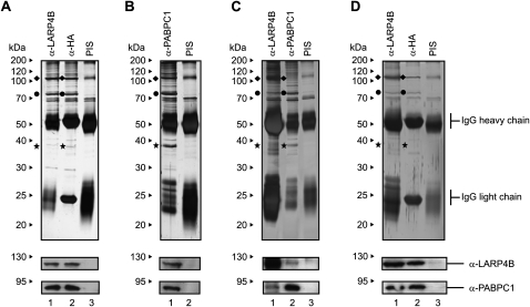
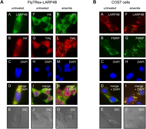
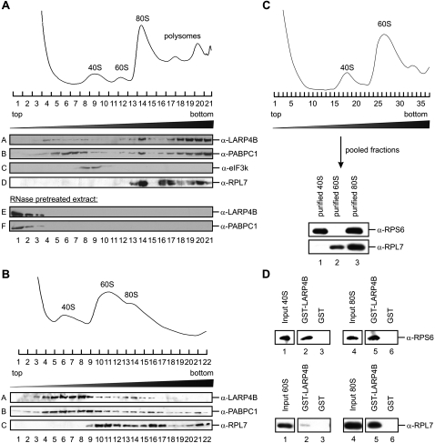
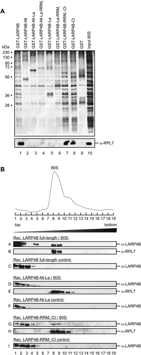
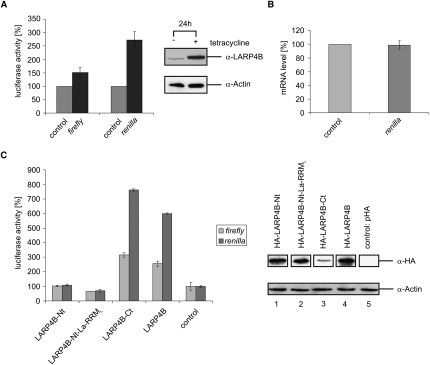
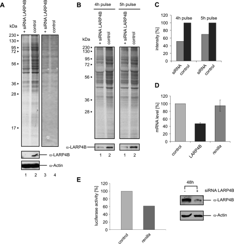
La-related proteins (LARPs) belong to an evolutionarily conserved family of factors with predicted roles in RNA metabolism. Here, we have analyzed the cellular interactions and function of LARP4B, a thus far uncharacterized member of the LARP family. We show that LARP4B is a cytosolic protein that accumulates upon arsenite treatment in cellular stress granules. Biochemical experiments further uncovered an interaction of LARP4B with the cytosolic poly(A) binding protein 1 (PABPC1) and the receptor for activated C Kinase (RACK1), a component of the 40S ribosomal subunit. Under physiological conditions, LARP4B co-sedimented with polysomes in cellular extracts, suggesting a role in translation. In agreement with this notion, overexpression of LARP4B stimulated protein synthesis, whereas knockdown of the factor by RNA interference impaired translation of a large number of cellular mRNAs. In sum, we identified LARP4B as a stimulatory factor of translation. We speculate that LARP4B exerts its function by bridging mRNA factors of the 3' end with initiating ribosomes.


Figures

FIGURE 1.
FIGURE 1.
Identification of PABPC1 and RACK1 as interaction partners of LARP4B. (A) FlpTRex-LARP4B cells were induced with tetracycline and immunoprecipitated using antibodies against LARP4B (lane 1), HA (lane 2) or a pre-immune serum (PIS) (lane 3). (B) Immunoprecipitation of FlpTRex-LARP4B cell extract using anti-PABPC1 antibody (lane 1) or a PIS (lane 2). (C) Extract from nontransfected HEK293 cells was immunoprecipitated with antibodies against LARP4B (lane 1), PABPC1 (lane 2), or a PIS (lane 3). (D) Immunoprecipitation of RNase treated FlpTRex-LARP4B cell extract using antibodies against LARP4B (lane 1), HA (lane 2), or a PIS (lane 3). The gels were analyzed by mass spectrometry or Western blotting using anti-LARP4B and anti-PABPC1 antibodies (see lower panels). (◆) LARP4B protein; (●) PABPC1; (★) RACK1.
FIGURE 2.
FIGURE 2.
Two separate segments of LARP4B mediate binding to PABPC1 and RACK1. (A) Schematic drawing of LARP4B fragments used for interaction mapping with PABPC1 and RACK1. (B) In vitro binding assay using the indicated immobilized GST-LARP4B fusions with in vitro translated [35S]-labeled HA-PABPC1. HA-PABPC1 was detected by autoradiography. (Lane 1) Five percent of labeled HA-PABPC1 input. (C) In vitro binding studies of in vitro translated [35S]-labeled His-RACK1 with indicated immobilized GST-LARP4B truncations. (Lane 1) Input, 5% of [35S]-labeled His-RACK1. (D) In vitro binding experiment of HA-LARP4B and HA-PABPC1 with immobilized GST-RACK1. (Lane 1) Five percent of [35S]-labeled HA-LARP4B; (lane 4) 5% of [35S]-labeled HA-PABPC1 input. (E) Schematic model of the interactions established in B and C.
FIGURE 3.
FIGURE 3.
LARP4B is a cytosolic protein and a component of SGs. (A) Tetracycline-induced FlpTRex-LARP4B cells were either mock-treated (panels A–J) or treated with arsenite (panels K–O). Immunofluorescence analyses of LARP4B were carried out using antibodies against HA-tag and TIAL as SG marker. (Panels A–E) Co-staining with anti-HA and anti-LARP4B antibodies. (Panels K–O) Stress-induced SG localization of TIAL and LARP4B. (Arrows) SGs. (B) Immunofluorescence studies in nontransfected COS7 cells using antibodies against LARP4B and FMRP as a SG marker protein. Cells were either mock-treated (panels A–E) or treated with arsenite (panels F–J).
FIGURE 4.
FIGURE 4.
LARP4B associates with the 80S complex and polysomes. (A) A 15%–45% sucrose gradient of cycloheximide-treated HeLa cell extract. Shown are the Western blot analyses of individual gradient fractions with antibodies against LARP4B, PABPC1, eIF3k, and RPL7 (panels A–D). (E,F) Sedimentations of LARP4B and PABPC1 in extract that has been treated with RNase A prior to centrifugation. (B) A 10%–30% sucrose gradient of EDTA-treated HeLa cell extract. The individual gradient fractions were analyzed by Western blotting using antibodies against LARP4B, PABPC1, and RPL7 (panels A–C). (Upper part) The A260 RNA profile of the gradient. (C) Purification of 40S and 60S ribosomal subunits. Ribosomal subunits were dissociated using puromycin and separated on a 10%–30% sucrose gradient. (Upper panel) A260 RNA profile. Purified ribosomal subunits were analyzed by Western blotting using anti-RPS6 and anti-RPL7 antibodies as markers for 40S and 60S subunits, respectively. (D) In vitro binding assays of purified 40S and 60S ribosomal subunits to immobilized GST-LARP4B. (Lanes 3,6) GST was used as a negative control. (Lanes 2,5) Bound ribosomal complexes were detected by Western blot analyses with antibodies against RPS6 and RPL7, respectively.
FIGURE 5.
FIGURE 5.
The association of LARP4B with 80S ribosomal complexes is mediated via its C terminus. (A) In vitro interaction of immobilized GST-LARP4B truncations with purified 80S complexes. Interactions were analyzed by silver staining (upper part) and Western blotting using anti-RPL7 antibody (lower part). (B) Determination of the ribosome binding site of LARP4B. Mixtures of the indicated LARP4B truncations and purified ribosomes were separated by a 5%–45% sucrose gradient. Shown are the A260 RNA profile (upper part) and Western blot analyses of individual gradient fractions with antibodies against LARP4B (A,D,G) and RPL7 (B,E,H). (C,F,I) As a control, migration of the LARP4B truncations in the absence of 80S complexes is shown.
FIGURE 6.
FIGURE 6.
Overexpression of LARP4B enhances translation in vivo. (A) Luciferase activity in tetracycline-induced FlpTRex-LARP4B cells (black bars) and noninduced control cells (gray bars) after 24 h of cell growth. Shown are the activity of firefly luciferase expressed from transfected pGL3 vector and of Renilla luciferase expressed from co-transfected pRL-TK vector. Tetracycline-induced overexpression of HA-LARP4B was analyzed by Western blotting using anti-LARP4B and anti-Actin antibodies. Presented data were normalized to global protein level by Bradford measurement. (B) Renilla luciferase mRNA level in tetracycline-induced FlpTRex-LARP4B cells measured by qRT-PCR (dark gray bar). (C) Influence on luciferase activity of the indicated pHA/LARP4B constructs transfected in HEK293 cells. Shown are the firefly (pGL3; gray bars) and Renilla luciferase activities (pRL-TK; dark gray bars) 48 h post-transfection. As a control, empty pHA plasmid was transfected (see two bars on the right). Transfection efficiency was determined by Western blot analysis using anti-HA and anti-Actin antibodies. Extracts were normalized by Bradford measurement.
FIGURE 7.
FIGURE 7.
Knockdown of LARP4B decreases translation levels in vivo. (A) HEK293 cells were transfected with LARP4B siRNA and incubated for 48 h at 37°C. After addition of medium containing [35S]-methionine and subsequent incubation for 5 h, cells were harvested and the extracts separated by SDS-PAGE. The gel was analyzed by autoradiography (lanes 1,2) and Coomassie staining (lanes 3,4). Western blots using anti-LARP4B and anti-Actin antibodies were performed to analyze the efficiency of RNAi-induced LARP4B knockdown. (B) Comparison of the global translation level for different time points of [35S]-methionine labeling. (C) Translation level after LARP4B knockdown and 4 h or 5 h of labeling with [35S]-methionine. (D) QRT-PCR analysis of mRNA levels of LARP4B (black bar) and Renilla luciferase (dark gray bar) in HEK293 cells treated with siRNA against LARP4B. (E) Influence of LARP4B siRNA transfection on Renilla luciferase activity. Shown is the activity of Renilla luciferase (pRL-TK; dark gray bar) 48 h post-transfection.

Similar articles

Cited by

References

    1. Aigner S, Cech TR 2004. The Euplotes telomerase subunit p43 stimulates enzymatic activity and processivity in vitro. RNA 10: 1108–1118 - PMC - PubMed
    1. Aigner S, Postberg J, Lipps HJ, Cech TR 2003. The Euplotes La motif protein p43 has properties of a telomerase-specific subunit. Biochemistry 42: 5736–5747 - PubMed
    1. Alfano C, Sanfelice D, Babon J, Kelly G, Jacks A, Curry S, Conte MR 2004. Structural analysis of cooperative RNA binding by the La motif and central RRM domain of human La protein. Nat Struct Mol Biol 11: 323–329 - PubMed
    1. Anderson P, Kedersha N 2008. Stress granules: The Tao of RNA triage. Trends Biochem Sci 33: 141–150 - PubMed
    1. Angenstein F, Evans AM, Settlage RE, Moran ST, Ling SC, Klintsova AY, Shabanowitz J, Hunt DF, Greenough WT 2002. A receptor for activated C kinase is part of messenger ribonucleoprotein complexes associated with poly(A)-mRNAs in neurons. J Neurosci 22: 8827–8837 - PMC - PubMed

Publication types

MeSH terms