. 2010 Sep;84(17):8871-87.
doi: 10.1128/JVI.00725-10. Epub 2010 Jun 23.

Identification of rep-associated factors in herpes simplex virus type 1-induced adeno-associated virus type 2 replication compartments

Affiliations

Identification of rep-associated factors in herpes simplex virus type 1-induced adeno-associated virus type 2 replication compartments

Armel Nicolas et al. J Virol. 2010 Sep.

Abstract

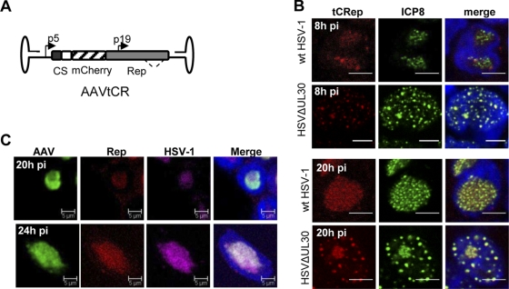
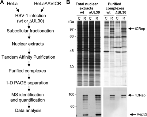
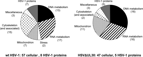
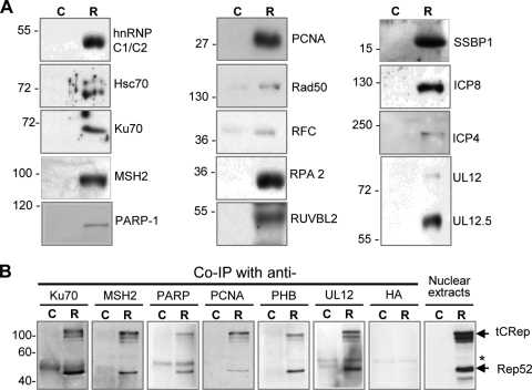
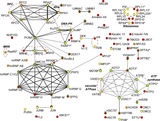
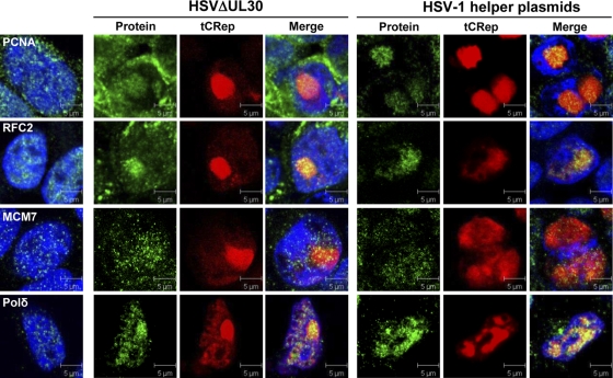
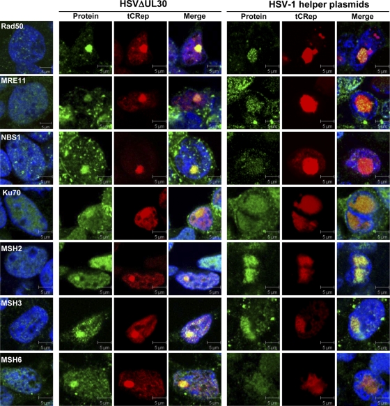
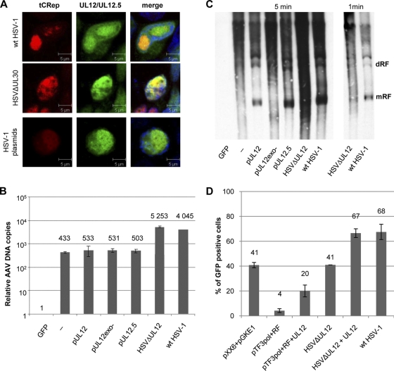
Adeno-associated virus (AAV) is a human parvovirus that replicates only in cells coinfected with a helper virus, such as adenovirus or herpes simplex virus type 1 (HSV-1). We previously showed that nine HSV-1 factors are able to support AAV rep gene expression and genome replication. To elucidate the strategy of AAV replication in the presence of HSV-1, we undertook a proteomic analysis of cellular and HSV-1 factors associated with Rep proteins and thus potentially recruited within AAV replication compartments (AAV RCs). This study resulted in the identification of approximately 60 cellular proteins, among which factors involved in DNA and RNA metabolism represented the largest functional categories. Validation analyses indicated that the cellular DNA replication enzymes RPA, RFC, and PCNA were recruited within HSV-1-induced AAV RCs. Polymerase delta was not identified but subsequently was shown to colocalize with Rep within AAV RCs even in the presence of the HSV-1 polymerase complex. In addition, we found that AAV replication is associated with the recruitment of components of the Mre11/Rad50/Nbs1 complex, Ku70 and -86, and the mismatch repair proteins MSH2, -3, and -6. Finally, several HSV-1 factors were also found to be associated with Rep, including UL12. We demonstrated for the first time that this protein plays a role during AAV replication by enhancing the resolution of AAV replicative forms and AAV particle production. Altogether, these analyses provide the basis to understand how AAV adapts its replication strategy to the nuclear environment induced by the helper virus.


Figures

FIG. 1.
FIG. 1.
(A) Schematic view of the AAVtCR genome. p5 and p19 are the wt AAV-2 promoters, and C and S refer to the sequences coding for the calmodulin and streptavidin binding peptide tags. The dotted line indicates the position of alternative splicing in the rep gene. (B) Immunofluorescence analysis of HSV-1-infected HeLaAAVtCR cells to examine colocalization of tCRep and HSV-1 DBP ICP8 proteins. Infected cells were fixed at 8 and 20 h p.i., stained with an anti-ICP8 antibody, and analyzed by confocal microscopy. Bars, 5 μm. (C) Immuno-FISH analysis of HSV-1-infected HeLaAAVtCR cells. HeLaAAVtCR cells were infected for 20 or 24 h with wt HSV-1 (5 PFU/cell) and then analyzed using a biotin-labeled HSV-1 probe and a DIG-labeled AAV probe. After hybridization and washes, cells were additionally stained with an anti-Rep antibody and DAPI (4′,6-diamidino-2-phenylindole) and were analyzed by confocal microscopy. Bars, 5 μm.
FIG. 2.
FIG. 2.
(A) Outline of the experimental procedure used to purify nuclear complexes associated with AAV Rep proteins. See Materials and Methods for details of the TAP steps with streptavidin and calmodulin resins. (B) Sequential analysis of the purified extracts. Proteins present either in total nuclear extracts or in the Rep-containing complexes after elution from the second calmodulin resin were resolved by SDS-PAGE, followed by silver staining (upper panels) or Western blotting using an anti-Rep antibody (lower panels). C, control infected HeLa cells; R, HeLaAAVtCR cells expressing Rep upon HSV-1 infection; wt, wt HSV-1; ΔUL30, HSVΔUL30.
FIG. 3.
FIG. 3.
Functional classification of proteins copurified with Rep from HeLaAAVtCR cells infected with wt HSV-1 or HSVΔUL30 and identified by mass spectrometry analysis. See Table 1 for definitions of the functional categories.
FIG. 4.
FIG. 4.
Western blot analyses of purified complexes. (A) Protein complexes were purified from HeLa (C) or HeLaAAVtCR (R) cells infected with wt HSV-1 and then analyzed by Western blotting, using antibodies against a panel of cellular and viral proteins. (B) Reverse coimmunoprecipitation of tCRep in purified complexes containing selected cellular and viral proteins. Control HeLa (C) cells and HeLaAAVtCR (R) cells were infected with wt HSV-1 for 20 h (MOI = 5 PFU/cell). Nuclear extracts from 2 × 10⁶ infected cells were subjected to coimmunoprecipitation, using UL12, Ku70, MSH2, PARP-1, PCNA, and PHB polyclonal antibodies as indicated. The anti-HA antibody was used as a negative control. The immunoprecipitates were then immunoblotted with an anti-Rep antibody (303.9). The arrows indicate the positions of the tCRep and Rep52 proteins, and the asterisk shows a nonspecific band.
FIG. 5.
FIG. 5.
In silico reconstitution of Rep-associated interaction networks. Likely protein-protein interactions were reconstituted using the cellular protein list from Table 1 and analysis tools provided by the VirHostNet interactome website (http://pbildb1.univ-lyon1.fr/virhostnet/login.php), complemented with additional information derived from the UniProtKB/SwissProt database and specialized literature. The color codes refer to proteins specifically identified from HeLaAAVtCR cells infected with wt HSV-1 (red), HSVΔUL30 (yellow), or both helper viruses (orange). Green circles, proteins for which a physical or functional interaction with a protein from another virus has already been reported; black lines, VirHostNet-derived interactions, with the link width correlating with the number of different reports describing the interaction found in the database (black circles correspond to self-interacting proteins); dashed brown arrows, UniProtKB/SwissProt-derived interactions; asterisks, proteins identified in Rep-containing complexes purified from adenovirus- and AAV-coinfected cells (42); dashed brown circles, proteins constituting a known functional complex.
FIG. 6.
FIG. 6.
Colocalization of cellular DNA replication factors with Rep in HSV-1-induced AAV RCs. HeLaAAVtCR cells were either infected with HSVΔUL30 (MOI = 5 PFU/cell) and fixed at 9 or 14 h p.i. or transfected with 0.25 pmol/35-mm well of each HSV-1 helper plasmid (pTF3pol and pRF) and fixed at 48 h posttransfection. Alexa Fluor 488-conjugated donkey anti-mouse, anti-rabbit, and anti-goat secondary antibodies were used. DNA was stained with TO-PRO-3 iodide (Invitrogen). Images were taken using an Axioplan2 LSM510 confocal microscope (Zeiss). Green, protein of interest; red, tCRep; blue, TO-PRO-3 staining. The left panels correspond to results for noninfected and nontransfected control HeLaAAVtCR cells. Bars, 5 μm. The cytoplasmic background visible for some antibodies in HSV-infected cells is due to antibody binding to the HSV-encoded Fc receptor (29, 83).
FIG. 7.
FIG. 7.
Colocalization of cellular DNA repair factors with Rep in AAV RCs. Cells were processed as described in the legend to Fig. 6 and then stained with antibodies recognizing the indicated cellular DNA repair factors. Green, protein of interest; red, tCRep; blue, TO-PRO-3 staining. The left panels indicate the staining observed in noninfected and nontransfected HeLaAAVtCR cells. Bars, 5 μm.
FIG. 8.
FIG. 8.
Involvement of HSV-1 UL12 protein in AAV replication. (A) Colocalization of UL12 with Rep within AAV RCs. HeLaAAVtCR cells were either infected with the indicated HSV-1 strain or transfected with the HSV-1 helper plasmids and pSAKUL12 plasmid and then stained with an anti-UL12 antibody and TO-PRO-3. Bars, 5 μm. (B) Analysis of the effect of UL12 on AAV DNA replication. Total DNA was extracted from HeLaAAVtCR cells either infected with wt HSV-1 or HSVΔUL12 or transfected with HSV-1 helper plasmids and a plasmid encoding either UL12, UL12.5, or an exonuclease-negative UL12 mutant (pUL12exo−) and then quantified by qPCR, using rep-specific primers. Data presented are means with error bars calculated for three independent experiments. (C) The same samples used in panel B were analyzed by Southern blotting, using a DIG-labeled rep probe. mRF, monomer replicative form; dRF, dimer replicative form. (D) Effect of UL12 on rAAV-GFP particle production. HeLa cells transfected with rAAV-GFP and rep-cap-expressing plasmids were either cotransfected with HSV-1 helper plasmids or infected with HSVΔUL12 in the absence or presence of a plasmid encoding UL12. As a control, the cells were cotransfected with adenoviral helper plasmids (pXX6 and pGKE1) or infected with wt HSV-1. Cell lysates were then used to infect HeLa cells in the presence of wt adenovirus, and GFP-positive cells were counted by flow cytometry at 24 h p.i. Mean values with error bars, calculated for at least three independent experiments, are presented.

Similar articles

Cited by

References

    1. Alazard-Dany, N., A. Nicolas, A. Ploquin, R. Strasser, A. Greco, A. L. Epstein, C. Fraefel, and A. Salvetti. 2009. Definition of herpes simplex virus type 1 helper activities for adeno-associated virus early replication events. PLoS Pathog. 5:e1000340. - PMC - PubMed
    1. Araujo, F. D., T. H. Stracker, C. T. Carson, D. V. Lee, and M. D. Weitzman. 2005. Adenovirus type 5 E4orf3 protein targets the Mre11 complex to cytoplasmic aggresomes. J. Virol. 79:11382-11391. - PMC - PubMed
    1. Baker, A., K. J. Rohleder, L. A. Hanakahi, and G. Ketner. 2007. Adenovirus E4 34k and E1b 55k oncoproteins target host DNA ligase IV for proteasomal degradation. J. Virol. 81:7034-7040. - PMC - PubMed
    1. Bevington, J. M., P. G. Needham, K. C. Verrill, R. F. Collaco, V. Basrur, and J. P. Trempe. 2007. Adeno-associated virus interactions with B23/nucleophosmin: identification of sub-nucleolar virion regions. Virology 357:102-113. - PMC - PubMed
    1. Blumel, J., S. Graper, and B. Matz. 2000. Structure of simian virus 40 DNA replicated by herpes simplex virus type 1. Virology 276:445-454. - PubMed

Publication types

MeSH terms