. 2010 Sep;84(17):8913-25.
doi: 10.1128/JVI.00169-10. Epub 2010 Jun 23.

Activation of the interferon response by human cytomegalovirus occurs via cytoplasmic double-stranded DNA but not glycoprotein B

Affiliations

Activation of the interferon response by human cytomegalovirus occurs via cytoplasmic double-stranded DNA but not glycoprotein B

Victor R DeFilippis et al. J Virol. 2010 Sep.

Abstract

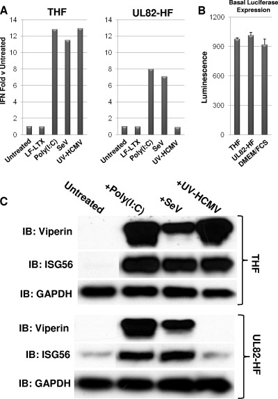
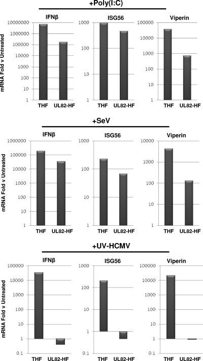
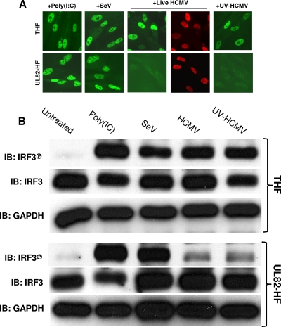
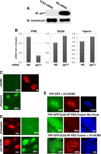
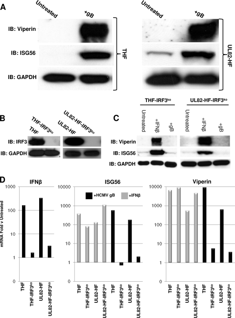
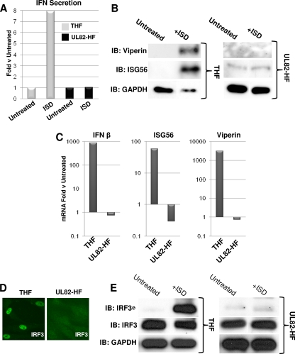
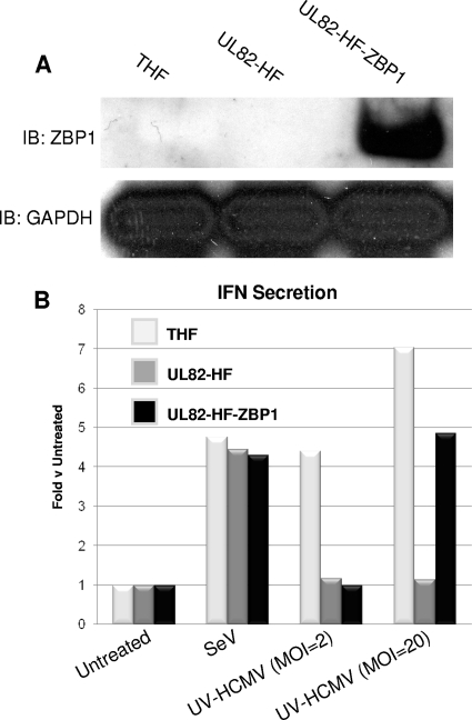
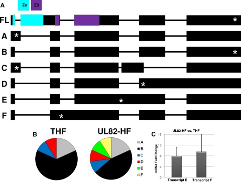
In vitro infection of cells with the betaherpesvirus human cytomegalovirus (HCMV) stimulates an innate immune response characterized by phosphorylation of the transcription factor interferon regulatory factor 3 (IRF3) and subsequent expression of IRF3-dependent genes. While previous work suggests that HCMV envelope glycoprotein B is responsible for initiating this reaction, the signaling pathways stimulated by virus infection that lead to IRF3 phosphorylation have largely been uncharacterized. Recently, we identified Z DNA binding protein 1 (ZBP1), a sensor of cytoplasmic DNA, as an essential protein for this response. We now describe a human fibroblast cell line exhibiting a recessive defect that results in the absence of activation of IRF3 following treatment with HCMV but not Sendai virus or double-stranded RNA. In addition, we show that while exposure of these cells to soluble HCMV glycoprotein B is capable of triggering IRF3-dependent gene transcription, transfection of the cells with double-stranded DNA is not. Furthermore, we show that overexpression of ZBP1 in these cells reestablishes their ability to secrete interferon in response to HCMV and that multiple ZBP1 transcriptional variants exist in both wild-type and mutant cells. These results have two major implications for the understanding of innate immune stimulation by HCMV. First, they demonstrate that HCMV glycoprotein B is not the essential molecular pattern that induces an IRF3-dependent innate immune response. Second, IRF3-terminal signaling triggered by HCMV particles closely resembles that which is activated by cytoplasmic double-stranded DNA.


Figures

FIG. 1.
FIG. 1.
Synthesis of IFN-β, ISG56, and viperin in THF and UL82-HF cells following exposure to poly(I:C), SeV, and UV-HCMV. (A) Expression of type I IFN-dependent luciferase from THF-ISRE cells exposed to medium collected at 24 h posttreatment from either THF or UL82-HF cells treated with LF-LTX, transfected poly(I:C), SeV, or UV-HCMV as described in the text. Values presented are normalized to untreated cells (set to 1) and are representative of duplicate experiments. (B) Samples (50 μl) of media from THF or UL82-HF cells grown in 9 wells of a 24-well dish were transferred to 4 wells each of THF-ISRE reporter cells grown to confluence in 96-well plates. IFN-dependent luciferase expression was measured as described in the text. Luciferase was also measured in THF-ISRE cells exposed only to fresh DMEM-FCS. (C) Immunoblots showing synthesis of ISG56, viperin, and GAPDH in untreated THF and UL82-HF cells or in THF and UL82-HF cells following 6 h of exposure to transfected poly(I:C), SeV, or UV-HCMV.
FIG. 2.
FIG. 2.
IFN-β, ISG56, and viperin mRNA accumulation in THF and UL82-HF cells following exposure to poly(I:C), SeV, and UV-HCMV. Values presented are fold changes of mRNA levels in treated versus untreated cells at 6 h posttreatment, as determined by semiquantitative RT-PCR as described in the text. Data are representative of duplicate experiments.
FIG. 3.
FIG. 3.
IRF3 activation in THF and UL82-HF cells following exposure to poly(I:C), SeV, HCMV, and UV-HCMV. (A) IFA showing subcellular localization of IRF3 (green) and HCMV UL123-encoded IE protein (red) in THF and UL82-HF cells following 6 h of treatment with transfected poly(I:C), SeV, live HCMV, or UV-HCMV as described in the text. (B) Immunoblots showing the presence of total IRF3 and IRF3 phosphorylated on Ser398, as well as GAPDH, in untreated THF and UL82-HF cells or in THF and UL82-HF cells following 6 h of exposure to poly(I:C), SeV, live HCMV, or UV-HCMV.
FIG. 4.
FIG. 4.
HCMV-triggered IRF3-dependent activity in UL82-HF cells following knockdown of UL82 and in THF and UL82-HF cell fusions. (A) Immunoblots showing siRNA-mediated knockdown of stably expressed pp71 in UL82-HF cells. (B) Accumulation of IFN-β, ISG56, and viperin mRNAs in UV-HCMV-exposed UL82-HF cells relative to those in unexposed cells following transfection with UL82-directed or nonspecific (NS) siRNA, as determined by semiquantitative qPCR. (C) Indirect IFA showing subcellular localization of IRF3 (green) and HCMV IE protein (red) in UL82-HF cells exposed to HCMV or poly(I:C) following transfection with UL82-directed siRNA. (D) Indirect IFA showing subcellular localization of IRF3 (green) and HA (red) in THF cells that were reconstructed to stably express HA-tagged pp71. Cells were left untreated or exposed to UV-HCMV. (E) Indirect IFA showing stably expressed GFP (green), subcellular localization of IRF3 (red), and DAPI staining (blue) or pp71-HA expression (blue) in THF-GFP and UL82-HF cells in the presence or absence of UV-HCMV and PEG, as described in the text.
FIG. 5.
FIG. 5.
IRF3-dependent mRNA accumulation in THF and UL82-HF cells following treatment with HCMV gB. (A) Immunoblots showing synthesis of viperin and ISG56 following exposure of THF and UL82-HF cells to 20 μg ml−1 HCMV gB. (B) Immunoblots showing NPro-mediated degradation of IRF3 from stably transfected THF and UL82-HF cells. (C) Immunoblots showing synthesis of viperin and ISG56 in THF-IRF3ko and UL82-HF-IRF3ko cells either left untreated or exposed to IFN-β or HCMV gB. (D) Accumulation of IFN-β, ISG56, and viperin mRNAs following exposure of THF, THF-IRF3ko, UL82-HF, and UL82-HF-IRF3ko cells to 20 μg ml−1 HCMV gB (IFN-β, ISG56, and viperin) or IFN-β (ISG56 and viperin). Values presented are fold changes relative to levels in untreated cells.
FIG. 6.
FIG. 6.
ISD does not activate IRF3-dependent gene expression in UL82-HF cells. (A) Expression of type I IFN-dependent luciferase from THF-ISRE cells exposed to medium collected at 24 h posttreatment from THF or UL82-HF cells either transfected with ISD or left untreated. Values presented are normalized to untreated cells (set to 1). (B) Immunoblots showing synthesis of viperin and ISG56 following 6 h of transfection of 5 μg ml−1 ISD into THF and UL82-HF cells. (C) Accumulation of IFN-β, ISG56, and viperin mRNAs following transfection of THF and UL82-HF cells with 5 μg ml−1 ISD. Values presented are fold changes relative to the levels in untreated cells. (D) IFA showing subcellular localization of IRF3 in THF and UL82-HF cells transfected with 5 μg ml−1 ISD for 6 h. (E) Immunoblots showing IRF3 Ser398 phosphorylation status in THF and UL82-HF cells transfected with 5 μg ml−1 ISD for 6 h.
FIG. 7.
FIG. 7.
Stable overexpression of ZBP1 reestablishes the ability of UL82-HF cells to secrete type I IFN in response to HCMV. (A) Immunoblots showing expression of ZBP1 in THF, UL82-HF, and UL82-HF-ZBP1 cells. (B) Expression of type I IFN-dependent luciferase from THF-ISRE cells exposed to medium collected at 24 h posttreatment from THF, UL82-HF, or UL82-HF-ZBP1 cells left untreated, exposed to SeV, or exposed to UV-HCMV at an MOI of 2 PFU/cell or 20 PFU/cell. Values presented are normalized to those for untreated cells (set to 1).
FIG. 8.
FIG. 8.
ZBP1 transcript variants identified in THF and UL82-HF cells. (A) Sequence maps and labels (A to F and FL [full length]) for ZBP1 mRNA transcript variants identified in THF and UL82-HF cells. Approximate locations of the Zα and Zβ DNA binding domains are color coded as indicated. Asterisks indicate approximate locations of initial predicted stop codons. (B) Cell type-specific distribution of transcript variants found in THF and UL82-HF cells. (C) Accumulation of ZBP1 transcripts E and F in UL82-HF cells versus THF cells, expressed as mean fold changes ± SEM, as determined by qPCR.

Similar articles

Cited by

References

    1. Ablasser, A., F. Bauernfeind, G. Hartmann, E. Latz, K. A. Fitzgerald, and V. Hornung. 2009. RIG-I-dependent sensing of poly(dA:dT) through the induction of an RNA polymerase III-transcribed RNA intermediate. Nat. Immunol. 10:1065-1072. - PMC - PubMed
    1. Alford, C. A., S. Stagno, R. F. Pass, and W. J. Britt. 1990. Congenital and perinatal cutomegalovirus infection. Rev. Infect. Dis. 12:745-753. - PubMed
    1. Ankel, H., D. F. Westra, S. Welling-Wester, and P. Lebon. 1998. Induction of interferon-alpha by glycoprotein D of herpes simplex virus: a possible role of chemokine receptors. Virology 251:317-326. - PubMed
    1. Boehme, K. W., J. Singh, S. T. Perry, and T. Compton. 2004. Human cytomegalovirus elicits a coordinated cellular antiviral response via envelope glycoprotein B. J. Virol. 78:1202-1211. - PMC - PubMed
    1. Boudinot, P., S. Riffault, S. Salhi, C. Carrat, C. Sedlik, N. Mahmoudi, B. Charley, and A. Benmansour. 2000. Vesicular stomatitis virus and pseudorabies virus induce a vig1/cig5 homologue in mouse dendritic cells via different pathways. J. Gen. Virol. 81:2675-2682. - PubMed

Publication types

MeSH terms