. 2010 Sep;84(17):8811-20.
doi: 10.1128/JVI.00496-10. Epub 2010 Jun 23.

Delaying the expression of herpes simplex virus type 1 glycoprotein B (gB) to a true late gene alters neurovirulence and inhibits the gB-CD8+ T-cell response in the trigeminal ganglion

Affiliations

Delaying the expression of herpes simplex virus type 1 glycoprotein B (gB) to a true late gene alters neurovirulence and inhibits the gB-CD8+ T-cell response in the trigeminal ganglion

Srividya Ramachandran et al. J Virol. 2010 Sep.

Abstract

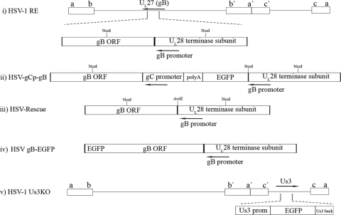
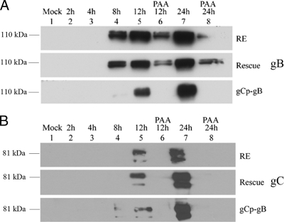
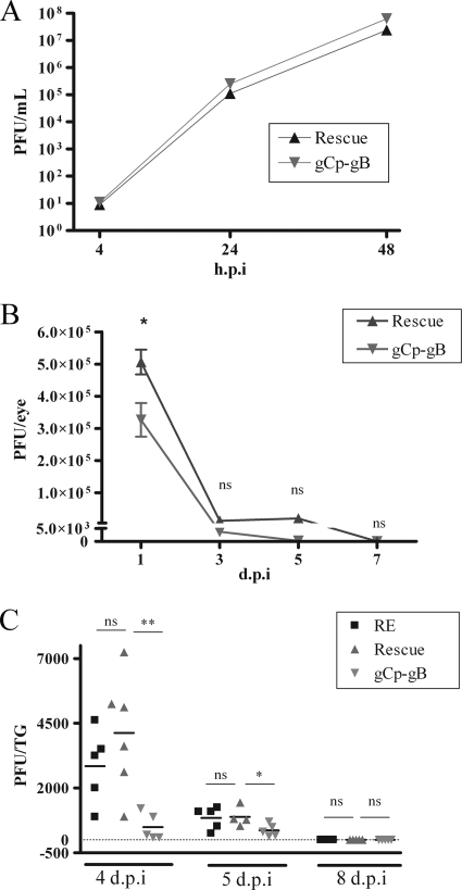
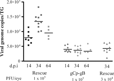
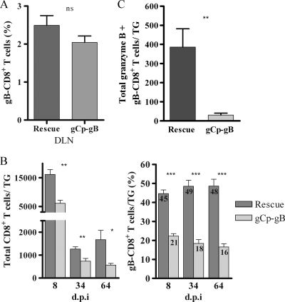
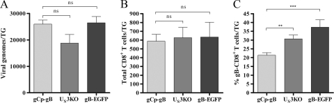
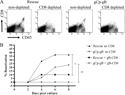
Following herpes simplex virus type 1 (HSV-1) ocular infection of C57BL/6 mice, activated CD8(+) T cells specific for an immunodominant epitope on HSV-1 glycoprotein B (gB-CD8 cells) establish a stable memory population in HSV-1 latently infected trigeminal ganglia (TG), whereas non-HSV-specific CD8(+) T cells are lost over time. The retention and activation of gB-CD8 cells appear to be influenced by persistent viral antigenic exposure within the latently infected TG. We hypothesized that the low-level expression of gB from its native promoter before viral DNA synthesis is critical for the retention and activation of gB-CD8 cells in the TG during HSV-1 latency and for their ability to block HSV-1 reactivation from latency. To test this, we created a recombinant HSV-1 in which gB is expressed only after viral DNA synthesis from the true late gC promoter (gCp-gB). Despite minor growth differences compared to its rescuant in infected corneas, gCp-gB was significantly growth impaired in the TG and produced a reduced latent genome load. The gCp-gB- and rescuant-infected mice mounted similar gB-CD8 effector responses, but the size and activation phenotypes of the memory gB-CD8 cells were diminished in gCp-gB latently infected TG, suggesting that the stimulation of gB-CD8 cells requires gB expression before viral DNA synthesis. Surprisingly, late gB expression did not compromise the capacity of gB-CD8 cells to inhibit HSV-1 reactivation from latency in ex vivo TG cultures, suggesting that gB-CD8 cells can block HSV-1 reactivation at a very late stage in the viral life cycle. These data have implications for designing better immunogens for vaccines to prevent HSV-1 reactivation.


Figures

FIG. 1.
FIG. 1.
Construction of HSV gCp-gB, HSV-rescue, and HSV-1 US3KO viruses. (i) Representation of the HSV-1 genome showing the UL27/UL28 locus, which contains the gB promoter (arrow) in the UL28 coding sequences upstream of the gB ORF. The restriction sites used to identify the insertion, as detailed in the text, are indicated on the gene locus at their approximate positions. (ii) Expansion of the corresponding region in the recombinant HSV-1 gCp-gB virus showing that in place of its native promoter, gB is driven by the well-characterized γ2 gC promoter in gCp-gB so that the protein is expressed only following DNA replication. The strategy required the maintenance of the gB promoter in the genome, because it is concurrent with the upstream UL28 ORF encoding an essential terminase subunit. As such, HSV-1 gCp-gB had the gB promoter driving EGFP followed by a polyadenylation motif to terminate RNA made from the gB promoter. (iii) Representative structure of the HSV-rescue virus showing that gB expression is restored to its native promoter but that a unique noncoding AvrII site distinguishes it from the parental strain. (iv) Representative structure of the HSV gB-EGFP virus showing that the gB protein is expressed as a fusion with EGFP at its C terminus. (v) Representation of the genome showing the position of the US3 locus, which was modified such that the US3 promoter drives the expression of EGFP, followed by an untranslated portion (amino acids 170 to 481) of the US3 ORF. If spurious transcription/translation allowed the expression of the remaining part of the US3 reading frame, that part would not be kinase functional, since it lacks the critical ATP binding domain and the first part of the catalytic domains.
FIG. 2.
FIG. 2.
HSV gCp-gB expresses gB with true late kinetics. Confluent monolayers of Vero cells were infected with HSV-1 RE, gCp-gB, or rescuant at an MOI of 10 with or without the addition of 350 μg/ml of phosphonoacetic acid (PAA). Total SDS-PAGE-separated proteins were analyzed by immunoblotting for gB (A) or gC (B) using pools of monoclonal antibodies. The times of harvest are shown above each figure and lane designation in hours. The top of each figure represents infection with HSV-1 RE, the middle represents infection with HSV-rescue virus, and the bottom represents infection with HSV gCp-gB. Only the regions corresponding to the main glycoprotein products are shown.
FIG. 3.
FIG. 3.
Viral replication titers are reduced in corneal tear films and TG of gCp-gB-infected mice but not in culture. (A) Vero cell monolayers were infected at an MOI of 0.01 with gCp-gB or rescue viruses. Cells and supernatants were collected at the designated hours p.i. and subjected to three freeze-thaw cycles, and PFU/ml of HSV-1 were measured on Vero cells. The difference between the viral titers of gCp-gB and rescue was not significantly different at any time tested (P > 0.01). The experiment was repeated two independent times, with similar results. (B) Mice were infected with rescue or gCp-gB virus at 1 × 10⁵ PFU/eye. Eye swabs were performed at the indicated days p.i., and the titers of HSV-1 were determined on Vero cells. The viral titers are shown as means ± standard errors of the means (SEM). An asterisk indicates that titers were significantly different as assessed by a Student's t test (P < 0.05). (C) Mice were infected with RE, rescue, or gCp-gB virus at 1 × 10⁵ PFU/eye. Infected TG were excised, homogenized, and subjected to three freeze-thaw cycles, and HSV-1 titers were determined on Vero cells. Each data point represents the mean viral titer from a single TG as determined by plaque assay. The data shown are combined data from two independent experiments. The significance of differences in TG titers was assessed by a Student's t test (**, P = 0.0008; *, P = 0.0573). At 8 days p.i., no infectious virus could be detected from TG infected with either virus. ns, not significant.
FIG. 4.
FIG. 4.
HSV gCp-gB establishes latency with fewer genome copies than the rescue virus. Corneas of mice were infected at an infectious dose of 1 × 10⁵ or 3 × 10⁴ PFU/eye. TG were excised at 14, 34, and 64 days p.i., and genome copy numbers were determined by real-time PCR. Each data point represents the viral genome copy numbers from a single TG. The data shown are combined data from two independent experiments. At a similar infectious dose (1 × 10⁵ PFU), the rescue virus induced a significantly higher (P < 0.05) latent viral load than the gCp-gB virus at all times tested. Reducing the infectious dose of rescue virus 3-fold relative to that of gCp-gB virus resulted in latent viral loads that were not significantly different (P > 0.05). Data were analyzed by a Student's t test.
FIG. 5.
FIG. 5.
HSV gCp-gB-infected mice contain fewer gB498-505-specific CD8+ T cells in their TG but not lymph nodes, and fewer gB498-505-specific CD8+ T cells in the TG of gCp-gB-infected mice are activated. TG or lymph nodes were excised; dispersed into single-cell suspensions; stained for CD45, CD8, gB498-505 T-cell receptor or intracellular granzyme B expression; and analyzed by flow cytometry. The data are represented as the means ± SEM. (A) Graph representing the percentage of gB-CD8+ T cells in draining lymph nodes (DLN) of mice infected with 1 × 10⁵ PFU/eye of either rescue or gCp-gB virus at 8 days p.i. The data shown are combined data from two independent experiments. The mean percentages of gB-CD8+ T cells are not significantly different between rescue and gCp-gB viruses as assessed by a Student's t test (P > 0.05). (B) Total number of CD8+ T cells (left) and percentage of gB-CD8+ T cells (right) per TG from mice infected with 1 × 10⁵ PFU/eye HSV. The mean for the percentage of gB-CD8+ T cells is shown within each bar graph. The data are combined data from three independent experiments. The mean percentages of total and gB498-505-specific CD8+ T cells between rescue and gCp-gB viruses are significantly different at all time points tested as assessed by a Student's t test (P < 0.05). (C) Graph representing the absolute number of gB-CD8+ T cells in infected TG expressing intracellular granzyme B at 34 days p.i. from mice infected with 3 × 10⁴ PFU/eye rescue virus and 1 × 10⁵ PFU/eye gCp-gB virus to yield equal genome loads. The data are combined data from two independent experiments. The differences between the absolute numbers are significantly different as assessed by a Student's t test (P = 0.0016).
FIG. 6.
FIG. 6.
Delaying gB contributes to the diminished gB-CD8+ T-cell response in the TG. Mice were infected at 1 × 10⁵ PFU/eye with gCp-gB or two other TG replication-impaired HSV-1 strains, one lacking the US3 kinase (US3KO) and the other expressing gB as a fusion protein with EGFP (gB-EGFP). The data shown are combined data from two independent experiments. (A) Viral genome copy numbers were determined by real-time PCR at 34 days p.i. The data are represented as the means ± SEM. The differences between gCp-gB and US3KO genome copy numbers and gCp-gB and gB-EGFP genome copy numbers are not significantly different as assessed by a Student's t test (P > 0.05). (B and C) Single-cell suspensions of TG infected for 34 days were stained for CD45, CD8, and gB498-505 TCR expression and analyzed by flow cytometry. The data are presented as the means ± SEM. (B) The total number of CD8+ T cells retained in TG of mice infected with gCp-gB or US3KO and gCp-gB or gB-EGFP is not significantly different as assessed by a Student's t test (P > 0/05). (C) The percentage of gB498-505-specific CD8+ T cells is significantly different between gCp-gB and US3KO (P = 0.0013) and gCp-gB and gB-EGFP (P = 0.0003) viruses at all time points tested as assessed by a Student's t test.
FIG. 7.
FIG. 7.
gB498-505-specific CD8 T cells can block gCp-gB reactivation. Mice were infected with 3 × 10⁴ PFU/eye rescue virus and 1 × 10⁵ PFU/eye gCp-gB virus to establish equal numbers of genome copies during latency. TG were excised at 34 days p.i., and single-cell suspensions were depleted of CD8 as described in Materials and Methods. Depleted TG were plated as one-fifth TG cultures, half of the cultures received an add-back of gB498-505-specific CD8 T cells (gB-CD8 T cells), and the other half did not. Reactivation was monitored by sampling supernatants for infectious virus via plaque assay. (A) Representative dot plots before and after CD8 depletion, showing depletion efficacy. (B) Representative graph showing reactivation frequencies of rescue and gCp-gB viruses with or without gB-CD8 cells added back. For both viruses, the difference in reactivation frequencies between cultures with gB-CD8 cells added back and without CD8+ T cells is statistically significant (*, P = 0.0114; **, P = 0.0017), as assessed by a survival curve analysis (log rank test). The experiment was repeated three independent times, with similar results.

Similar articles

Cited by

References

    1. Ahn, K., T. H. Meyer, S. Uebel, P. Sempe, H. Djaballah, Y. Yang, P. A. Peterson, K. Fruh, and R. Tampe. 1996. Molecular mechanism and species specificity of TAP inhibition by herpes simplex virus ICP47. EMBO J. 15:3247-3255. - PMC - PubMed
    1. Bedoui, S., P. G. Whitney, J. Waithman, L. Eidsmo, L. Wakim, I. Caminschi, R. S. Allan, M. Wojtasiak, K. Shortman, F. R. Carbone, A. G. Brooks, and W. R. Heath. 2009. Cross-presentation of viral and self antigens by skin-derived CD103+ dendritic cells. Nat. Immunol. 10:488-495. - PubMed
    1. Decman, V., P. R. Kinchington, S. A. K. Harvey, and R. L. Hendricks. 2005. Gamma interferon can block herpes simplex virus type 1 reactivation from latency, even in the presence of late gene expression. J. Virol. 79:10339-10347. - PMC - PubMed
    1. Feldman, L. T., A. R. Ellison, C. C. Voytek, L. Yang, P. Krause, and T. P. Margolis. 2002. Spontaneous molecular reactivation of herpes simplex virus type 1 latency in mice. Proc. Natl. Acad. Sci. U. S. A. 99:978-983. - PMC - PubMed
    1. Freeman, M. L., B. S. Sheridan, R. H. Bonneau, and R. L. Hendricks. 2007. Psychological stress compromises CD8+ T cell control of latent herpes simplex virus type 1 infections. J. Immunol. 179:322-328. - PMC - PubMed

Publication types

MeSH terms

Substances