. 2010 Sep;84(17):8460-9.
doi: 10.1128/JVI.00902-10. Epub 2010 Jun 23.

Visualizing the replication cycle of bunyamwera orthobunyavirus expressing fluorescent protein-tagged Gc glycoprotein

Affiliations

Visualizing the replication cycle of bunyamwera orthobunyavirus expressing fluorescent protein-tagged Gc glycoprotein

Xiaohong Shi et al. J Virol. 2010 Sep.

Abstract

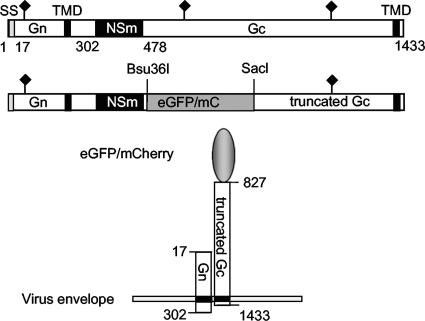
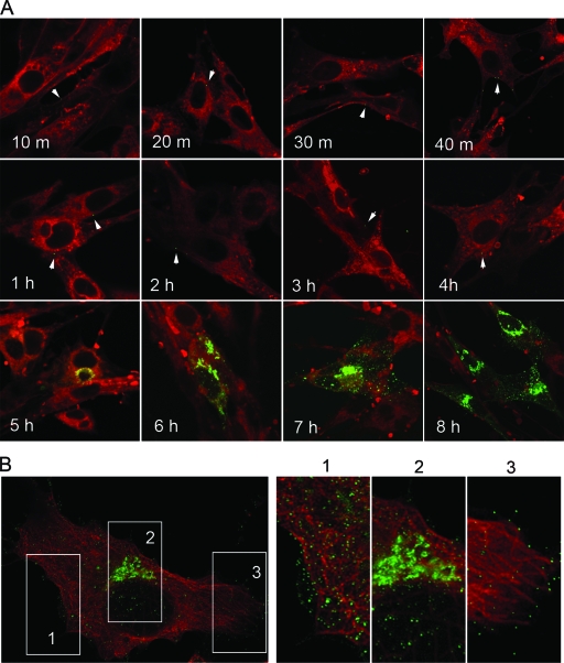
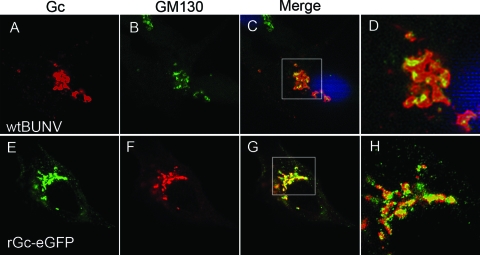
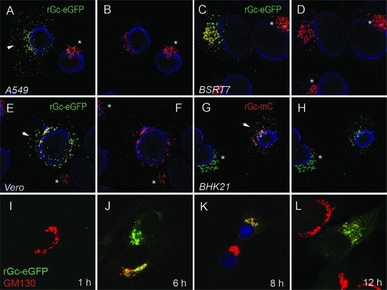
The virion glycoproteins Gn and Gc of Bunyamwera virus (BUNV), the prototype of the Bunyaviridae family and also of the Orthobunyavirus genus, are encoded by the medium (M) RNA genome segment and are involved in both viral attachment and entry. After their synthesis Gn and Gc form a heterodimer in the endoplasmic reticulum (ER) and transit to the Golgi compartment for virus assembly. The N-terminal half of the Gc ectodomain was previously shown to be dispensable for virus replication in cell culture (X. Shi, J. Goli, G. Clark, K. Brauburger, and R. M. Elliott, J. Gen. Virol. 90:2483-2492, 2009.). In this study, the coding sequence for a fluorescent protein, either enhanced green fluorescent protein (eGFP) or mCherry fluorescent protein, was fused to the N terminus of truncated Gc, and two recombinant BUNVs (rBUNGc-eGFP and rBUNGc-mCherry) were rescued by reverse genetics. The recombinant viruses showed bright autofluorescence under UV light and were competent for replication in various mammalian cell lines. rBUNGc-mCherry was completely stable over 10 passages, whereas internal, in-frame deletions occurred in the chimeric Gc-eGFP protein of rBUNGc-eGFP, resulting in loss of fluorescence between passages 5 and 7. Autofluorescence of the recombinant viruses allowed visualization of different stages of the infection cycle, including virus attachment to the cell surface, budding of virus particles in Golgi membranes, and virus-induced morphological changes to the Golgi compartment at later stages of infection. The fluorescent protein-tagged viruses will be valuable reagents for live-cell imaging studies to investigate virus entry, budding, and morphogenesis in real time.


Figures

FIG. 1.
FIG. 1.
Schematic diagrams of eGFP- and mCherry-tagged BUNV glycoproteins. The layout of the wt BUNV glycoprotein precursor (Gn, NSm, and Gc) is shown at the top, with positions of amino acid residues marking the protein boundaries indicated. Below is shown the structure of the chimeric Gc protein, with substitution of the N terminus of Gc (residues 500 to 826) with the coding sequence of either enhanced green fluorescent protein (eGFP) or mCherry fluorescent protein (mC) attached to truncated Gc. The predicted topology of Gn and eGFP/mCherry-tagged Gc on the viral envelope is shown at the bottom. SS, signal peptide; TMD, transmembrane domain. The filled diamonds indicate glycosylation sites.
FIG. 2.
FIG. 2.
Plaque morphology, growth kinetics, and autofluorescence of recombinant viruses. (A) Comparison of the plaque sizes on BHK-21 cells. Cells were fixed at 6 days postinfection with 4% formaldehyde and stained with Giemsa solution. (B) Virus growth curves on BHK-21 cells at 33°C. Cells were infected with wt BUNV virus or the rBUNGc-eGFP (rGc-eGFP) or rBUNGc-mCherry (rGc-mC) recombinant virus at an MOI of 0.1 PFU/cell. Virus was harvested at the indicated times after infection and titrated by plaque formation on BHK-21 cells. Results are shown as the mean of the duplicate titrations. (C) Autofluorescence of recombinant viruses. The supernatant from infected cells containing released virus particles was mixed with an equal volume of Mowiol mounting medium and observed in the Deltavision microscope. Left and middle panels show the autofluorescence of rBUNGc-eGFP (green) and rBUNGc-mCherry (red) viruses, and the right panel shows the image when green and red viruses were mixed. The enlarged inset shows aggregates of green and red viruses. rGcΔ-7, rBUNGcΔ7.
FIG. 3.
FIG. 3.
Characterization of recombinant BUNV expressing chimeric Gc proteins. (A) Comparison of protein profiles of the recombinant viruses rBUNGc-eGFP (rGc-eGFP), rBUNGc-mCherry (rGc-mC), rBUNGcΔ7 (rGcΔ-7), and wt BUNV. BHK-21 cells were infected with wt or mutant viruses at an MOI of 1.0 PFU/cell or were mock infected, and cells were then labeled with [35S]methionine at 24 h postinfection. Viral proteins were precipitated with anti-BUN antibodies and analyzed on 4 to 12% polyacrylamide NuPAGE gels (Invitrogen) under reducing conditions. The positions of viral proteins and protein molecular mass markers are indicated. (B) Western blot analysis to detect eGFP. BHK-21 cells were infected with wt BUNV, rBUNGcΔ7, or rBUNGc-eGFP stock from passage 2 (P2) or passage 10 (P10) or mock infected, as indicated, and cell lysates were fractionated by SDS-PAGE. After transfer to PVDF membrane, the blots were probed with anti-GFP, anti-N, and anti-tubulin antibodies as shown. (C) Glycosylation analysis of viral Gc proteins. Radiolabeled viral proteins were immunoprecipitated with anti-BUN antibodies, and then precipitates were subjected to digestion with PNGase F (F), as indicated, before fractionation on 4 to 12% polyacrylamide NuPAGE gels (Invitrogen) under reducing conditions. The positions of viral proteins are indicated.
FIG. 4.
FIG. 4.
Stability of fluorescent protein-tagged Gc in recombinant viruses. (A and B) Protein analysis. BHK-21 cells were infected with wt BUNV, rBUNGcΔ7, and stocks of rBUNGc-eGFP (rGc-eGFP) or rBUNGc-mCherry (rGc-mC) from passages 2 to 9 or 10, as indicated, at an MOI of 1.0 PFU/cell and labeled with [35S]methionine at 24 h postinfection. Viral proteins were precipitated with anti-BUN antibodies and analyzed on 4 to 12% polyacrylamide NuPAGE gels (Invitrogen) under reducing conditions. The positions of viral proteins are indicated. (C) Sequence analysis. Alignment of the deduced amino acid sequences of the NSm-Gc region from wt BUNV, rBUNGc-eGFP passage 9, wt MAGV, and MAGV R2. Above is shown a schematic of this region, with amino acid positions indicated.
FIG. 5.
FIG. 5.
Time course of rBUNGc-eGFP infection. (A) BHK-21 cells were infected with rBUNGc-eGFP at an MOI of 1 PFU/cell and incubated at 37°C for 10 min to 8 h, as indicated, prior to fixation with 4% paraformaldehyde. Cells were counterstained in red with CellMask Deep Red Plasma Membrane Stain (Invitrogen). Representative green virus particles that were either attached to or internalized into the cells at early stages of infection (1 h to 4 h) are marked by arrows though in some cells multiple particles can be seen attached. (B) BSR-T7/5 cells were infected for 8 h with rBUNGc-eGFP at an MOI of 1 PFU/cell, fixed, and costained with anti-tubulin antibody (in red). Enlarged images of selected regions 1, 2, and 3 show progeny virus particles and the virus assembly site as described in the text.
FIG. 6.
FIG. 6.
Visualization of virus budding at the Golgi complex. BSRT7/5 cells were infected with either wt BUNV (A to D) or rBUNGc-eGFP (E to H) and costained with antibodies to the Golgi marker GM130 (red). BUNV Gc protein in wt BUNV-infected cells was detected with BUNV Gc-specific MAb742. Colocalization between Gc proteins and the Golgi protein are shown in yellow in the merged images (C and G), and enlarged images are shown in panels D and H.
FIG. 7.
FIG. 7.
Morphological changes to the Golgi complex in BUNV-infected cells. A549 (A and B), BSR-T7/5 (C and D), Vero (E and F), and BHK-21 (G and H) cells were infected with either rBUNGc-eGFP (A to F) or rBUNGc-mCherry (G and H). After fixation, cells were stained with anti-GM130 antibodies to detect the Golgi complex, and 3D images were reconstructed from Z-stack images using Huygens deconvolution and surface rendering software. The Golgi complexes in uninfected cells are marked with an asterisk, and those in infected cells are indicated with an arrow. Nuclei were stained in blue with 4′,6′-diamidino-2-phenylindole (DAPI). Panels I to L show the time course of Golgi complex disruption in BSR-T7/5 cells infected with rBUNGc-eGFP. Cells were fixed at 1, 6, 8, and 12 h postinfection, and the Golgi complex was stained in red with antibodies to GM130.
FIG. 8.
FIG. 8.
Live-cell imaging of BHK-21 cells infected with rBUNGc-eGFP. BHK-21 cells grown on microscope chamber slides were infected with rBUNGc-eGFP at an MOI of 1 PFU/cell and incubated at 33°C in an environmental chamber mounted on the Deltavision restoration microscope. Images were collected at 10-min intervals from 12 h postinfection with a Z-section setting of 1 μm (11 images from top to bottom of the cell). Images taken at each hour are shown in the figure (for the complete video clip, see Video S1 in the supplemental material).

Similar articles

Cited by

References

    1. Bosch, B. J., C. A. M. de Haan, and P. J. M. Rottier. 2004. Coronavirus spike glycoprotein, extended at the carboxy terminus with green fluorescent protein, is assembly competent. J. Virol. 78:7369-7378. - PMC - PubMed
    1. Brandenburg, B., and X. Zhuang. 2007. Virus trafficking—learning from single-virus tracking. Nat. Rev. Microbiol. 5:197-208. - PMC - PubMed
    1. Bridgen, A., and R. M. Elliott. 1996. Rescue of a segmented negative-strand RNA virus entirely from cloned complementary DNAs. Proc. Natl. Acad. Sci. U. S. A. 93:15400-15404. - PMC - PubMed
    1. Buchholz, U. J., S. Finke, and K. K. Conzelmann. 1999. Generation of bovine respiratory syncytial virus (BRSV) from cDNA: BRSV NS2 is not essential for virus replication in tissue culture, and the human RSV leader region acts as a functional BRSV genome promoter. J. Virol. 73:251-259. - PMC - PubMed
    1. Cureton, D. K., R. H. Massol, S. Saffarian, T. L. Kirchhausen, and S. P. J. Whelan. 2009. Vesicular stomatitis virus enters cells through vesicles incompletely coated with clathrin that depend upon actin for internalization. PLoS Pathog. 5:e1000394. - PMC - PubMed

Publication types

Substances

LinkOut - more resources