. 2010 Jun 23;30(25):8353-66.
doi: 10.1523/JNEUROSCI.0546-10.2010.

Aplysia cell adhesion molecule and a novel protein kinase C activity in the postsynaptic neuron are required for presynaptic growth and initial formation of specific synapses

Affiliations

Aplysia cell adhesion molecule and a novel protein kinase C activity in the postsynaptic neuron are required for presynaptic growth and initial formation of specific synapses

Jiang-Yuan Hu et al. J Neurosci. .

Abstract

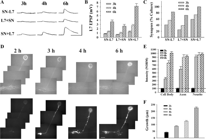
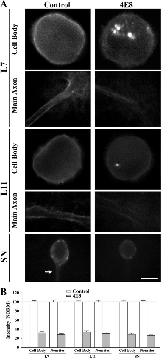
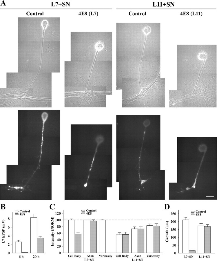
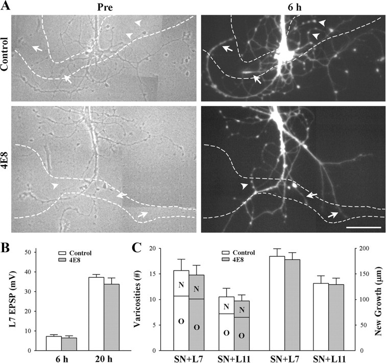
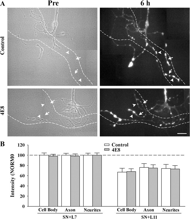
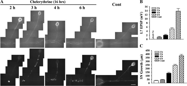
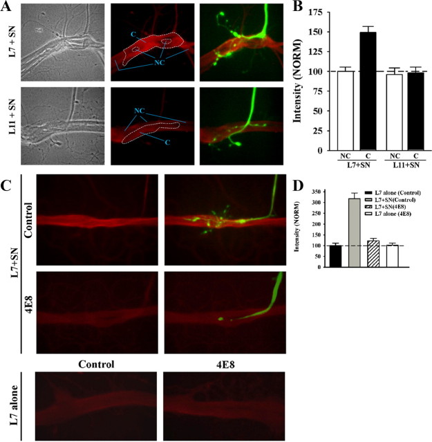
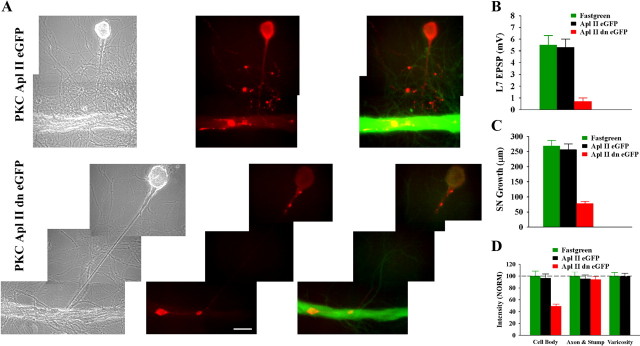
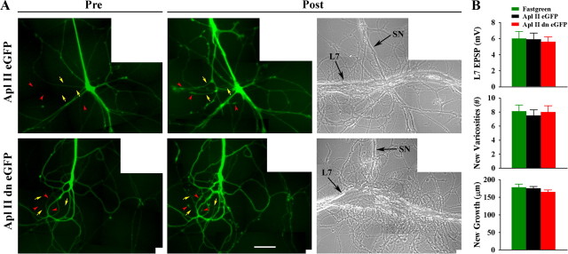
To explore the role of both Aplysia cell adhesion molecule (ApCAM) and activity of specific protein kinase C (PKC) isoforms in the initial formation of sensory neuron synapses with specific postsynaptic targets (L7 but not L11), we examined presynaptic growth, initial synapse formation, and the expression of the presynaptic neuropeptide sensorin following cell-specific reduction of ApCAM or of a novel PKC activity. Synapse formation between sensory neurons and L7 begins by 3 h after plating and is accompanied by a rapid accumulation of a novel PKC to sites of synaptic interaction. Reducing ApCAM expression specifically from the surface of L7 blocks presynaptic growth and initial synapse formation, target-induced increase of sensorin in sensory neuron cell bodies and the rapid accumulation of the novel PKC to sites of interaction. Selective blockade of the novel PKC activity in L7, but not in sensory neurons, with injection of a dominant negative construct that interferes with the novel PKC activity, produces the same actions as downregulating ApCAM; blockade of presynaptic growth and initial synapse formation, and the target-induced increase of sensorin in sensory neuron cell bodies. The results indicate that signals initiated by postsynaptic cell adhesion molecule ApCAM coupled with the activation of a novel PKC in the appropriate postsynaptic neuron produce the retrograde signals required for presynaptic growth associated with initial synapse formation, and the target-induced expression of a presynaptic neuropeptide critical for synapse maturation.


Figures

Figure 1.
Figure 1.
Time course for synapse formation, sensorin expression, and axon growth of sensory neurons when interacting with L7. A–C, Time course for synapse formation. EPSPs evoked in L7 at 3–6 h of interaction for the three types of cell culture: simultaneous plating (SN-L7); L7 first, then sensory neuron added the next day (L7 + SN); and sensory neuron first and L7 added the next day (SN + L7). EPSP amplitudes increased with time (A). Calibration: Vertical bar, 10 mV; horizontal bar, 25 ms. Plating sensory neuron first leads to stronger synapses (B) and a higher proportion of cultures with detectable synapses (C). In B the height of each bar is the mean ± SEM of EPSP amplitude at each time point for the three culture conditions. ANOVA indicated a significant difference with condition and time (df = 4, 36; F = 4.032, p < 0.02). Individual comparisons (Scheffe F) indicated that EPSP amplitudes for SN + L7 cultures were significantly greater than the amplitudes in the other two culture conditions at each time point (p < 0.05 at 3 h and p < 0.01 at both 4 and 6 h). In addition to EPSP amplitudes, the proportion of cultures with synapses was higher for SN + L7 cultures (C). D, E, Sensorin expression in each cellular compartment increases over time for SN-L7 cultures. Phase contrast and corresponding epifluorescent images for sensorin immunostaining depict changes in sensorin expression at the various time points (D). Sensorin expression is low at 2 h (n = 6) but increases significantly at distal sites by 3 h (n = 8). At 4 h (n = 8), sensorin staining peaked in the axon, while peak intensity in the cell body occurred at 6 h (n = 10). Scale bar, 50 μm. In E the height of each bar is the mean ± SEM staining intensity for sensorin in each compartment at each time point after normalizing to the 2 h intensity values for each compartment (100% ± SEM). The peak intensity is first achieved in the distal neurites, while the peak intensity in the cell body is achieved last. ANOVA indicated a difference with time in each compartment (df = 2, 28, F = 78.785, p < 0.001). Individual comparisons indicated that staining intensity in the cell body increased with each time point (p < 0.01), staining intensity in the axon increased at each time point (p < 0.01) up to 4 h, and staining intensity in the distal neurites increased at 3 h (p < 0.01) and remained relatively constant afterward. F, Axon growth by sensory neurons increased continuously from 2 to 6 h. Height of each bar is the mean ± SEM extent of sensory neuron axon growth in contact with the major processes of L7 based on detectable sensorin immunochemistry in the distal neurites.
Figure 2.
Figure 2.
Treatment with anti-ApCAM antibody 4E8 downregulates ApCAM expression on the cell surface membrane of L7, L11, and sensory neurons. A, Epifluorescent images of ApCAM immunostaining of cell body and processes of L7 (n = 7 for each treatment), L11 (n = 7 for each treatment), or SN (n = 9 for each treatment) following overnight preincubation (14 h) with control or 4E8 (anti-ApCAM antibody). Midlevel focal planes (between substrate and top of each structure) are presented here. After antibody washout, cells were fixed 3 h later and processed for ApCAM immunochemistry of the surface membrane (no detergent added). Treatment with 4E8 significantly reduced surface membrane staining throughout the cells. Scale bar, 50 μm. B, Treatment with 4E8 reduced ApCAM on the surface of cells by ∼70%. The height of each bar is the mean ± SEM percentage for ApCAM staining on the surface of the cell body or processes following treatment with 4E8 normalized to the staining levels measured on the surface of each compartment following treatment with controls (100% ± SEM). ANOVA indicated a significant effect of treatment (df = 5, 40; F = 177.02, p < 0.001). Individual comparisons indicated that treatment with 4E8 significantly reduced ApCAM levels from the cell bodies (p < 0.01) and processes (p < 0.01) of all cells compared with controls. There were no significant differences in surface membrane staining for ApCAM for the control cells (see Results).
Figure 3.
Figure 3.
Downregulating ApCAM from the surfaces of L7 blocks initiation of synapse formation, L7-induced regulation of sensorin expression in the cell body of the sensory neurons and synapse-associated growth. A, Phase contrast and epifluorescent images of sensorin immunostaining in sensory neurons after 6 h of contact with L7 (n = 10 for control and n = 12 for 4E8) or L11 (n = 8 for each treatment) that were plated the previous day and preincubated with control or 4E8 antibody. Antibodies were washed out and sensory neurons plated ∼1 h later. Sensorin staining in the sensory neuron cell body was low following treatment of L7 with 4E8. Also note that there was no growth from the axon stump of the sensory neuron. Treatment of L11 with 4E8 did not interfere with sensorin expression in the cell body, axon or neurites of the sensory neurons. Scale bar, 50 μm. B, Downregulating ApCAM on L7 with 4E8 blocked synapse formation at 6 h, and significantly reduced synapse strength when EPSP were recorded again at 20 h. The height of each bar is the mean ± SEM of the EPSP amplitude measured at 6 and 20 h for control and 4E8 treatment of L7 before the addition of the sensory neuron. ANOVA indicated a significant effect of treatment (df = 1, 26; F = 7.979, p < 0.01). Individual comparisons indicated a significant reduction in EPSP with treatment with 4E8 at both 6 and 20 h (p < 0.01). C, Downregulating ApCAM on L7 with 4E8 blocked the increase in sensorin expression, especially in the cell body, while treatment of L11 with 4E8 did not interfere with sensorin expression throughout the sensory neuron. The height of each bar is the mean ± SEM staining intensity in the various compartments of the sensory cell normalized to the staining intensity in each compartment for control L7 + SN cultures (100%). Since no varicosities formed with pretreatment of L7 with 4E8, there is no bar for that compartment. ANOVA indicated a significant effect of treatment (df = 6, 72; F = 195.526, p < 0.001). Individual comparisons indicated that treatment with 4E8 resulted in a significant reduction in sensorin expression in the cell body of sensory neurons contacting L7 (p < 0.01), but had no effect on expression in sensory neuron axons. There was no significant difference in the treatment of L11 with 4E8; both 4E8 and control treatments did not alter the significant reduction (p < 0.05) in sensorin staining in the cell body and original axons. D, Downregulating ApCAM on the surface of L7 significantly blocked neuritic growth by sensory neurons, but downregulating ApCAM on the surface of L11 did not affect neuritic growth by sensory neurons. The height of each bar is the mean ± SEM in the extent of neuritic growth of sensory neurons in contact with L7 or L11 based on detectable sensorin immunochemistry in the distal neurites of the sensory neurons. Treatment of L7 with 4E8 blocked neuritic growth by sensory neurons. This was also verified by injection with carboxyfluorescein (n = 3, data not shown). ANOVA indicated a significant effect of treatment (df = 3, 36; F = 62.015, p < 0.001). Individual comparisons indicated that pretreatment of L7 with 4E8 significantly reduced growth (p < 0.01) while pretreatment of L11 with 4E8 did not affect growth by sensory neurons contacting L11.
Figure 4.
Figure 4.
Reducing ApCAM from the surface of the sensory neurons does not affect synapse formation and growth. Sensory neurons were plated and treated overnight with either control or anti-ApCAM antibody 4E8 and washed, and then axon growth was imaged before plating L7 or L11 (only SN + L7 cultures are shown here). After 6 h, EPSPs were recorded, and in some cultures, sensory neurons were injected with carboxyfluorescein to map out new growth by the sensory neurons. Other cultures were maintained overnight and EPSP amplitudes were reexamined. A, Phase contrast images (left) of axon growth from sensory neurons treated overnight with control IgG or 4E8. The dashed outline represents the position of the motor neuron L7 (axon hillock and original axon) that was plated with each sensory neuron after taking the images. Treatment with 4E8 does not impact axon growth by sensory neuron. On the right are epifluorescent images of neuritic growth following injection with carboxyfluorescein from the same sensory neurons after 6 h of interacting with the motor neurons. Arrowheads point to some of the varicosities and arrows point to some of the new branches that had grown over the 6 h of interaction. The white dashed lines represent the borders of the L7 motor neurons. Treatment with 4E8 failed to block new growth and formation of new varicosities. Scale bar, 75 μm. B, Reducing ApCAM from the surface of the sensory neurons did not interfere with synapse formation. The height of each bar is the mean ± SEM of the EPSP amplitudes recorded at 6 and 20 h. ANOVA indicated no significant effect of treatment with 4E8. At 20 h both control- and 4E8-treated cultures had formed strong synapses. C, Reducing ApCAM from the surface of the sensory neurons did not interfere with growth and formation of new varicosities. The height of each bar is the mean ± SEM of the total number of varicosities contacting L7 or L11 (left bars) and the length of new sensory neuron axon branches contacting L7 or L11 (right bars). For each treatment and culture (n = 6 for each treatment of SN + L7 or SN + L11 cultures), sensory neuron varicosities contacting each target were divided into two groups: old varicosities (O) on preexisting sensory neuron axon branches when the motor neuron was first plated, and new varicosities (N) formed by new axon growth. Regardless of target (L7 or L11), treatment with 4E8 had no impact on growth. Growth and varicosity number were higher for sensory neurons contacting L7 then sensory neurons contacting L11.
Figure 5.
Figure 5.
Reducing ApCAM from the surface of the sensory neurons does not affect target-induced regulation of sensorin expression. Cultures were processed as described above, except at 6 h the cocultures were fixed and processed for sensorin immunochemistry. A, Phase contrast images (left) of axon growth from the stump of sensory neurons treated overnight with control IgG (n = 7 for SN + L7 or SN + L11) or 4E8 (n = 7 for SN + L7 or SN + L11). The dashed outline represents the position of the motor neuron L7 (axon hillock and major processes) that was plated with each sensory neuron after taking the images (only SN + L7 cultures are shown here). Treatment with 4E8 did not affect sensorin expression in the regenerated neurites, especially the new branches and varicosities. On the right are epifluorescent images of sensorin immunostaining from the same sensory neuron at 6 h of interacting with the motor neuron. In both images, arrowheads point to the location of some of the varicosities and arrows point to the location of some of the new branches that had grown over the 6 h of interaction. Scale bar, 40 μm. B, Reducing ApCAM from the surface of the sensory neurons did not block target-induced regulation of sensorin expression throughout the sensory neurons. The height of each bar is mean ± SEM of the staining intensity in the 4E8-treated cultures after normalization with the staining intensity for each compartment of sensory neurons treated with controls and interacting with L7 after 6 h. Treatment with 4E8 failed to block the strong staining in sensory neurons contacting L7, and failed to block the reduction in staining in each compartment for sensory neurons contacting L11.
Figure 6.
Figure 6.
Blocking PKC activity in sensory neuron and L7 with chelerythrine interferes with initial synapse formation, synapse-associated growth, and sensorin expression. A–C, Initial synapse formation, growth, and sensorin expression were affected differently when chelerythrine was bath applied to SN-L7 cultures at the indicated times after plating both cells. Cocultures were imaged after 16 h of drug treatment (18–22 h in culture) after processing for sensorin immunoreactivity (A). Note that the time at which chelerythrine was added to the coculture had a significant impact on sensorin expression. The differences in synapse strength and sensorin expression in control cultures at 18 and 22 h were not significant. An overall ANOVA indicated a significant effect of treatment on sensorin expression in the different compartments (df = 4, 43, F = 82.193, p < 0.001). Individual comparisons indicated that expression in the axon and distal sites was reduced only when inhibitor was added at 2 h (F = 3.825 p < 0.05). Expression in the cell body remained suppressed when inhibitor was added at 3 and 4 h (F = 32.145, p < 0.01, and F = 30.762, p < 0.01) but reached control levels at 6 h. EPSP amplitude detected after drug treatment was significantly affected (B). Compared with control, adding drug at each time point significantly reduced EPSPs (F = 19.894–38.278; p < 0.01). Applying drug after 2 h blocked synapse formation, while adding drug after 6 h allowed cultures to form stronger synapses than those detected when drug was added at 3 or 4 h (p < 0.05). Extent of growth after drug treatment was also influenced by the time of application (C). For each time point, growth was significantly reduced compared with control (F = 13.802–92.526; p < 0.01).
Figure 7.
Figure 7.
ApCAM on the surface of L7 is required for local targeting of PKC Apl II to sites of sensory neuron contact. A, B, PKC Apl II staining intensity is enhanced at contact sites of sensory neuron with L7 (n = 12), but not with L11 (n = 8). In A, phase contrast (left), epifluorescent images of PKC Apl II immunostaining (red; middle), and combined epifluorescent images of PKC Apl II staining and dye-filled neurites and varicosities of the sensory neuron (green; right) contacting either L7 (L7 + SN) or L11 (L11 + SN) are compared. Note the enhanced staining at the contact sites (within the large dashed zone in the middle micrographs) between sensory neuron and L7 compared with nearby sites on L7 without contacts (such as areas within the NC zone). Contact sites on the surface of L11 showed little or no differences in staining compared with neighboring NC sites. In B, staining intensity at sites of contact with the target by the axon stump, neurites, and varicosities of sensory neurons are normalized to neighboring non-contact sites on the surface of L7 (normalized to 100%). An overall ANOVA indicated a significant effect of the target on staining intensity at contact sites (df = 3, 36, F = 14.516, p < 0.001). Individual comparisons indicated that staining intensity at sensory neuron contact sites (C) with L7 were significantly greater than that in neighboring non-contact sites (NC) on L7 (F = 9.645, p < 0.01), and contact sites between sensory neurons and L11 (F = 13.461, p < 0.01). Intensity at contact sites between sensory neurons and L11 were not significantly different from intensity at the non-contact sites on L11. C, D, Reducing ApCAM on the surface of L7 by incubating with anti-ApCAM antibody 4E8 abolishes the increase in PKC Apl II at L7 contact sites with sensory neurons. Preplated L7 was incubated overnight either with control (n = 16) or 4E8 antibodies (n = 16). After antibody washout, sensory neurons were added to some of the preplated L7 (n = 10 per treatment). In C, epifluorescent views of PKC Apl II immunostaining for both L7 + SN cultures and L7-alone cultures (n = 6 per treatment) treated with both antibodies are compared. In the two upper right panels, dye-filled staining of the sensory axon, stump, and neurites (green) is superimposed on PKC Apl II immunostaining (red) for control- and 4E8-treated L7 and compared. We also compared the overall staining of sections of L7's axon that were not contacted by sensory neurons (200–300 μm sections) in L7 + SN cultures (C, top left) to the overall staining of sections of L7 axons plated alone (C, bottom left). No significant difference was detected in the immunostaining for the novel PKC. In D, staining intensities at sites of contact with L7 by axon, axon stump, and varicosities of the sensory neuron are compared with normalized staining for the PKC Apl II on the surface of control-treated L7 axon (300 μm portion of the axon normalized to 100%). An overall ANOVA indicated a significant effect of antibody treatment on staining at contact sites (df = 3, 28, F = 33.371, p < 0.001). Individual comparisons indicated that sensory neuron contact with L7 increased staining for PKC Apl II at contact sites compared with overall staining along the axon of control L7 plated alone (F = 19.944, p < 0.01). Treatment with the anti-ApCAM antibody abolished the increase in PKC Apl II staining at contact sites in L7 + SN cultures compared with control-treated L7 + SN cultures (F = 10.447, p < 0.01). Staining at contact sites between sensory neurons and L7 when ApCAM was downregulated from L7 was not significantly different from overall staining for L7 plated alone. Treatment of L7 alone with anti-ApCAM 4E8 did not change significantly overall staining for PKC Apl II compared with control-treated L7 alone.
Figure 8.
Figure 8.
Downregulating activity of PKC Apl II in the motor neuron L7 by injecting a dominant negative construct blocks initial synapse formation, synapse-associated growth, and increased expression of sensorin. A, Phase contrast (left), rhodamine epifluorescent images of sensorin immunostaining in sensory neuron cell bodies, axons, and varicosities (red; middle) and double label for sensorin immunostaining (red) in sensory neurons and eGFP-expression (green) epifluorescent images indicating expression of the construct in L7 (right panel) of the same view areas showing sensory neuron interaction with L7. Growth of sensory neuron and sensorin expression, especially in the cell body, are significantly reduced following injection and expression of the dominant negative PKC (PKC Apl II dn eGFP; bottom) compared with the normal PKC Apl II (PKC Apl II eGFP; top). B, Initial synapse formation is blocked following injection of the dominant negative construct. An ANOVA indicated a significant effect of construct injection (df = 2, 28, F = 19.968, p < 0.001). Individual comparisons indicated that injection of the dominant negative construct (n = 11) significantly reduced EPSP amplitude compared with fast green (F = 15.356, p < 0.01; n = 10) or to injections with the control construct (F = 14.096, p < 0.01; n = 10). Injection of the normal gene into L7 did not affect synapse formation compared with injection of fast green alone. C, Sensory neuron growth was significantly reduced when L7 was injected with the dominant negative construct. An ANOVA indicated a significant effect of construct injection (df = 2, 28, F = 47.128, p < 0.001). Individual comparisons indicated that injection with the dominant negative construct significantly reduced growth compared with fast green injection (F = 37.016, p < 0.01) or injection of construct for the normal gene (F = 32.415, p < 0.01). Growth following injection of construct for the normal gene was not different from the growth after fast green injection. D, Sensorin expression, particularly in the cell body of the sensory neuron, was significantly reduced by injection of the dominant negative construct. An overall ANOVA indicated a significant effect of treatment (df = 4, 56, F = 20.703, p < 0.001). Individual comparisons indicated that injection of the dominant negative construct in L7 significantly reduced sensorin expression in the cell body of the sensory neuron compared with injection of fast green (F = 15.119, p < 0.01) or the normal gene (F = 13.008, p < 0.01). Sensorin staining in the axons was not affected by injections. Since there were no stained varicosities after injection of the dominant negative construct, differences in growth were indeterminate. There were no significant differences in the staining in all compartments between injection of fast green and injection of the construct for the normal gene.
Figure 9.
Figure 9.
Downregulating activity of PKC Apl II in the sensory neuron by injecting a dominant negative construct does not interfere with initial synapse formation, and new synapse-associated growth. A, Injection of the dominant negative construct into the sensory neuron (green) does not interfere with growth of new sensory neuron branches that form new varicosities. The left images are epifluorescent views of eGFP expression in the preplated sensory neuron before adding L7 (Pre). The eventual locations of some of the new branches (yellow arrows) and new varicosities (red arrowheads) are indicated. The right images (Post) show epifluorescent views of the same area after 8 h of interaction with L7 (n = 8 per treatment). Arrows and arrowheads indicate some new branches and varicosities, respectively. Note that expression of the dominant negative construct does not interfere with new growth and varicosity formation. The right image is a phase contrast view of the same area 8 h after adding L7. Arrows point to the axon of the SN and the major axon of L7. B, EPSP amplitude (top), formation of new varicosities (middle), and growth of new branches (bottom) are unaffected by injection of the dominant negative construct. An overall ANOVA indicated no significant effect of treatment.

Similar articles

Cited by

References

    1. Ashley J, Packard M, Ataman B, Budnik V. Fasciclin II signals new synapse formation through amyloid precursor protein and the scaffolding protein dX11/Mint. J Neurosci. 2005;25:5943–5955. - PMC - PubMed
    1. Bailey CH, Chen M, Keller F, Kandel ER. Serotonin-mediated endocytosis of ApCAM: an early step of learning-related synaptic growth in Aplysia . Science. 1992;256:645–649. - PubMed
    1. Bailey CH, Kaang BK, Chen M, Martin KC, Lim CS, Casadio A, Kandel ER. Mutation in the phosphorylation sites of MAP kinase blocks learning-related internalization of ApCAM in Aplysia sensory neurons. Neuron. 1997;18:913–924. - PubMed
    1. Baines RA, Seugnet L, Thompson A, Salvaterra PM, Bate M. Regulation of synaptic connectivity: levels of Fasciclin II influence synaptic growth in the Drosophila CNS. J Neurosci. 2002;22:6587–6595. - PMC - PubMed
    1. Bank M, Schacher S. Segregation of presynaptic inputs on an identified target neuron in vitro: structural remodeling visualized over time. J Neurosci. 1992;12:2960–2972. - PMC - PubMed

Publication types

LinkOut - more resources