. 2010 Jun 23;30(25):8529-40.
doi: 10.1523/JNEUROSCI.0032-10.2010.

Astn2, a novel member of the astrotactin gene family, regulates the trafficking of ASTN1 during glial-guided neuronal migration

Affiliations

Astn2, a novel member of the astrotactin gene family, regulates the trafficking of ASTN1 during glial-guided neuronal migration

Perrin M Wilson et al. J Neurosci. .

Abstract

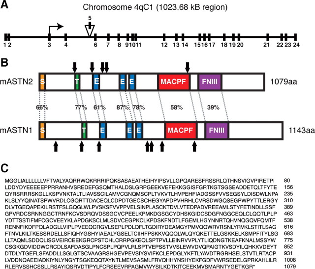
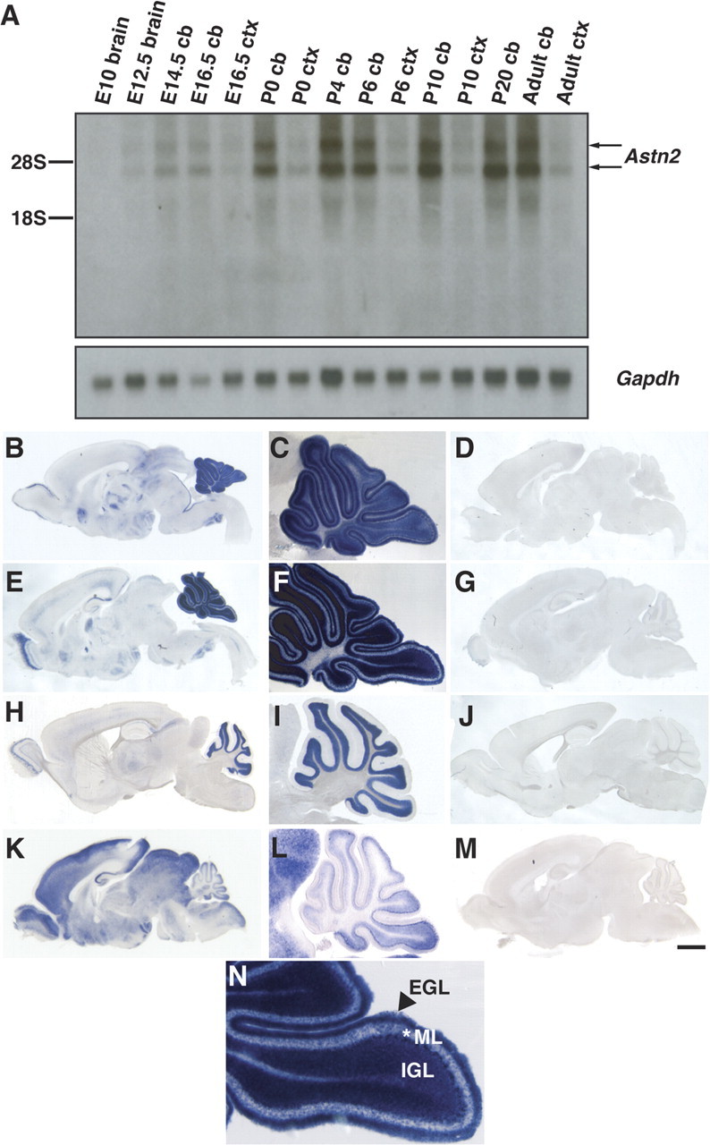
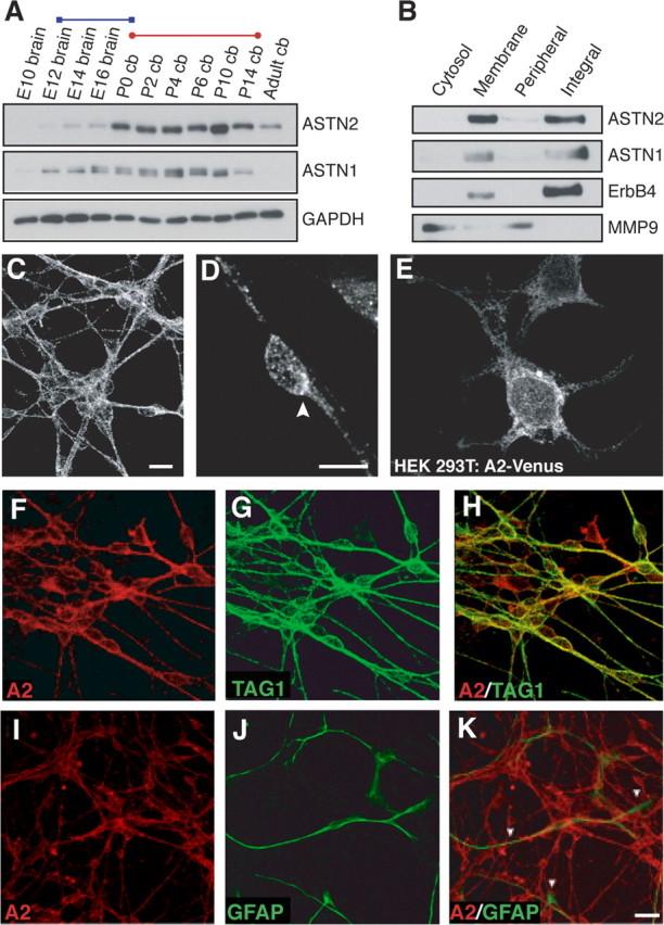
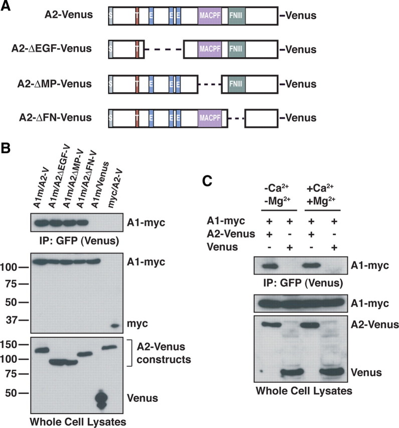
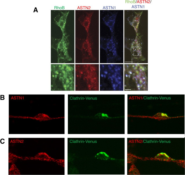
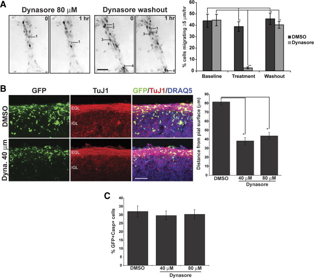
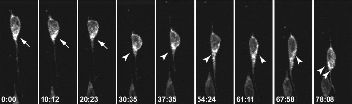
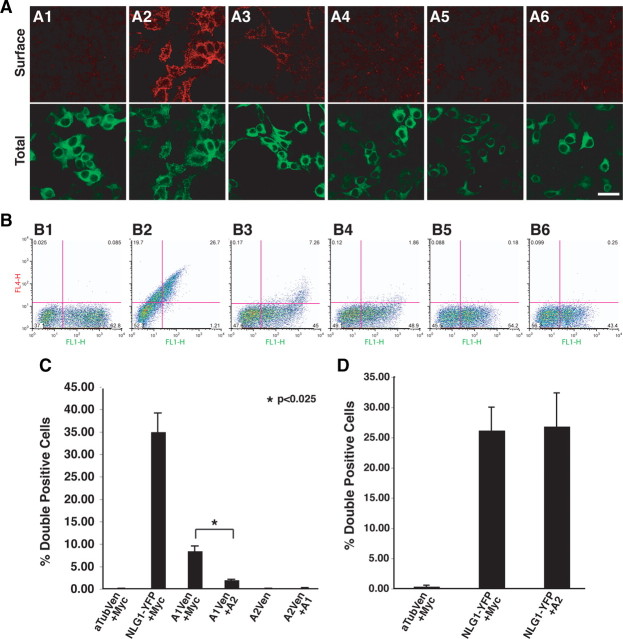
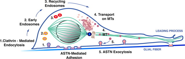
Glial-guided neuronal migration is a key step in the development of laminar architecture of cortical regions of the mammalian brain. We previously reported that neuronal protein astrotactin (ASTN1) functions as a neuron-glial ligand during CNS glial-guided migration. Here, we identify a new Astn family member, Astn2, that is expressed at high levels in migrating, cerebellar granule neurons, along with Astn1, at developmental stages when glial-guided migration is ongoing. Biochemical and flow cytometry experiments show that ASTN2 forms a complex with ASTN1 and regulates surface expression of ASTN1. Live imaging of Venus-tagged ASTN1 in migrating cerebellar granule cells reveals the intracellular trafficking of ASTN1-Venus, with ASTN1-Venus accumulating in the forward aspect of the leading process where new sites of adhesion will form. Treatment of migrating neurons with Dynasore, a soluble noncompetitive inhibitor of Dynamin, rapidly arrests the migration of immature granule cells in a reversible manner, suggesting the critical importance of receptor trafficking to neuronal locomotion along Bergmann glial fibers in the developing cerebellum. Together, these findings suggest that ASTN2 regulates the levels of ASTN1 in the plasma membrane and that the release of neuronal adhesions to the glial fiber during neuronal locomotion involves the intracellular trafficking of ASTN1.


Figures

Figure 1.
Figure 1.
Astn2 genomic and protein structure. A, Schematic showing genomic structure of Astn2 on mouse chromosome 4qC1. Astn2 contains 24 exons and spans 1023.68 kb. Isoform a lacks exon 5 (156 nt). B, Schematic illustrating homology between mouse ASTN2 and ASTN1, arrows indicate N-linked glycosylation sites. ASTN2 contains a signal sequence (S), a transmembrane domain (T), three EGF repeats (E), a MAC/Perforin domain (MACPF), and a fibronectin III domain (FNIII). C, ASTN2 isoform a amino acid sequence.
Figure 2.
Figure 2.
Astn2 is expressed in the developing and adult brain. A, Northern blot of Astn2 from brain, cerebellum (cb), or cortex (ctx) RNA. Astn2 expression is enriched in the cerebellum; Gapdh control is shown in the bottom panel. Northern blotting reveals that there are two Astn2 transcripts (4.2 and 5.8 kb), which is likely due to alternative splicing in an untranslated region. B–J, N, In situ hybridization of sagittal brain sections reveals that Astn2 is expressed in the cerebellum, cortex, olfactory bulb, hindbrain, and hippocampus at P6 (B), P10 (E), and adult (H) stages. Magnification (5×) of the cerebellum reveals that at P6 (C) and P10 (F, N), Astn2 is expressed in granule neurons in the EGL, IGL, and the molecular layer (ML), and Purkinje neurons. At adult stages (H, I), Astn2 is localized to Purkinje neurons and granule neurons in the IGL. K–M, In situ hybridization of Astn1 at P6. D, G, J, M, Corresponding sense controls. Scale bar: (in M) B, D, K, M, 1000 μm; C, F, I, L, 400 μm; E, G, 1200 μm; H, J, 1500 μm; N, 200 μm.
Figure 3.
Figure 3.
ASTN2 is an integral membrane protein expressed in cerebellar neurons. A, Expression of ASTN2 and ASTN1 in the brain is developmentally regulated. ASTN2 protein is expressed in the embryonic, postnatal, and adult brain, and peak expression occurs during the radial migration of the granule neurons into the IGL (red bar), whereas ASTN1 expression overlaps with both the tangential migration of the granule cell precursors in the cerebellar anlage (blue bar) and the migration of granule neurons (red bar). GAPDH immunostaining was performed as a loading control. B, Cell fractionation experiments show that ASTN2 and ASTN1 are enriched in membranes and are integral membrane proteins. Immunoblotting was performed for integral membrane (ErbB4) and peripheral membrane (MMP9) proteins as controls. C, D, Immunocytochemistry reveals ASTN2 is localized in a punctate pattern in cerebellar granule neurons with an accumulation of protein at one pole of the cell (arrowhead in D). E, Venus-tagged ASTN2 (A2-Venus) is localized in a punctate pattern in HEK293T cells. F–K, Immunocytochemistry shows that ASTN2 (A2) is expressed in cerebellar granule neurons, but not astroglia. The anti-ASTN2 antibody double labels Tag-1-positive cerebellar granule neurons (H), but not GFAP-positive astroglia (K). Scale bars: (in C) C, E–H, 10 μm; (in D) D, 5 μm; (in K) I–K, 20 μm. cb, Cerebellum.
Figure 4.
Figure 4.
ASTN2 interacts with ASTN1. A, Schematic representation of Venus-tagged ASTN2 domain deletions. Dashed lines represent deleted regions. B, Coimmunoprecipitation reveals Venus-tagged ASTN2 (A2-V and A2-Venus) interacts with Myc-tagged ASTN1 (A1m and A1-myc). Coimmunoprecipitation with Venus-tagged ASTN2 constructs lacking the EGF (A2ΔEGF-V), MACPF (A2ΔMP-V), and FNIII (A2ΔFN-V) domains show that the ASTN2/ASTN1 interaction does not depend on any individual conserved domain. Anti-GFP antibody was used to immunoprecipitate and immunoblot Venus-tagged proteins. Anti-Myc and Anti-GFP (to recognize Venus) antibodies were used for immunoblotting. C, Coimmunoprecipitation demonstrates that the ASTN1/ASTN2 interaction is calcium independent.
Figure 5.
Figure 5.
ASTN1 and ASTN2 colocalize with endosomal markers. A, Coexpression of ASTN1 and ASTN2-mCherry with the endosomal marker GFP-Endo (RhoB) in HEK293T cells demonstrates that the ASTN proteins partially colocalize with early and late endosomes. B, C, Endogenous ASTN1 and ASTN2 proteins colocalize with Venus-tagged Clathrin light chain (Clathrin-Venus, expressed via retrovirus infection) in migrating cerebellar granule neurons. Images shown represent single optical confocal sections. Scale bars: (in A) A, 10 μm; B, C, 5 μm.
Figure 6.
Figure 6.
The Dynamin inhibitor Dynasore inhibits glial-guided neuronal migration. A, A granule cell culture migration assay demonstrates that the effects of a soluble noncompetitive inhibitor of Dynamin, Dynasore, on neuronal migration are reversible. Dissociated cerebellar granule neurons, labeled with a Venus encoding retrovirus, were imaged for three 60 min periods; vehicle (DMSO) or DMSO and either 40 or 80 μm Dynasore were added during the second imaging period. To remove the Dynasore, we washed the cells three times with granule cell medium, and imaged migrating neurons as above for 1 h. The images in A are inverted for better resolution and show migrating granule neurons labeled with Venus. In this study, we selected neurons with the features of actively migrating neurons, i.e., an elongated cell soma that was apposed to and flattened against Bergmann-like glial fibers. Migrating neurons are labeled by number in the 0 h panel and their final position is indicated by the same number in the 1 h panel. The number of cells that migrated >5 μm/h was counted and expressed as a percentage of total labeled cells.(*p <0.001, **p <0.0001).B, Cerebellar slices transfected with Venus-expressing retrovirus were treated with Dynasore (Dyna.) at 40 or 80 μm concentration, or with control vehicle (DMSO). TuJ1 staining reveals the parallel fiber layer of differentiated granule neurons in the inner EGL. In slices incubated with Dynasore, Venus-expressing neurons seem to arrest in the EGL compared with DMSO control cells. DRAQ5 was used to counterstain nuclei. Quantitation of neurons in organotypic cerebellar slices shows that neurons remain significantly closer to the pial surface in the presence of 40 or 80 μm Dynasore, relative to controls (*p < 0.001). C, In slices double immunostained with antibodies to GFP and caspase 3, 48 h incubation with Dynasore did not significantly alter the percentage of GFP-positive cells that also were caspase 3 positive. Significance was assessed via Student's t test; values are mean ± SEM (*p < 0.001). Scale bars, 50 μm.
Figure 7.
Figure 7.
Live imaging of ASTN1-Venus dynamics in a migrating cerebellar granule neuron. Purified granule neurons were electroporated with Venus-tagged ASTN1 and mCherry-tagged ASTN2, and Venus-tagged ASTN1 was imaged throughout a migration cycle. Live imaging revealed dynamic changes in the localization of ASTN1-Venus protein during migration. As the neuron moved, ASTN1-Venus accumulated at the anterior pole of the neuronal soma and in the base of the leading process during migration (arrow). At the beginning of the migration cycle, when the neuron adheres to the glial fiber, ASTN1-Venus levels were high in the base of the cell soma. The forward flow of ASTN1-Venus protein continued during the stationary phase of the movement cycle, resulting in an accumulation of ASTN1-Venus at the front of the neuronal soma. In the third step of the cycle, with the release of the interstitial junction, ASTN1-Venus accumulated in the proximal domain of the leading process where a new adhesion site will form (double arrowheads).
Figure 8.
Figure 8.
Coexpression with ASTN2 reduces the cell surface localization of ASTN1. HEK293T cells were transfected with a Venus or EYFP fusion protein, and a second construct (Myc, ASTN2, or ASTN1). A, The top row shows a live cell stain using a rabbit anti-GFP primary and an Alexa Fluor 555-conjugated secondary antibody indicating expression of the Venus/EYFP-conjugated protein on the surface of the cell; while the bottom row shows the total cell expression of the fusion protein. A1, α-Tubulin-Venus and Myc; A2, neuroligin-EYFP and Myc; A3, ASTN1-Venus and Myc; A4, ASTN1-Venus and ASTN2; A5, ASTN2-Venus and Myc; A6, ASTN2-Venus and ASTN1. In the absence of ASTN2, Venus-tagged ASTN1 localizes on the cell surface (A3). Live cell surface staining of transfected HEK293T cells demonstrates that Venus-tagged ASTN1 is no longer exposed on the cell surface when coexpressed with ASTN2 (A4). These experiments also demonstrated that ASTN2-Venus is not exposed on the cell surface in the absence (A5, B5) or presence (A6, B6) of ASTN1. B, For the flow cytometry experiments, an AlexaFluor-647 secondary antibody is substituted for the AlexaFluor-555 antibody, and the data are presented as dot plots of surface labeling with the axes as FL1-H (Venus/EYFP signal on x-axis) versus FL4-H (Alexa 647 signal on y-axis). Lower right quadrant values represent singly labeled Venus/EYFP-positive cells, and upper right quadrant values represent doubly labeled Venus/EYFP and Alexa Fluor 647 live stain-positive cells. B1, α-Tubulin-Venus and Myc; B2, neuroligin-EYFP and Myc; B3, ASTN1-Venus and Myc; B4, ASTN1-Venus and ASTN2; B5, ASTN2-Venus and Myc; B6, ASTN2-Venus and ASTN1. C, Quantitation of surface labeling from B. Average surface labeling from three experiments was calculated and Student's t test was performed to determine significance. Values are mean ± SEM. D, Flow cytometry analysis reveals that coexpression of ASTN2 does not alter the surface localization of EYFP-tagged NLG1. Graphs represents average surface labeling from three experiments and significance was assessed via Student's t test; values are mean ± SEM. αTub, α-Tubulin; A1, ASTN1; A2, ASTN2; Ven, Venus. Scale bar, 20 μm.
Figure 9.
Figure 9.
Model for endocytosis and receptor recycling in glial-guided neuronal migration. Integrated model for neuronal migration demonstrates that Par6α signaling at the neuronal centrosome (blue) functions to maintain integrity of the perinuclear tubulin cage (green) and drives somal translocation. At the beginning of the movement cycle, the ASTN-mediated interstitial adhesion beneath the neuronal cell soma (step 1, purple circles) is disassembled and released via clathrin-mediated endocytosis, after which the neuron glides along the glial fiber. During movement, ASTN receptor recycling continues as ASTN1-containing vesicles traffic through early (step 2, orange circles) and late endosomes (step 3, bright pink circles), through intracellular compartments, including the trans-Golgi network (step 4, pink circles), before being transported along microtubules (green) and inserted onto the surface membrane via exocytosis (step 5, pink circles), to generate new adhesion sites at the front of the migrating neuron.

References

    1. Adams NC, Tomoda T, Cooper M, Dietz G, Hatten ME. Mice that lack astrotactin have slowed neuronal migration. Development. 2002;129:965–972. - PubMed
    1. Anton ES, Marchionni MA, Lee KF, Rakic P. Role of GGF/neuregulin signaling in interactions between migrating neurons and radial glia in the developing cerebral cortex. Development. 1997;124:3501–3510. - PubMed
    1. Barkovich AJ, Kuzniecky RI, Dobyns WB, Jackson GD, Becker LE, Evrard P. Malformations of the cortical development. Neuropediatrics. 1996;27:59–63. - PubMed
    1. Bellion A, Baudoin JP, Alvarez C, Bornens M, Métin C. Nucleokinesis in tangentially migrating neurons comprises two alternating phases: forward migration of the Golgi/centrosome associated with centrosome splitting and myosin contraction at the rear. J Neurosci. 2005;25:5691–5699. - PMC - PubMed
    1. Belvindrah R, Graus-Porta D, Goebbels S, Nave KA, Müller U. Beta1 integrins in radial glia but not in migrating neurons are essential for the formation of cell layers in the cerebral cortex. J Neurosci. 2007;27:13854–13865. - PMC - PubMed

Publication types

MeSH terms