. 2010 Jun 23;30(25):8551-65.
doi: 10.1523/JNEUROSCI.5489-09.2010.

Dipeptidyl peptidase-like protein 6 is required for normal electrophysiological properties of cerebellar granule cells

Affiliations

Dipeptidyl peptidase-like protein 6 is required for normal electrophysiological properties of cerebellar granule cells

Brian M Nadin et al. J Neurosci. .

Abstract

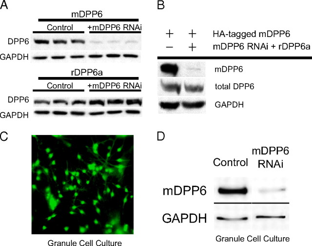
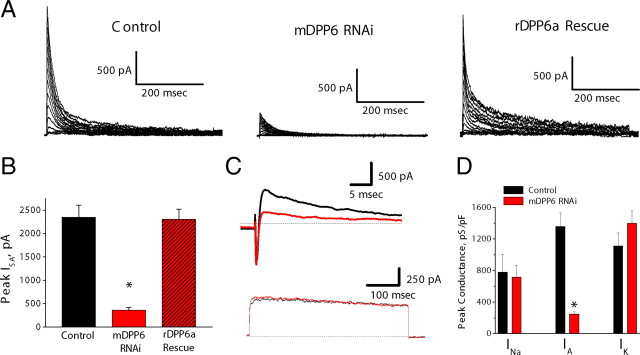
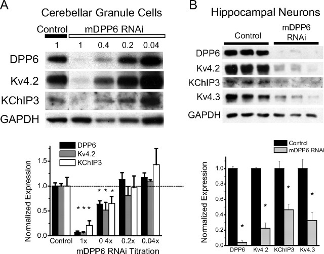
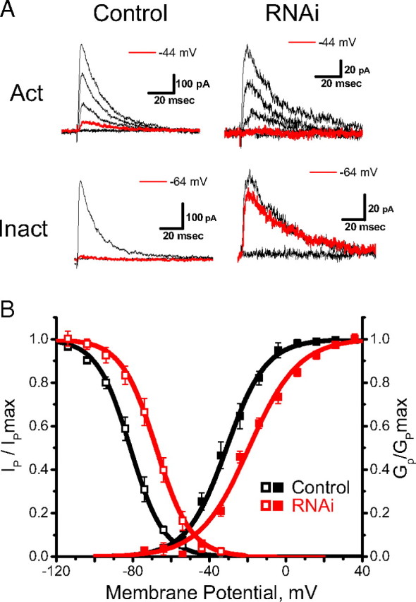
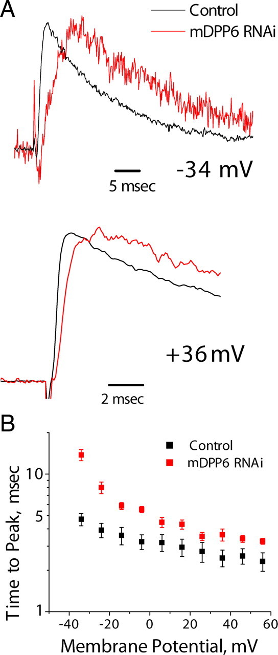
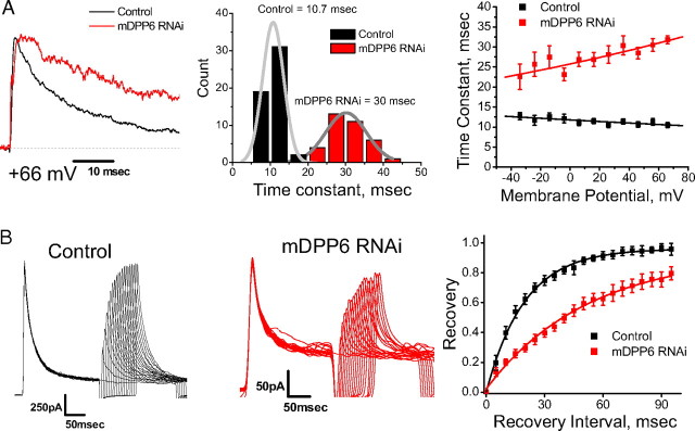
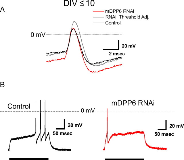
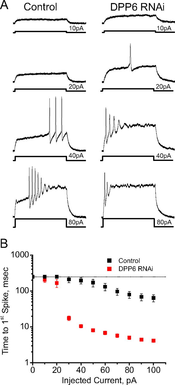
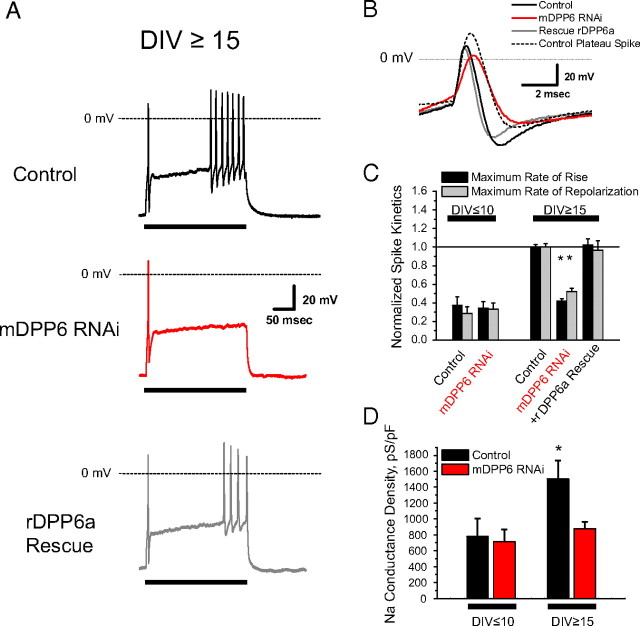
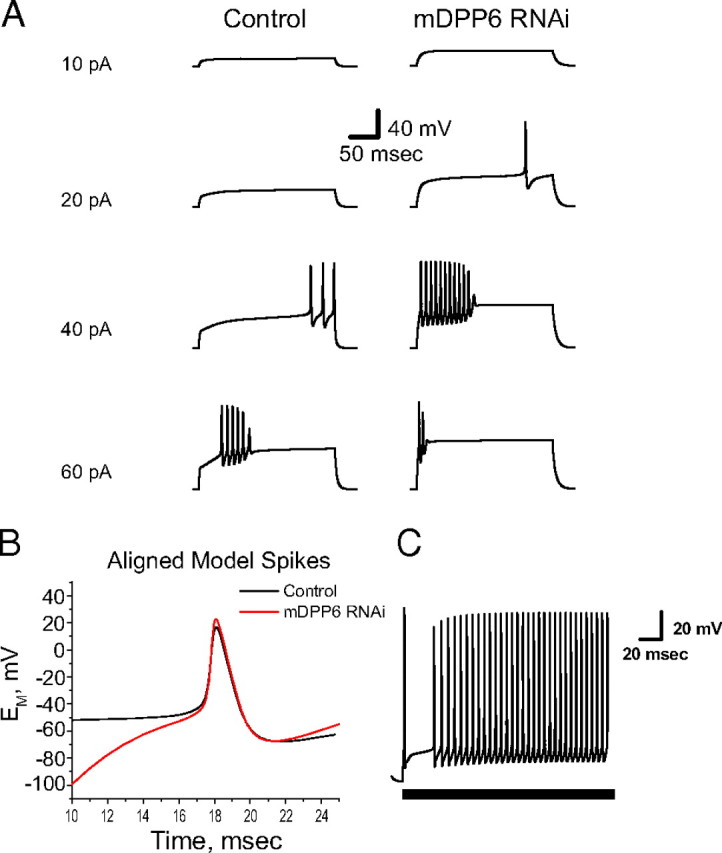
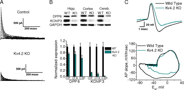
In cerebellar granule (CG) cells and many other neurons, A-type potassium currents play an important role in regulating neuronal excitability, firing patterns, and activity-dependent plasticity. Protein biochemistry has identified dipeptidyl peptidase-like protein 6 (DPP6) as an auxiliary subunit of Kv4-based A-type channels and thus a potentially important regulator of neuronal excitability. In this study, we used an RNA interference (RNAi) strategy to examine the role DPP6 plays in forming and shaping the electrophysiological properties of CG cells. DPP6 RNAi delivered by lentiviral vectors effectively disrupts DPP6 protein expression in CG cells. In response to the loss of DPP6, I(SA) peak conductance amplitude is reduced by >85% in parallel with a dramatic reduction in the level of I(SA) channel protein complex found in CG cells. The I(SA) channels remaining in CG cells after suppression of DPP6 show alterations in gating similar to Kv4 channels expressed in heterologous systems without DPP6. In addition to these effects on A-type current, we find that loss of DPP6 has additional effects on input resistance and Na(+) channel conductance that combine with the effects on I(SA) to produce a global change in excitability. Overall, DPP6 expression seems to be critical for the expression of a high-frequency electrophysiological phenotype in CG cells by increasing leak conductance, A-type current levels and kinetics, and Na(+) current amplitude.


Figures

Figure 1.
Figure 1.
A mouse DPP6 lentiviral-based RNAi suppression and specific rescue system. A, HEK cells were transfected with the indicated DPP6 expression vector, and then treated with control or mDPP6 RNAi vectors. Triplicate Western blots show that mDPP6 RNAi has a strong selective suppression of mDPP6 (n = 9) with no effect on rDPP6a (n = 3). B, Specific rescue vector expressing rDPP6a along with mDPP6 RNAi was coexpressed with mouse HA-DPP6 in HEK cells. Western blots show that the rescue vector suppresses mDPP6, producing a loss of HA-tag immunoreactivity, and replaces it with a similar level of rat DPP6 expression (n = 3). C, CG cells were infected with lentiviral particles generated from the mDPP6 RNAi vector. At optimal dose, >95% of CG cells are expressing eGFP indicative of infection with lentiviral vector and show the expected morphology even after 3 weeks in culture. D, Western blots on CG cells 10 DIV infected with mDPP6 RNAi lentivirus show that level of suppression of DPP6 protein in neurons is similar to that seen in control studies in HEK cells (n = 6).
Figure 2.
Figure 2.
Suppression of CG cell ISA by mDPP6 RNAi and specific rescue by coexpression of rat DPP6a. A, Representative voltage-clamp recordings of ISA isolated from CG cells after infection with indicated lentiviral vector. Infection with mDPP6 RNAi lentiviral vector strongly suppresses ISA and can be specifically rescued by coexpression of the rat DPP6a protein. B, Quantitation of the results shows that mDPP6 RNAi suppresses ISA by >85% compared with control. This effect is reversed when the mDPP6 suppression is compensated by coexpression of rat DPP6a (control, n = 5; mDPP6 RNAi, n = 9; rDPP6a rescue, n = 8). C, Voltage clamp of total membrane currents without pharmacological blockers shows that mDPP6 RNAi (red) selectively suppresses the amplitude of the A-type current compared with control (black). Top, Use of variable prepulse to isolate inactivation sensitive inward Na currents (INa) and outward A-type current (IA). Comparing control (black) with mDPP6 RNAi (red) shows no significant change in INa but a clear reduction in IA. Bottom, No significant changes in outward Kv currents (IK) isolated by a strong test pulse after a prepulse to −34 mV to inactivate INa and IA. D, Quantitation of results from recordings such as those shown in C for INa and IA, and the non-inactivating IK current components in control and mDPP6 RNAi-treated CG cells. Comparison of peak conductance densities for these components reveals that a conductance underlying A-type current is selectively suppressed by mDPP6 RNAi (control, n = 7; mDPP6 RNAi, n = 8). Error bars indicate SEM.
Figure 3.
Figure 3.
ISA channel proteins are reduced in neurons infected with mDPP6 RNAi expressing lentiviral vectors. A, CG cells were infected with a series of dilutions of purified virus to determine an optimal titer for mDPP6 suppression. A series of dilutions (0.4, 0.2, and 0.04×) of the highest titer (1×) were tested by Western blot for the ability to suppress mDPP6 expression. Control granule cells (1×) were infected with the same titer as mDPP6 RNAi-treated cells of the same label (1×). Westerns at 10 DIV show that, along with DPP6, other protein components of the ISA channel complex (Kv4.2 and KChIP3) are specifically lost in a dose-dependent manner. Quantitation of Western blots after normalization using GAPDH shows that at the optimal titer >95% of DPP6 protein is lost from CG cells, with similar suppression of Kv4.2 and KChIP3. Shown are summary data from experiments performed in triplicate on three separate cultures. B, Triplicate Western blots for mouse hippocampal neuron cultures infected with control and mDPP6 RNAi-expressing lentiviral vectors. Westerns for DPP6, Kv4.2, KChIP3, and Kv4.3 all show significant protein reductions after mDPP6 RNAi treatment. A control protein, GAPDH, is not sensitive to mDPP6 RNAi expression. Quantitation of Western blots after normalization to GAPDH signal shows a >95% suppression of DPP6 and 55–80% loss of other ISA associated proteins. Summary data from experiments performed in triplicate on three separate cultures. Error bars indicate SEM.
Figure 4.
Figure 4.
Steady-state inactivation curves and peak activation curves for ISA in control and mDPP6 RNAi-infected CG cells. A, Representative traces for control and mDPP6 RNAi CG cells showing changes in voltage-dependent gating produced by loss of mDPP6. Activation shows currents in response to six voltage steps from −64 to −4 mV. Highlighted trace from −44 mV shows clearly that ISA in control cells activates at more negative potentials than in cells treated with mDPP6 RNAi. Inactivation shows test currents after prepulses to −114, −64, and −34 mV. Intermediate level of inactivation produced by a pulse to −64 mV shows that control ISA inactivates more completely at this potential than ISA in cells treated with mDPP6 RNAi. B, Summary results show that mDPP6 RNAi shifts gating curves to more positive potentials. Scatter plots show average values for activation and inactivation gating parameters for ISA recorded from control and mDPP6 RNAi-treated CG cells. Smooth curves show Boltzmann plots generated using midpoint and slope values obtained by the averages of fits to individual neurons. Curves verify that the average of the fits provides a good representation of the average data values at different voltages. Error bars indicate SEM.
Figure 5.
Figure 5.
Residual ISA after mDPP6 RNAi suppression of DPP6 shows slowed activation kinetics. A, Peak normalized currents for ISA in control and mDPP6 RNAi-treated CG cells in response to voltage steps to the indicated potentials are shown. At both small and large depolarizations, the rate of rise is slower after loss of DPP6. B, Cumulative data showing that time to reach peak current is slower for ISA at all potentials after suppression of DPP6 (control, n = 5; mDPP6 RNAi n = 8). Two-way ANOVA shows that time to peak is significantly slower for mDPP6 RNAi-treated CG cells compared with control. Error bars indicate SEM.
Figure 6.
Figure 6.
Inactivation and recovery kinetics of ISA are slowed after loss of DPP6 from CG cells. A, Peak normalized currents for recordings from control and mDPP6 RNAi-infected CG cells recorded at a test potential of +66 mV show slower decay kinetics after suppression of DPP6. Histogram of all inactivation time constants for fits measured from currents evoked in response to voltage steps between +46 and +66 mV. Control and mDPP6 RNAi inactivation time constants separate into two distinct distributions with loss of DPP6 slowing inactivation by approximately three times. ISA inactivation time constant shows an altered voltage dependence after suppression of DPP6. In control CG cells (n = 9), inactivation time constant accelerates as the test potential is made more positive. After infection with mDPP6 RNAi (n = 13), inactivation time constant slows as the test potential is made more positive. Time constants are significantly slower at all potentials after mDPP6 RNAi treatment by ANOVA. B, Recovery from inactivation for ISA is slower after suppression of DPP6 by RNAi. Currents for control and mDPP6 RNAi-infected neurons recorded using a two-pulse recovery protocol show that the recovery time is longer for ISA in the second pulse after CG cells have been treated with mDPP6 RNAi. Summary recovery curves show recovery from inactivation is approximately three times longer after suppression of DPP6 [control τ, 19.9 ± 2.2 ms (n = 4); DPP6 RNAi τ, 62.0 ± 12.6 ms (n = 8); p < 0.05 by t test]. Error bars indicate SEM.
Figure 7.
Figure 7.
Current-clamp analysis of effects of mDPP6 RNAi on excitability of CG cells at <10 d in culture. A, First spike properties for control and mDPP6 RNAi-treated CG cells show that neurons lacking DPP6 fire from a lower potential. When traces are adjusted for change in threshold, then the mDPP6 RNAi-treated cells are seen to produce spikes with a larger amplitude, but similar sized afterhyperpolarization. B, Sustained current injection at a level 10 pA above the threshold current injection show that control CG cells have a significant delay before firing the first spike. After suppression of DPP6, the delay is dramatically reduced and the CG cells fire during the initial depolarization.
Figure 8.
Figure 8.
Control and mDPP6 RNAi-treated CG cells show distinctive changes in firing properties as sustained current injections are increased in amplitude. Shown are recordings from control and mDPP6 RNAi-expressing CG cells after 10 d or less in culture. A, Control CG cells require higher current injections to reach threshold and maintain a significant delay before firing the first spike at all current amplitudes. CG cells infected with mDPP6 RNAi fire at lower current injections and the delay to first spike gets dramatically shorter as the amplitude of the current injection increases. B, Summary data show that the time to first spike is dramatically shortened with increasing current injections after suppression of mDPP6. The dotted line at 250 ms indicates the maximum duration of the current injection, and thus values at this level indicate a failure to fire a spike (control, n = 8; mDPP6 RNAi, n = 9). Differences are significant for all current injections above threshold. Error bars indicate SEM.
Figure 9.
Figure 9.
Action potential waveforms are different after suppression of DPP6 at later times in culture. Recordings from CG cells cultured for 15 d or more after treatment with the indicated lentiviral vectors. A, At DIV ≥ 15, control CG cells can fire a spike in response to the initial depolarization. In 80% of cells, there is a significant delay between the first spike and the start of repetitive firing. Firing is thus similar to control CG cells at 10 d, except a spike can be generated early and repetitive firing is more regular. For mDPP6 RNAi-treated CG cells, there is an early spike but the cells fail to sustain repetitive firing. Rescue with rDPP6a suggests the loss of repetitive firing is specifically attributable to the loss of DPP6 expression in these cells. B, Analysis of first spike properties shows that, in CG cells treated with mDPP6 RNAi, the spikes are much slower and fail to reach the same height as control cells. This effect is completely reversed by rescue with rDPP6a expression. In control cells, the first plateau spike that initiates the repetitive firing has a larger amplitude but smaller afterhyperpolarization than the initial spike. Rate of rise of the plateau spike is similar to the first spike. The reduced size of the AHP for control neuron plateau spikes is similar to what is seen with mDPP6 RNAi possibly because ISA inactivates during the delay before the start of plateau firing. This analysis suggests that, in control cells, the initial spike amplitude is smaller and afterhyperpolarization is larger because of ISA activation. C, Summary data from phase plot analysis to characterize maximal rate of rise and fall for first spikes recorded under different conditions. At 10 d in culture, spike properties are similar between control and mDPP6 RNAi-treated CG cells. At later times, action potentials in control CG cells become much faster, but in mDPP6 RNAi-treated cells the action potential kinetics remain slow. Spike kinetics can be rescued by coexpression of rDPP6a with mDPP6 RNAi, showing this failure of rapid spike kinetics maturation is a specific effect caused by the loss of DPP6. Maximum rate of rise and maximum rate of repolarization of all conditions were normalized to mean values of control spikes at DIV ≥15 (data from analysis of 6–10 recordings for each condition). D, Na+ channel conductance density is significantly greater in control neurons after 15 d in culture. In comparison, in mDPP6 RNAi-treated CG cells, the Na+ channel conductance density is not significantly changed compared with that observed at 10 d in culture. Error bars indicate SEM.
Figure 10.
Figure 10.
NEURON model of CG cell firing properties shows that most effects of mDPP6 RNAi can be reproduced by simply reducing the amplitude of A-type current. A, Single-compartment model for a CG cell reproduces the basic firing properties of the representative CG cell shown in Figure 8A. Maintaining all the current parameters from the control model and only reducing the amplitude of the ISA components reproduces the altered firing properties of the mDPP6 RNAi-treated neurons. B, Model-generated first spikes at a current injection of 40 pA for control CG cells are peak aligned with spikes for mDPP6 RNAi CG cells. Differences in prepotentials are attributable to the first spike occurring later during the current injection in control CG cell model. Reduction of ISA in mDPP6 RNAi-treated CG cell model only produces a small increase in peak amplitude and spike width, and a slight decrease in threshold potential. AHP is not significantly different between control and mDPP6 RNAi model neurons because of the inactivation of ISA channel in control CG cells before spike initiation during the plateau. C, Firing properties of CG cells at later times in culture can be reproduced by increasing A-type current somatic amplitude and the amplitude of Na+ current in the axon hillock.
Figure 11.
Figure 11.
Kv4.2 KO reduces ISA amplitude, maintains DPP6 expression, and selectively affects action potential repolarization but not action potential rise. A, Representative current traces from wild-type control and Kv4.2 KO cultured CG cells illustrate the dramatic reduction in the levels of ISA in Kv4.2 KO neurons. B, Top, Western blots of hippocampus, cortex, and cerebellum from P4 wild-type and Kv4.2 KO mice for DPP6, KChIP3, and GAPDH. Bottom, Quantitation of DPP6 and KChIP3 immunoreactivity by densitometry and normalization to GAPDH levels reveals a 90–99% decrease in the levels of KChIP3 but only a 25–40% decrease in the levels of DPP6 in brains of Kv4.2 KO animals relative to control. Kv4.2 KO animals were identified by PCR genotyping and the absence of Kv4.2 confirmed by Western blot (data not shown). C, Top, Representative action potentials from wild-type and Kv4.2 KO cerebellar granule cells reveal similar action potential threshold and peaks but wider spike width and shallower AHPs for Kv4.2 KO CG cells relative to control. Bottom, Representative phase plots of CG cell action potentials from control and Kv4.2 KO animals illustrates that only the rate of repolarization but not the rate of rise of the action potential is affected by elimination of Kv4.2. Error bars indicate SEM.

Similar articles

Cited by

References

    1. Aamodt SM, Constantine-Paton M. The role of neural activity in synaptic development and its implications for adult brain function. Adv Neurol. 1999;79:133–144. - PubMed
    1. Alders M, Koopmann TT, Christiaans I, Postema PG, Beekman L, Tanck MW, Zeppenfeld K, Loh P, Koch KT, Demolombe S, Mannens MM, Bezzina CR, Wilde AA. Haplotype-sharing analysis implicates chromosome 7q36 harboring DPP6 in familial idiopathic ventricular fibrillation. Am J Hum Genet. 2009;84:468–476. - PMC - PubMed
    1. Allen M, Heinzmann A, Noguchi E, Abecasis G, Broxholme J, Ponting CP, Bhattacharyya S, Tinsley J, Zhang Y, Holt R, Jones EY, Lench N, Carey A, Jones H, Dickens NJ, Dimon C, Nicholls R, Baker C, Xue L, Townsend E, et al. Positional cloning of a novel gene influencing asthma from chromosome 2q14. Nat Genet. 2003;35:258–263. - PubMed
    1. Amarillo Y, De Santiago-Castillo JA, Dougherty K, Maffie J, Kwon E, Covarrubias M, Rudy B. Ternary Kv4.2 channels recapitulate voltage-dependent inactivation kinetics of A-type K+ channels in cerebellar granule neurons. J Physiol. 2008;586:2093–2106. - PMC - PubMed
    1. An WF, Bowlby MR, Betty M, Cao J, Ling HP, Mendoza G, Hinson JW, Mattsson KI, Strassle BW, Trimmer JS, Rhodes KJ. Modulation of A-type potassium channels by a family of calcium sensors. Nature. 2000;403:553–556. - PubMed

Publication types

MeSH terms

Substances