. 2010 Jun 23;30(25):8602-12.
doi: 10.1523/JNEUROSCI.0633-10.2010.

Disease-modifying effects of phenobarbital and the NKCC1 inhibitor bumetanide in the pilocarpine model of temporal lobe epilepsy

Affiliations

Disease-modifying effects of phenobarbital and the NKCC1 inhibitor bumetanide in the pilocarpine model of temporal lobe epilepsy

Claudia Brandt et al. J Neurosci. .

Abstract

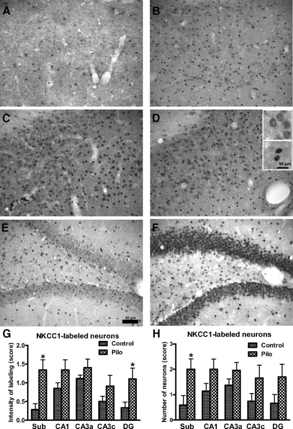
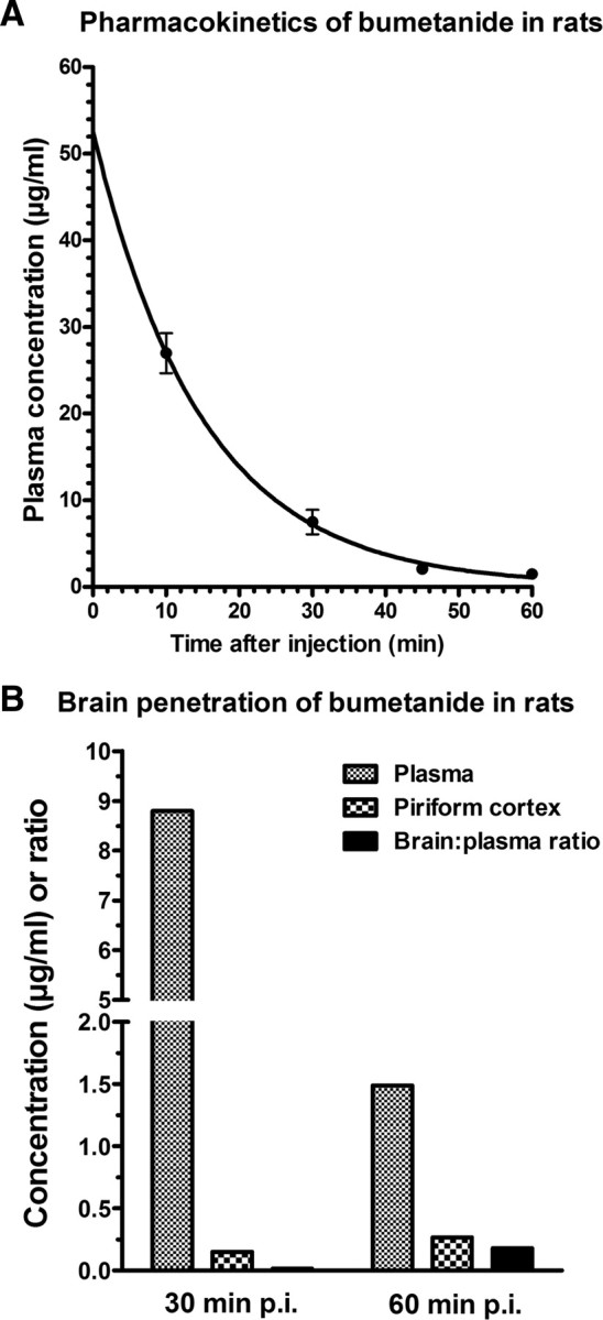
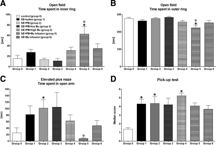
Accumulating evidence suggests that changes in neuronal chloride homeostasis may be involved in the mechanisms by which brain insults induce the development of epilepsy. A variety of brain insults, including status epilepticus (SE), lead to changes in the expression of the cation-chloride cotransporters KCC2 and NKCC1, resulting in intracellular chloride accumulation and reappearance of immature, depolarizing synaptic responses to GABA(A) receptor activation, which may critically contribute to the neuronal hyperexcitability underlying epileptogenesis. In the present study, it was evaluated whether prolonged administration of the selective NKCC1 inhibitor, bumetanide, after a pilocarpine-induced SE modifies the development of epilepsy in adult female rats. The antiepileptic drug phenobarbital, either alone or in combination, was used for comparison. Based on pharmacokinetic studies with bumetanide, which showed extremely rapid elimination and low brain penetration of this drug in rats, bumetanide was administered systemically with different dosing protocols, including continuous intravenous infusion. As shown by immunohistochemistry, neuronal NKCC1 expression was markedly upregulated shortly after SE. Prophylactic treatment with phenobarbital after SE reduced the number of rats developing spontaneous seizures and decreased seizure frequency, indicating a disease-modifying effect. Bumetanide did not exert any significant effects on development of spontaneous seizures nor did it enhance the effects of phenobarbital. However, combined treatment with both drugs counteracted several of the behavioral consequences of SE, which was not observed with single drug treatment. These data do not indicate that bumetanide can prevent epilepsy after SE, but the disease-modifying effect of this drug warrants further studies with more lipophilic prodrugs of bumetanide.


Figures

Figure 1.
Figure 1.
Schematic illustration of the experimental protocol used in this study. With respect to the time windows shown, it should be noted that histological analysis of neuronal damage was only performed in part of the animals.
Figure 2.
Figure 2.
NKCC1-immunostained hippocampal sections of control rats (A, B, E) and rats 24 h (C, F) and 4 d (D) after a pilocarpine-induced SE. Sections shown in A–D are from the subiculum, while sections shown in E and F are from the dentate gyrus. The control section in A does not exhibit any NKCC1 immunostained neurons, which was seen in the majority (6/9) of controls in this region. The few intensively stained cells in A are presumably glial cells. The control section in B shows weakly stained neurons and more intensively stained (presumably glial) cells. Following SE, many cells in the subiculum (C, D) exhibited intensive NKCC1 immunostaining, including both neurons (see upper inset in D for higher magnification) and glial cells (see lower inset in D). Markedly increased NKCC1 immunostaining was also seen in the dentate granule cell layer (F), but not the hilus. No obvious difference in increased NKCC1 staining was determined at 24 h and 4 d following SE, so that data from both time points were put together for the semiquantitative evaluation shown in G (intensity of NKCC1 labeling) and H (number of NKCC1-stained neurons). Data shown in G and H are means ± SEM of 9 controls and 8 SE rats. Analysis of data by two-way ANOVA indicated significant differences between means (p < 0.001). Significant differences between controls and SE rats are indicated by asterisk (p < 0.05). Scale bar in E indicates 50 μm (for A–F), while scale bar in the lower inset in D indicates 10 μm for the two insets.
Figure 3.
Figure 3.
Plasma pharmacokinetics and brain penetration of bumetanide in female Sprague Dawley rats. A shows average (±SEM) plasma concentrations of bumetanide in four rats after intraperitoneal administration of a single dose of 15 mg/kg. Assuming a one-phase exponential decay, best-fit values were calculated by the GraphPad Prism software, resulting in an elimination constant of 0.06694 min−1 and an elimination half-life of 10.36 min. B shows average bumetanide levels in plasma and piriform cortex and the brain:plasma ratio 30 and 60 min after intraperitoneal administration of 15 mg/kg. Data are means of two rats per time point.
Figure 4.
Figure 4.
SRS recorded by continuous video/EEG monitoring 8–9 weeks following SE. Group size was 16 for the group treated 2 weeks with saline after SE (group 1), 8 for the group treated 2 weeks with phenobarbital after SE (group 2), 8 for the group treated 2 weeks with phenobarbital and low-dose bumetanide after SE (group 3), 10 for the group treated 2 weeks with phenobarbital and high-dose bumetanide after SE (group 4), 15 for the group treated 5 d with phenobarbital and continuous infusion of bumetanide after SE (group 5), and 16 for the group treated 5 d by continuous infusion of bumetanide after SE (group 6), respectively. A illustrates seizure incidence, i.e., the percentage rats per group exhibiting spontaneous seizures during the 1 week of continuous video/EEG monitoring. B illustrates the frequency of spontaneous seizures, C seizure severity, and D seizure duration. Data in C and D are shown as means ± SEM, while individual values and median seizure frequency are shown in B. Analysis of data in A by Fisher's exact test indicated that groups 2 and 3 differed significantly from group 1 (p = 0.0139 and 0.0366, respectively; indicated by asterisk), while p was 0.0581 for group 4, thus just failing to reach statistical significance. Analysis of data in B–D by ANOVA indicated significant differences between means for the data on seizure frequency shown in B (p = 0.02615). Post hoc analysis indicated that groups 2, 3, and 5 differed significantly from group 1 (p < 0.05; indicated by asterisk). For group 6, p was 0.05505.
Figure 5.
Figure 5.
Behavioral alterations of epileptic rats (groups 1–6) vs nonepileptic controls (group 0; n = 9). See Figure 4 legend for group size of groups 1–6. All data are shown as means ± SEM. A illustrates the time that rats spent in the aversive inner ring of an open field. B illustrates the time that rats spent in the outer ring of the open field. C illustrates the time rats spent in the aversive open arms of an elevated plus maze. D illustrates behavioral hyperexcitability of epileptic rats in the pick-up test. Analysis of data by ANOVA indicated significant differences between means for A (p = 0.0172), B (p = 0.0063), C (p = 0.0360), and D (p = 0.0007), respectively. Significant differences to controls (group 0) are indicated by asterisk (p < 0.05), significant differences to the SE plus saline group (group 1) by circle (p < 0.05).

References

    1. Armstrong C, Morgan RJ, Soltesz I. Pursuing paradoxical proconvulsant prophylaxis for epileptogenesis. Epilepsia. 2009;50:1657–1669. - PMC - PubMed
    1. Bankstahl JP, Löscher W. Resistance to antiepileptic drugs and expression of P-glycoprotein in two rat models of status epilepticus. Epilepsy Res. 2008;82:70–85. - PubMed
    1. Bankstahl JP, Hoffmann K, Bethmann K, Löscher W. Glutamate is critically involved in seizure-induced overexpression of P-glycoprotein in the brain. Neuropharmacology. 2008;54:1006–1016. - PubMed
    1. Baulac M. Phenobarbital and other barbiturates: clinical efficacy and use in epilepsy. In: Levy RH, Mattson RH, Meldrum BS, Perucca E, editors. Antiepileptic drugs. Ed 5. Philadelphia: Lippincott Williams and Wilkins; 2002. pp. 514–521.
    1. Ben-Ari Y, Holmes GL. The multiple facets of gamma-aminobutyric acid dysfunction in epilepsy. Curr Opin Neurol. 2005;18:141–145. - PubMed

Publication types

MeSH terms