. 2010 Aug 20;285(34):26719-26.
doi: 10.1074/jbc.M110.134619. Epub 2010 Jun 23.

Neurotoxic mutants of the prion protein induce spontaneous ionic currents in cultured cells

Affiliations

Neurotoxic mutants of the prion protein induce spontaneous ionic currents in cultured cells

Isaac H Solomon et al. J Biol Chem. .

Abstract

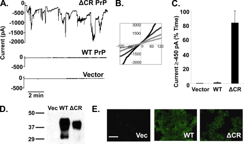
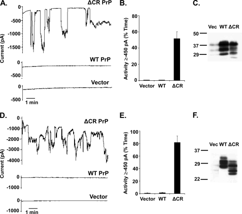
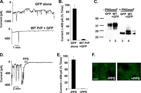
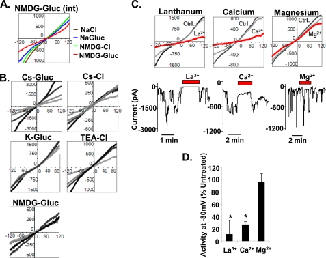
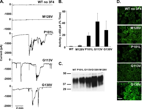
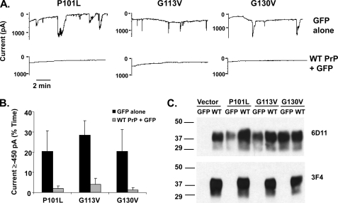
The mechanisms by which prions kill neurons and the role of the cellular prion protein in this process are enigmatic. Insight into these questions is provided by the neurodegenerative phenotypes of transgenic mice expressing prion protein (PrP) molecules with deletions of conserved amino acids in the central region. We report here that expression in transfected cells of the most toxic of these PrP deletion mutants (Delta105-125) induces large, spontaneous ionic currents that can be detected by patch-clamping techniques. These currents are produced by relatively non-selective, cation-permeable channels or pores in the cell membrane and can be silenced by overexpression of wild-type PrP, as well as by treatment with a sulfated glycosaminoglycan. Similar currents are induced by PrP molecules carrying several different point mutations in the central region that cause familial prion diseases in humans. The ionic currents described here are distinct from those produced in artificial lipid membranes by synthetic peptides derived from the PrP sequence because they are induced by membrane-anchored forms of PrP that are synthesized by cells and that are found in vivo. Our results indicate that the neurotoxicity of some mutant forms of PrP is attributable to enhanced ion channel activity and that wild-type PrP possesses a channel-silencing activity. Drugs that block PrP-associated channels or pores may therefore represent novel therapeutic agents for treatment of patients with prion diseases.


Figures

FIGURE 1.
FIGURE 1.
Δ CR PrP induces spontaneous inward currents in HEK cells. A, whole-cell patch clamp recordings were made from HEK cells expressing ΔCR PrP, WT PrP, or vector at a holding potential of −80 mV. B, I-V plots collected at four different times during recording from HEK cells expressing ΔCR PrP. C, current activity recorded from HEK cells expressing vector, WT PrP, or ΔCR PrP was plotted as the percentage of total time the cells exhibited inward current ≥450 pA (mean ± S.E., n = 5 cells) at a holding potential of −80 mV. D, Western blot showing levels of PrP in HEK cells expressing vector (Vec), WT PrP, or ΔCR PrP. Molecular size markers are given in kDa. E, surface immunofluorescence staining of PrP (green) on HEK cells expressing vector, WT PrP, or ΔCR PrP. Scale bar = 50 μm.
FIGURE 2.
FIGURE 2.
Expression of ΔCR PrP in N2a mouse neuroblastoma cells and Sf9 insect cells produces spontaneous inward currents. A, whole-cell patch clamp recordings at −80 mV of N2a cells 24 h after transfection with plasmids encoding ΔCR PrP, WT PrP, or vector. Transfected cells were recognized by expression of GFP, encoded in a co-transfected plasmid. B, quantitation of the currents shown in panel A, plotted as the percentage of total time the cells exhibited inward current ≥450 pA (mean ± S.E., n = 5 cells). C, Western blot to detect PrP in the N2a cells used in panel A. Vec, vector. D, whole-cell patch clamp recordings at −80 mV of Sf9 cells 36 h after transfection with plasmids encoding ΔCR PrP, WT PrP, or vector. Transfected cells were recognized by expression of GFP, encoded in a co-transfected plasmid. E, quantitation of the currents shown in panel D, plotted as the percentage of total time the cells exhibited inward current ≥450 pA (mean ± S.E., n = 5 cells). F, Western blot to detect PrP in the Sf9 cells used in panel D. PrP expressed in Sf9 cells migrates with a lower Mr than in mammalian cells (HEK or N2a) due to differences between insect and mammalian cells in N-linked glycosylation.
FIGURE 3.
FIGURE 3.
ΔCR PrP-induced current is eliminated by overexpression of WT PrP and by treatment with PPS. A, whole-cell patch clamp recordings were made from HEK cells expressing ΔCR PrP at 48 h after infection with lentivirus encoding GFP alone (upper trace) or WT PrP plus GFP (lower trace). The holding potential was −80 mV. B, quantitation of the currents recorded in panel A, plotted as the percentage of total time the cells exhibited inward current ≥450 pA (mean ± S.E., n = 5 cells). C, cells in panel A were analyzed for PrP by Western blotting. Samples in lanes 3 and 4 were enzymatically deglycosylated with N-glycosidase F (PNGaseF), whereas those in lanes 1 and 2 were not. Deglycosylated WT and ΔCR PrP are identified by white and black arrowheads, respectively. D, whole-cell patch clamp recordings were made from HEK cells expressing ΔCR PrP. During the period indicated by the bar (60 s), the cell was perfused with PPS at 100 μg/ml. E, quantitation of the currents recorded in panel D plotted as the percentage of total time the cells exhibited inward current ≥450 pA (mean ± S.E., n = 5 cells). F, surface immunofluorescence staining of PrP (green) on HEK cells expressing ΔCR PrP, with or without a 2-min treatment with PPS. Scale bar = 50 μm.
FIGURE 4.
FIGURE 4.
Biophysical properties of ΔCR PrP-induced currents. A, I-V relationships were recorded from HEK cells expressing ΔCR PrP with a constant internal solution containing NMDG-glucuronate (NMDG-Glu (int)) and four different external solutions: NaCl (brown), sodium glucuronate (NaGluc, blue), NMDG-Cl (green), and NMDG-glucuronate (red). The graph shows a set of I-V plots from a representative single cell. B, I-V relationships were recorded from HEK cells expressing ΔCR PrP with a constant external solution containing NaCl and five different internal solutions: cesium glucuronate (Cs-Gluc), potassium glucuronate (K-Gluc), NMDG-glucuronate, Cs-Cl, and TEA-Cl. Each graph shows a representative single cell at four different time points during the recording. C, I-V relationships (upper graphs) were recorded from HEK cells expressing ΔCR PrP under control (Ctrl) solutions (light and dark gray) or in the presence of various multivalent cations (red): La3+ (15 μm), Ca2+ (12 mm), or Mg2+ (10 mm). The lower traces show patch clamp recordings during application of the multivalent cations. D, quantitation of the current activity recorded in the presence of the indicated cations, expressed as the percentage of activity under control conditions (mean ± S.E., n ≥ 3 cells; *, p < 0.05, significantly different from control).
FIGURE 5.
FIGURE 5.
Disease-associated point mutants in the central region of PrP induce spontaneous inward currents. A, whole-cell patch clamp recordings at a holding potential of −80 mV were made from HEK cells expressing WT, M128V, P101L, G113V, or G130V PrP. PrP molecules did not carry a 3F4 tag. B, quantitation of the currents recorded in panel A, plotted as the percentage of total time the cells exhibited inward current ≥450 pA (mean ± S.E., n = 5 cells). C, Western blot showing relative PrP expression levels of each construct. D, surface immunofluorescence staining of PrP on HEK cells expressing the indicated constructs. Scale bar = 50 μm.
FIGURE 6.
FIGURE 6.
Currents induced by disease-associated point mutants are silenced by overexpression of WT PrP. A, whole-cell patch clamp recordings at a holding potential of −80 mV were made from HEK cells expressing P101L, G113V, or G130V PrP 48 h after infection with lentivirus encoding GFP alone (upper traces) or WT PrP plus GFP (lower traces). B, quantitation of the currents recorded in panel A, plotted as the percentage of total time the cells exhibited inward current ≥450 pA (mean ± S.E., n ≥ 4 cells). Black bars represent GFP alone, and gray bars represent WT PrP plus GFP. C, Western blot showing relative PrP expression levels of each stably transfected cell line after transduction with either WT PrP-encoding or control lentiviruses. Antibody 6D11 recognizes both mutant and WT PrP, whereas antibody 3F4 specifically recognizes WT PrP (which contains the 3F4 epitope).

References

    1. Prusiner S. B. (1998) Proc. Natl. Acad. Sci. U.S.A. 95, 13363–13383 - PMC - PubMed
    1. Prusiner S. B. (ed) (2004) Prion Biology and Diseases, Second Ed., Cold Spring Harbor Laboratory Press, Cold Spring Harbor, New York
    1. Chiesa R., Harris D. A. (2001) Neurobiol. Dis. 8, 743–763 - PubMed
    1. Harris D. A., True H. L. (2006) Neuron 50, 353–357 - PubMed
    1. Büeler H., Fischer M., Lang Y., Bluethmann H., Lipp H. P., DeArmond S. J., Prusiner S. B., Aguet M., Weissmann C. (1992) Nature 356, 577–582 - PubMed

Publication types