

NLRP3 plays a critical role in the development of experimental autoimmune encephalomyelitis by mediating Th1 and Th17 responses
- PMID: 20574004
- PMCID: PMC3593010
- DOI: 10.4049/jimmunol.0904145
NLRP3 plays a critical role in the development of experimental autoimmune encephalomyelitis by mediating Th1 and Th17 responses
Abstract
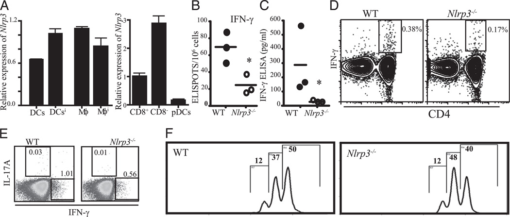
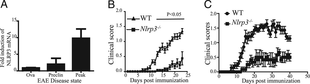
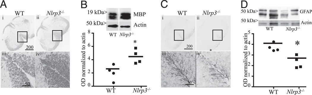
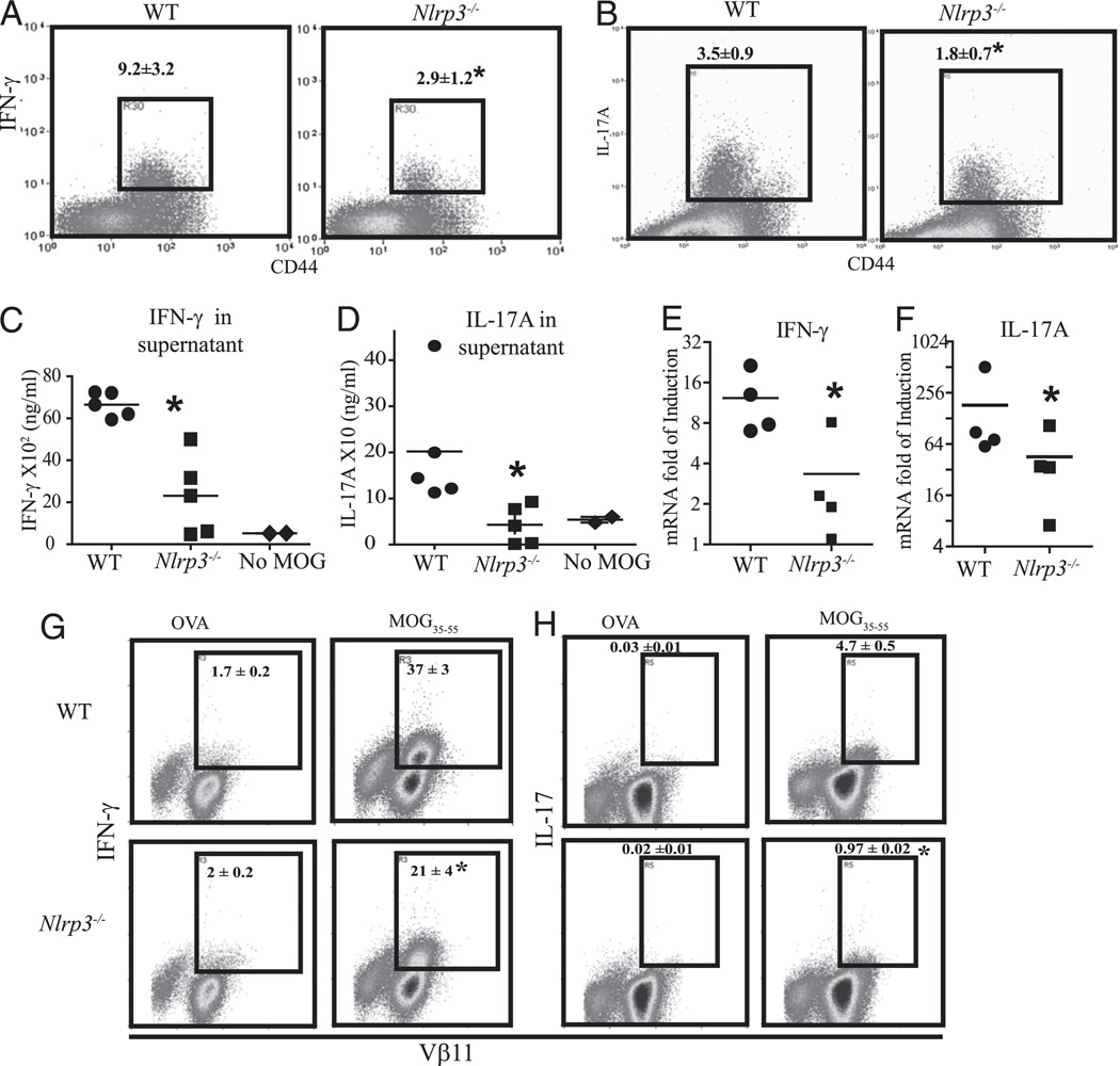
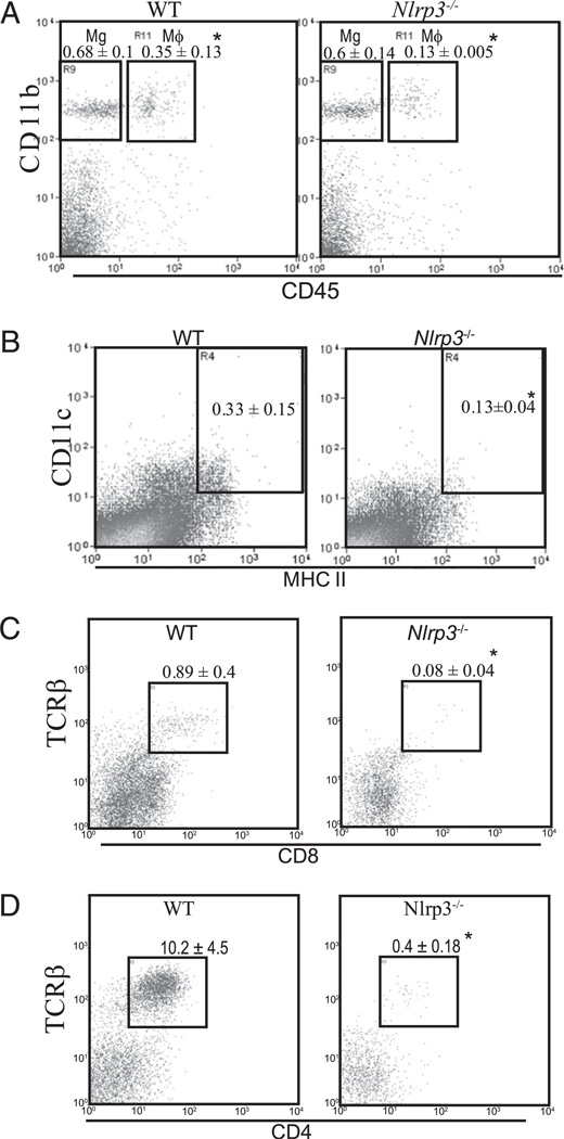
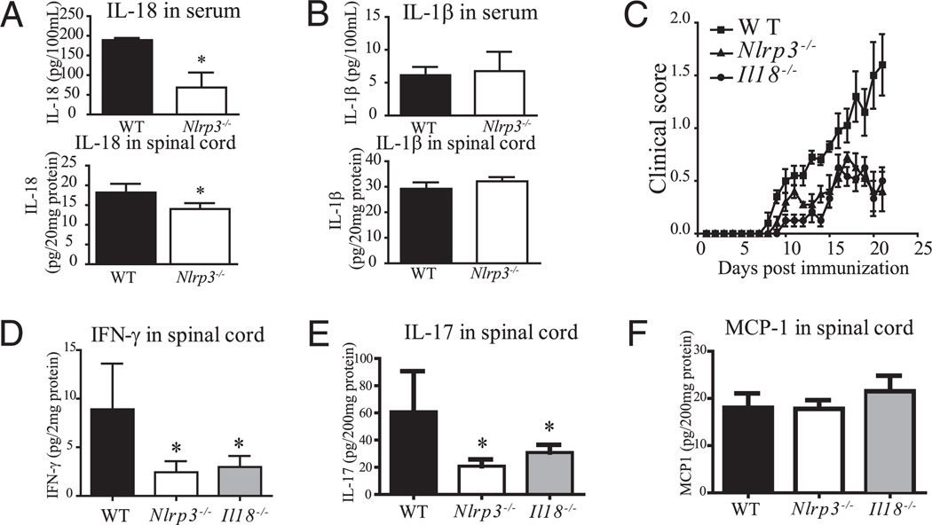
The interplay between innate and adaptive immunity is important in multiple sclerosis (MS). The inflammasome complex, which activates caspase-1 to process pro-IL-1beta and pro-IL-18, is rapidly emerging as a pivotal regulator of innate immunity, with nucleotide-binding domain, leucine-rich repeat containing protein family, pyrin domain containing 3 (NLRP3) (cryopyrin or NALP3) as a prominent player. Although the role of NLRP3 in host response to pathogen associated molecular patterns and danger associated molecular patterns is well documented, its role in autoimmune diseases is less well studied. To investigate the role of NLRP3 protein in MS, we used a mouse model of MS, experimental autoimmune encephalomyelitis (EAE). Nlrp3 expression was elevated in the spinal cords during EAE, and Nlrp3(-/-) mice had a dramatically delayed course and reduced severity of disease. This was accompanied by a significant reduction of the inflammatory infiltrate including macrophages, dendritic cells, CD4, and CD8(+) T cells in the spinal cords of the Nlrp3(-/-) mice, whereas microglial accumulation remained the same. Nlrp3(-/-) mice also displayed improved histology in the spinal cords with reduced destruction of myelin and astrogliosis. Nlrp3(-/-) mice with EAE produced less IL-18, and the disease course was similar to Il18(-/-) mice. Furthermore, Nlrp3(-/-) and Il18(-/-) mice had similarly reduced IFN-gamma and IL-17 production. Thus, NLRP3 plays a critical role in the induction of the EAE, likely through effects on capase-1-dependent cytokines which then influence Th1 and Th17.
Conflict of interest statement
The authors have no financial conflicts of interest.
Figures
Similar articles
-
Nicotinamide adenine dinucleotide treatment alleviates the symptoms of experimental autoimmune encephalomyelitis by activating autophagy and inhibiting the NLRP3 inflammasome.Int Immunopharmacol. 2021 Jan;90:107092. doi: 10.1016/j.intimp.2020.107092. Epub 2020 Dec 4. Int Immunopharmacol. 2021. PMID: 33290962
-
NLRP3 gain-of-function in CD4+ T lymphocytes ameliorates experimental autoimmune encephalomyelitis.Clin Sci (Lond). 2019 Sep 13;133(17):1901-1916. doi: 10.1042/CS20190506. Print 2019 Sep 13. Clin Sci (Lond). 2019. PMID: 31471462
-
Infiltration of Th1 and Th17 cells and activation of microglia in the CNS during the course of experimental autoimmune encephalomyelitis.Brain Behav Immun. 2010 May;24(4):641-51. doi: 10.1016/j.bbi.2010.01.014. Epub 2010 Feb 6. Brain Behav Immun. 2010. PMID: 20138983
-
Inflammasome activation in multiple sclerosis and experimental autoimmune encephalomyelitis (EAE).Brain Pathol. 2017 Mar;27(2):213-219. doi: 10.1111/bpa.12477. Brain Pathol. 2017. PMID: 27997058 Free PMC article. Review.
-
T cells in multiple sclerosis and experimental autoimmune encephalomyelitis.Clin Exp Immunol. 2010 Oct;162(1):1-11. doi: 10.1111/j.1365-2249.2010.04143.x. Clin Exp Immunol. 2010. PMID: 20682002 Free PMC article. Review.
Cited by
-
Interferon-β therapy against EAE is effective only when development of the disease depends on the NLRP3 inflammasome.Sci Signal. 2012 May 22;5(225):ra38. doi: 10.1126/scisignal.2002767. Sci Signal. 2012. PMID: 22623753 Free PMC article.
-
Inflammasome-derived IL-1β regulates the production of GM-CSF by CD4(+) T cells and γδ T cells.J Immunol. 2012 Apr 1;188(7):3107-15. doi: 10.4049/jimmunol.1103308. Epub 2012 Feb 17. J Immunol. 2012. PMID: 22345669 Free PMC article.
-
Ghrelin Attenuates Neuroinflammation and Demyelination in Experimental Autoimmune Encephalomyelitis Involving NLRP3 Inflammasome Signaling Pathway and Pyroptosis.Front Pharmacol. 2019 Nov 6;10:1320. doi: 10.3389/fphar.2019.01320. eCollection 2019. Front Pharmacol. 2019. PMID: 31780940 Free PMC article.
-
PIP-EL: A New Ensemble Learning Method for Improved Proinflammatory Peptide Predictions.Front Immunol. 2018 Jul 31;9:1783. doi: 10.3389/fimmu.2018.01783. eCollection 2018. Front Immunol. 2018. PMID: 30108593 Free PMC article.
-
The inflammasome NLRs in immunity, inflammation, and associated diseases.Annu Rev Immunol. 2011;29:707-35. doi: 10.1146/annurev-immunol-031210-101405. Annu Rev Immunol. 2011. PMID: 21219188 Free PMC article. Review.
References
-
- World Health Organization. Atlas Multiple Sclerosis Resources in the World. Geneva, Switzerland: World Health Organization Press; 2008.
-
- Bettelli E. Building different mouse models for human MS. Ann. N. Y. Acad. Sci. 2007;1103:11–18. - PubMed
-
- Ito A, Matejuk A, Hopke C, Drought H, Dwyer J, Zamora A, Subramanian S, Vandenbark AA, Offner H. Transfer of severe experimental autoimmune encephalomyelitis by IL-12– and IL-18–potentiated T cells is estrogen sensitive. J. Immunol. 2003;170:4802–4809. - PubMed
Publication types
MeSH terms
Substances
Grants and funding
LinkOut - more resources
Full Text Sources
Other Literature Sources
Medical
Molecular Biology Databases
Research Materials
Miscellaneous
