

Diffusion-weighted imaging improves the diagnostic accuracy of conventional 3.0-T breast MR imaging
- PMID: 20574085
- PMCID: PMC2897691
- DOI: 10.1148/radiol.10091367
Diffusion-weighted imaging improves the diagnostic accuracy of conventional 3.0-T breast MR imaging
Abstract
Purpose: To evaluate the incremental value of diffusion-weighted (DW) imaging and apparent diffusion coefficient (ADC) mapping in relation to conventional breast magnetic resonance (MR) imaging in the characterization of benign versus malignant breast lesions at 3.0 T.
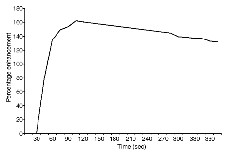
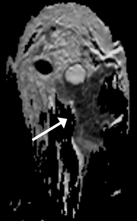
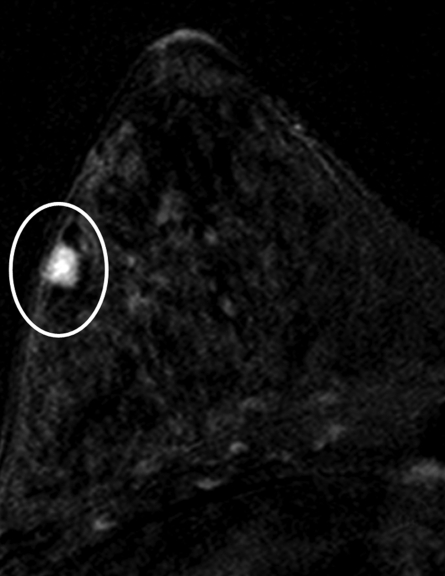
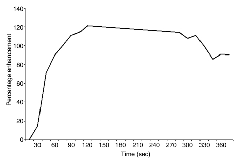
Materials and methods: This retrospective HIPAA-compliant study was approved by the institutional review board, with the requirement for informed patient consent waived. Of 550 consecutive patients who underwent bilateral breast MR imaging over a 10-month period, 93 women with 101 lesions met the following study inclusion criteria: They had undergone three-dimensional (3D) high-spatial-resolution T1-weighted contrast material-enhanced MR imaging, dynamic contrast-enhanced MR imaging, and DW imaging examinations at 3.0 T and either had received a pathologic analysis-proven diagnosis (96 lesions) or had lesion stability confirmed at more than 2 years of follow-up (five lesions). DW images were acquired with b values of 0 and 600 sec/mm(2). Regions of interest were drawn on ADC maps of breast lesions and normal glandular tissue. Morphologic features (margin, enhancement pattern), dynamic contrast-enhanced MR results (semiquantitative kinetic curve data), absolute ADCs, and glandular tissue-normalized ADCs were included in multivariate models to predict a diagnosis of benign versus malignant lesion.
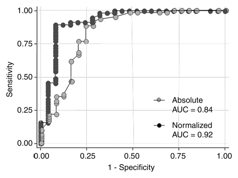
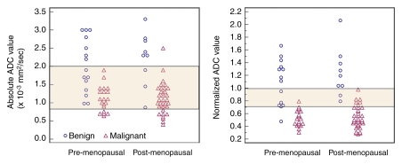
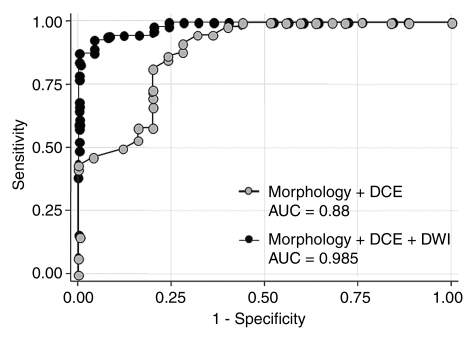
Results: Forty-one (44%) of the 93 patients were premenopausal, and 52 (56%) were postmenopausal. Thirty-three (32.7%) of the 101 lesions were benign, and 68 (67.3%) were malignant. Normalized ADCs were significantly different between the benign (mean ADC, 1.1 x 10(-3) mm(2)/sec +/- 0.4 [standard deviation]) and malignant (mean ADC, 0.55 x 10(-3) mm(2)/sec +/- 0.16) lesions (P < .001). Adding normalized ADCs to the 3D T1-weighted and dynamic contrast-enhanced MR data improved the diagnostic performance of MR imaging: The area under the receiver operating characteristic curve improved from 0.89 to 0.98, and the false-positive rate decreased from 36% (nine of 25 lesions) to 24% (six of 25 lesions).
Conclusion: DW imaging with glandular tissue-normalized ADC assessment improves the characterization of breast lesions beyond the characterization achieved with conventional 3D T1-weighted and dynamic contrast-enhanced MR imaging at 3.0 T.
Conflict of interest statement
Authors stated no financial relationship to disclose.
Figures
References
-
- Flickinger FW, Allison JD, Sherry RM, Wright JC. Differentiation of benign from malignant breast masses by time-intensity evaluation of contrast enhanced MRI. Magn Reson Imaging 1993;11(5):617–620 - PubMed
-
- Huang W, Fisher PR, Dulaimy K, Tudorica LA, O’Hea B, Button TM. Detection of breast malignancy: diagnostic MR protocol for improved specificity. Radiology 2004;232(2):585–591 - PubMed
-
- Kneeshaw PJ, Lowry M, Manton D, Hubbard A, Drew PJ, Turnbull LW. Differentiation of benign from malignant breast disease associated with screening detected microcalcifications using dynamic contrast enhanced magnetic resonance imaging. Breast 2006;15(1):29–38 - PubMed
-
- Bluemke DA, Gatsonis CA, Chen MH, et al. Magnetic resonance imaging of the breast prior to biopsy. JAMA 2004;292(22):2735–2742 - PubMed
MeSH terms
Substances
Grants and funding
LinkOut - more resources
Full Text Sources
Other Literature Sources
Medical
Miscellaneous
