

Detectability of urinary stones on virtual nonenhanced images generated at pyelographic-phase dual-energy CT
- PMID: 20574095
- PMCID: PMC3968072
- DOI: 10.1148/radiol.10091411
Detectability of urinary stones on virtual nonenhanced images generated at pyelographic-phase dual-energy CT
Abstract
Purpose: To evaluate the detectability of urinary stones on virtual nonenhanced images generated at pyelographic-phase dual-energy computed tomography (CT).
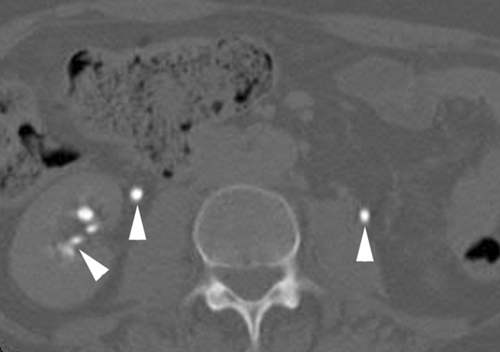
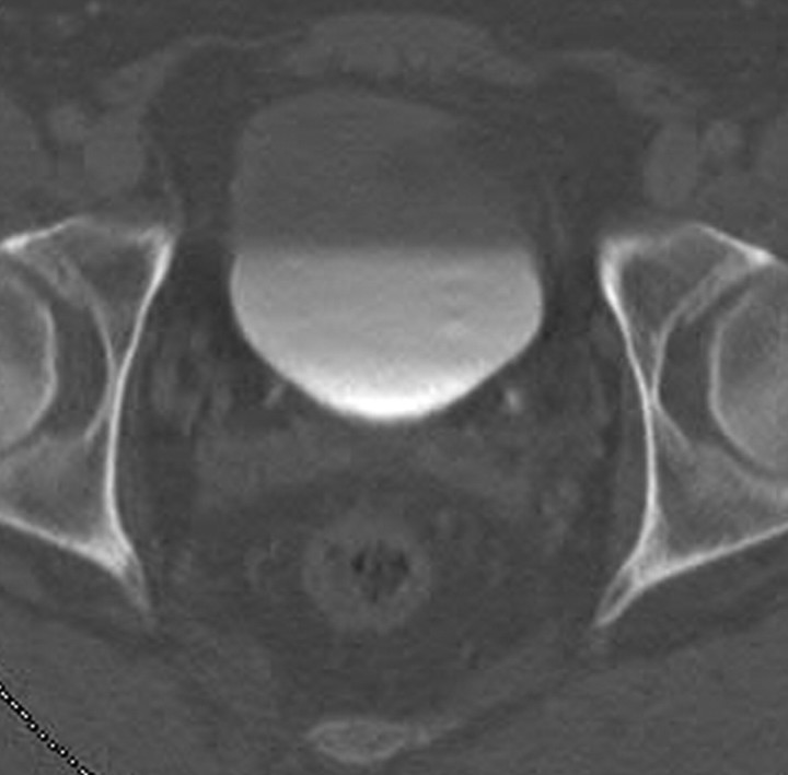
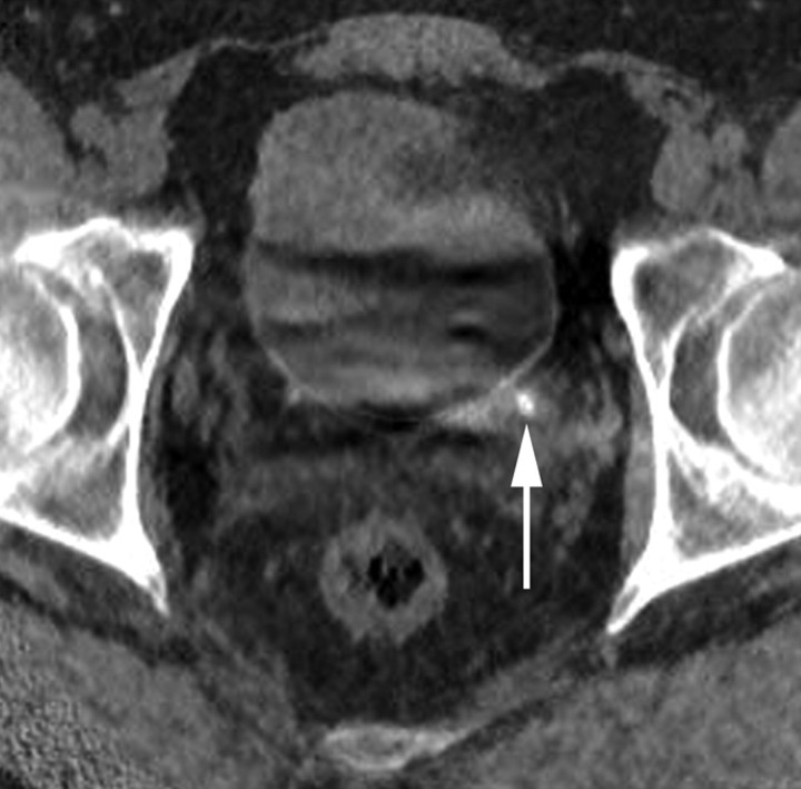
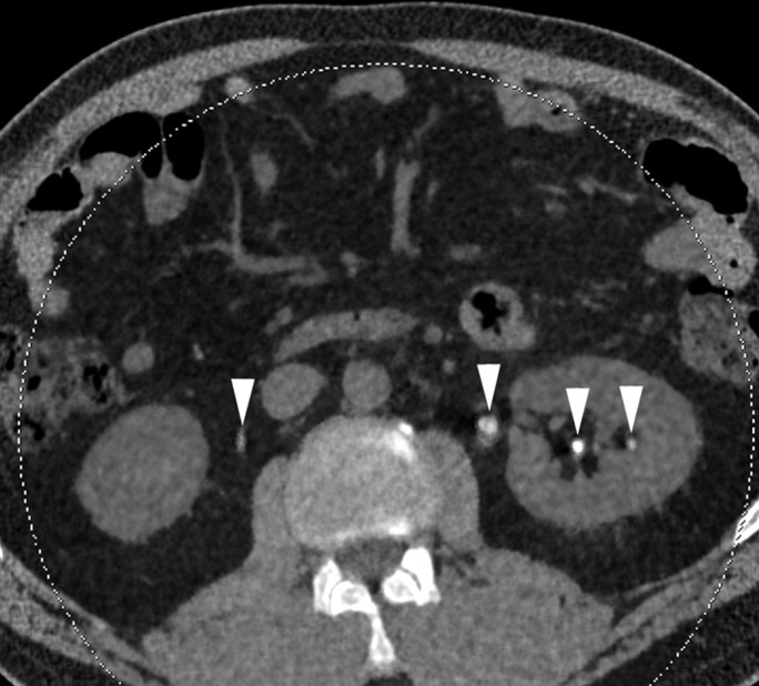
Materials and methods: This retrospective HIPAA-compliant study was institutional review board approved. All included patients had previously consented to the use of their medical records for research. Sixty-two patients (38 men, 24 women; age range, 35-91 years) had undergone CT urography, which consisted of nonenhanced and pyelographic-phase dual-energy CT performed by using a dual-source scanner. Commercial software was used to create virtual nonenhanced images by suppressing the iodine signal from the pyelographic-phase dual-energy CT scans. Two radiologists, in consensus, evaluated the virtual nonenhanced images for the presence of stones. Sensitivity for detecting stones was calculated on a per-stone basis. Sensitivity, specificity, and accuracy were also calculated on a per-renal unit (defined as the intrarenal collecting system and ureter of one kidney) basis. The true nonenhanced scan was considered the reference standard. A jackknife method was used because any patient may have multiple stones.
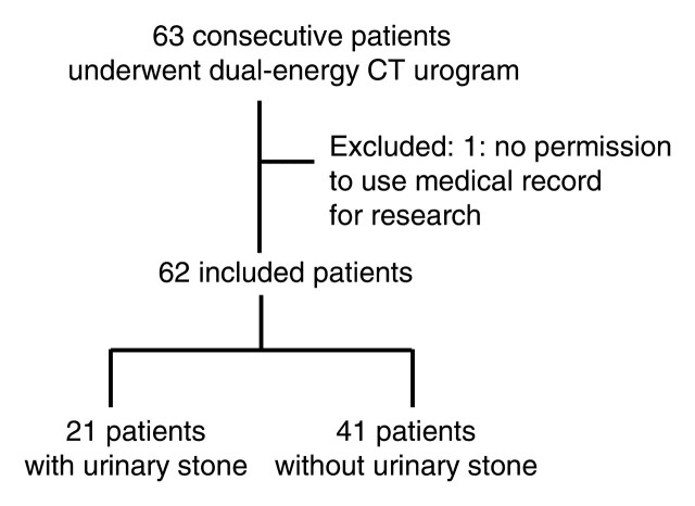
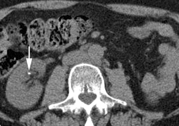
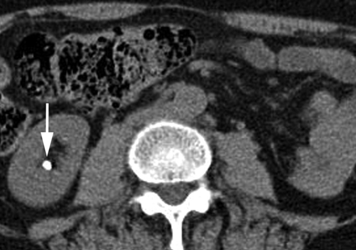
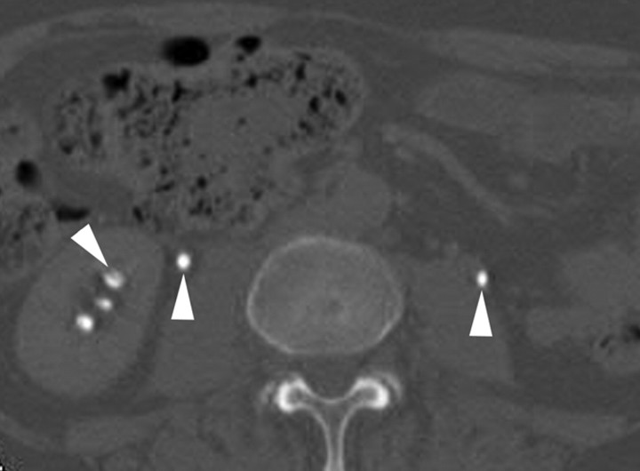
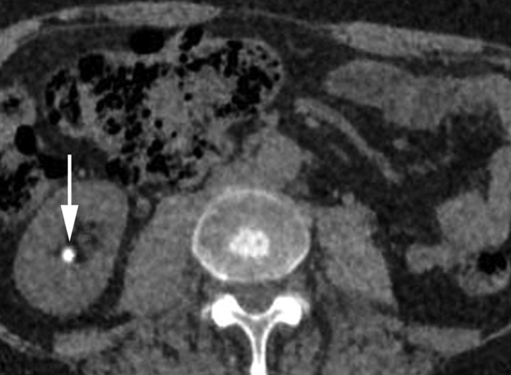
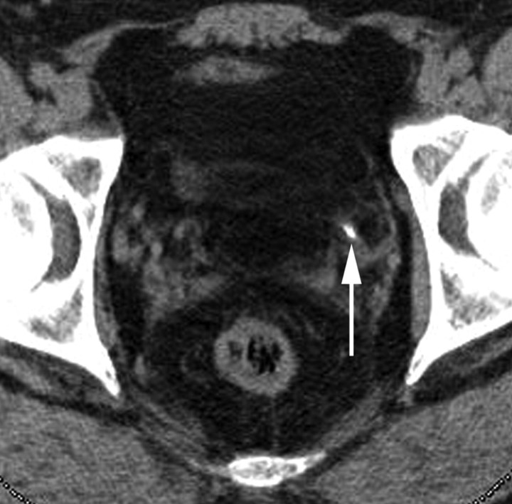
Results: Of 62 patients with 122 renal units, 21 patients with 25 renal units had a total of 43 stones (maximal transverse diameter range, 1-24 mm; median, 3 mm). The overall sensitivity for detecting stones was 63% (27 of 43 stones) per stone. Sensitivities were 29% (four of 14 stones) for 1-2-mm stones, 64% (nine of 14 stones) for 3-4-mm stones, 83% (five of six stones) for 5-6-mm stones, and 100% (nine of nine stones) for 7-mm or larger (7, 7, 7, 8, 8, 9, 11, 15, and 24 mm) stones. All three ureteral stones (3, 4, and 8 mm) were correctly identified. The sensitivity, specificity, and accuracy for detecting stones on a per-renal unit basis were 65% (17 of 26 renal units), 92% (88 of 96 renal units), and 86% (105 of 122 renal units), respectively.
Conclusion: Virtual nonenhanced images generated at pyelographic-phase dual-energy CT enabled the detection of urinary stones with moderate accuracy. The detection of small (1-2-mm) stones was limited.
Figures
References
-
- Millner MR, McDavid WD, Waggener RG, Dennis MJ, Payne WH, Sank VJ. Extraction of information from CT scans at different energies. Med Phys 1979;6(1):70–71. - PubMed
-
- Chiro GD, Brooks RA, Kessler RM, et al. . Tissue signatures with dual-energy computed tomography. Radiology 1979;131(2):521–523. - PubMed
-
- Johnson TR, Nikolaou K, Wintersperger BJ, et al. . Dual-source CT cardiac imaging: initial experience. Eur Radiol 2006;16(7):1409–1415. - PubMed
-
- Johnson TR, Krauss B, Sedlmair M, et al. . Material differentiation by dual energy CT: initial experience. Eur Radiol 2007;17(6):1510–1517. - PubMed
MeSH terms
Substances
Grants and funding
LinkOut - more resources
Full Text Sources
Medical
