. 2004 May;18(2):157-73.
doi: 10.1055/s-2004-829050.

Breast reconstruction in private practice

Affiliations

Breast reconstruction in private practice

Steven M Pisano et al. Semin Plast Surg. 2004 May.

Abstract

Comprehensive breast reconstruction can be performed in private practice. Our practice philosophy is that autogenous tissue provides the best substrate for breast reconstruction; the deep inferior epigastric perforator flap is our primary method of breast reconstruction. Microsurgical training and a group practice model permit routine use of all autogenous tissue techniques. Office, operating room, and hospital teams must be assembled; these teams follow clinical pathways, which make the execution of reconstructive procedures consistent and efficient. The practice must implement a plan for physician and patient education. The practice must review clinical outcomes, making adjustments in operative techniques and pre- and postoperative clinical pathways so that the best results can be achieved with a low complication rate. Breast reconstruction is a core service of our practice. We have accrued an economy of scale including these features: intraoperative and clinical efficiency, low practice overhead costs, and a high patient satisfaction rate.

Keywords: Autogenous tissue; clinical pathways; efficiency.


Figures

Figure 1
Figure 1
A 33-year-old woman 2 years after immediate right breast reconstruction with a muscle-sparing free TRAM flap and left breast mastopexy.
Figure 2
Figure 2
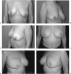
A 51-year-old woman 1 year after immediate right breast reconstruction with a muscle-sparing free TRAM flap and right breast mastopexy. (A, B, C) Preoperative views. (D, E, F) Postoperative views.
Figure 3
Figure 3
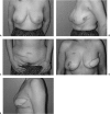
A 46-year-old woman with a failed tissue expander implant 5 months after delayed left breast reconstruction with a muscle-sparing free TRAM flap and right breast mastopexy. (A, B) Preoperative views. (C) Preoperative view of abdominal wall; patient has a history of multiple surgical procedures. (D, E) Postoperative views.
Figure 4
Figure 4
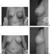
A 38-year-old woman 6 months after delayed left breast reconstruction with a muscle-sparing free TRAM flap. (A, B) Preoperative views; note the radiation dermatitis of the left and central chest wall areas. (C, D) Postoperative views.
Figure 5
Figure 5
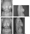
A 51-year-old woman 9 months after bilateral breast reconstruction with free DIEP flaps. (A, B) Preoperative views. (C, D) Postoperative views; the patient is 1 month status post—nipple-areola reconstruction.
Figure 6
Figure 6
(A–D) A 65-year-old woman 5 months after bilateral capsulectomy-implant removal with bilateral free DIEP flap reconstruction. The patient presented with capsular pain and contracture; she underwent bilateral subcutaneous mastectomies, through a Wise reduction pattern, followed by multiple implant reconstructions. Note the thin soft tissue envelope.
Figure 7
Figure 7
A patient with a left malignant phyllodes tumor who underwent a mastectomy, including a large portion of breast and chest wall skin, followed by immediate reconstruction with a muscle-sparing free TRAM flap. (A, B) Preoperative views. (C, D) Postoperative views.
Figure 8
Figure 8
(A) Preoperative and (B) 2-year postoperative views of a 38-year-old patient with left breast ductal carcinoma in situ (DCIS); the preoperative view shows the breast boundaries and skin-sparing pattern; the postoperative result shows the reconstruction with a free TRAM flap.
Figure 9
Figure 9
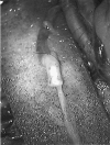
The venous microanastomosis set-up: the internal mammary and deep inferior epigastric vessels are immobilized in a double aneurysm clip; a running anastomosis is performed.
Figure 10
Figure 10
The microneurorrhaphy between the anterior branch of the fourth intercostal nerve and 11th intercostal branch of a DIEP flap; an nerve intubation technique using an arterial segment is shown.
Figure 11
Figure 11
A unilateral muscle-sparing free TRAM flap reconstruction complicated by necrosis of the native breast skin. (A) Preoperative view. (B) Postoperative view depicting native skin loss. (C, D) Long-term postoperative results following TRAM flap and native skin revision and nipple-areola reconstruction.
Figure 12
Figure 12
Partial dehiscence of a free TRAM donor site.
Figure 13
Figure 13
Decreases in reconstructive operative times follows implementation of surgical and operating room clinical pathways.
Figure 14
Figure 14
Decreases in flap ischemia times for three groups: 1996, 1999, and 2002.
Figure 15
Figure 15
A 28-year-old woman with severe symptomatic fibrocystic disease who underwent nipple-areola–sparing mastectomies and bilateral free DIEP flap reconstruction. (A) Preoperative view. (B) Postoperative view.

References

    1. Kroll S S. Why autologous tissue? Clin Plast Surg. 1998;25:135–143. - PubMed
    1. Miller M J. Immediate breast reconstruction. Clin Plast Surg. 1998;25:145–156. - PubMed
    1. Craigie J E, Allen R J, DellaCroce F J, Sullivan S K. Autogenous breast reconstruction with the deep inferior epigastric perforator flap. Clin Plast Surg. 2003;30:359–369. - PubMed
    1. Asko-Seljavaara S. Delayed breast reconstruction. Clin Plast Surg. 1998;25:157–166. - PubMed
    1. Schusterman M A. The free TRAM flap. Clin Plast Surg. 1998;25:191–195. - PubMed