. 2010 Jun 21;5(6):e11222.
doi: 10.1371/journal.pone.0011222.

Low doses of ionizing radiation promote tumor growth and metastasis by enhancing angiogenesis

Affiliations

Low doses of ionizing radiation promote tumor growth and metastasis by enhancing angiogenesis

Inês Sofia Vala et al. PLoS One. .

Abstract

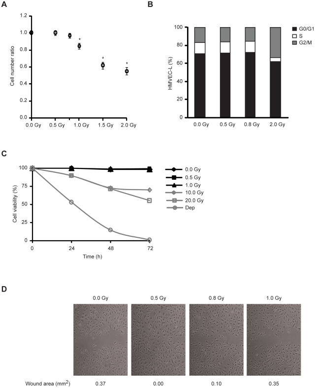
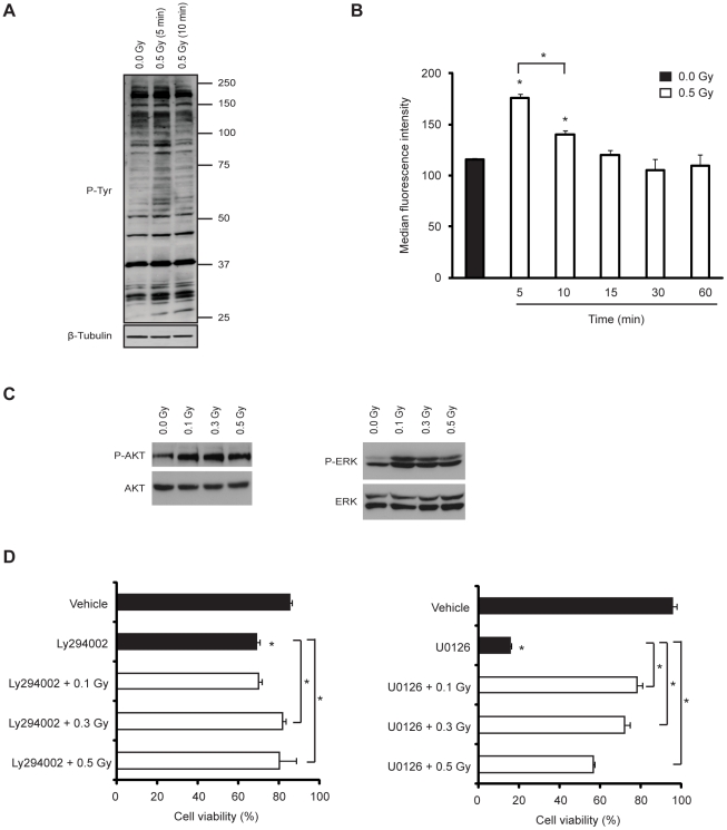
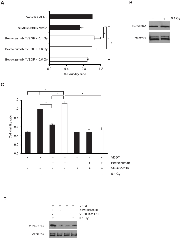
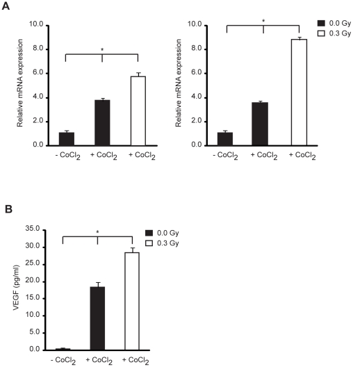
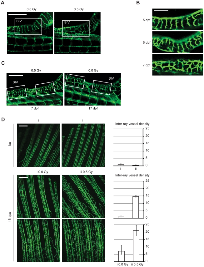
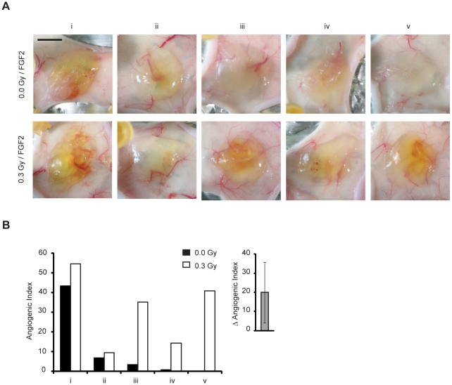
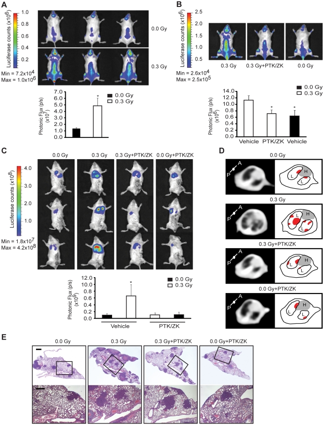
Radiotherapy is a widely used treatment option in cancer. However, recent evidence suggests that doses of ionizing radiation (IR) delivered inside the tumor target volume, during fractionated radiotherapy, can promote tumor invasion and metastasis. Furthermore, the tissues that surround the tumor area are also exposed to low doses of IR that are lower than those delivered inside the tumor mass, because external radiotherapy is delivered to the tumor through multiple radiation beams, in order to prevent damage of organs at risk. The biological effects of these low doses of IR on the healthy tissue surrounding the tumor area, and in particular on the vasculature remain largely to be determined. We found that doses of IR lower or equal to 0.8 Gy enhance endothelial cell migration without impinging on cell proliferation or survival. Moreover, we show that low-dose IR induces a rapid phosphorylation of several endothelial cell proteins, including the Vascular Endothelial Growth Factor (VEGF) Receptor-2 and induces VEGF production in hypoxia mimicking conditions. By activating the VEGF Receptor-2, low-dose IR enhances endothelial cell migration and prevents endothelial cell death promoted by an anti-angiogenic drug, bevacizumab. In addition, we observed that low-dose IR accelerates embryonic angiogenic sprouting during zebrafish development and promotes adult angiogenesis during zebrafish fin regeneration and in the murine Matrigel assay. Using murine experimental models of leukemia and orthotopic breast cancer, we show that low-dose IR promotes tumor growth and metastasis and that these effects were prevented by the administration of a VEGF receptor-tyrosine kinase inhibitor immediately before IR exposure. These findings demonstrate a new mechanism to the understanding of the potential pro-metastatic effect of IR and may provide a new rationale basis to the improvement of current radiotherapy protocols.


Conflict of interest statement

Competing Interests: The authors have declared that no competing interests exist.

Figures

Figure 1
Figure 1. Low-dose IR promotes endothelial cell migration without causing cell cycle arrest or apoptosis.
(A) HMVEC-L were plated at equal densities and, after 12 h, left untreated or exposed to 0.5, 0.8, 1.0, 1.5 and 2.0 Gy. After 72 h, the cells were counted using a Nucleocounter. The values (means ± s.d.) represent the ratio between cell number of irradiated and non-irradiated conditions and are derived from four independent experiments. * P<0.02. (B) HMVEC-L were exposed or not to 0.5, 0.8 and 2.0 Gy. Cell cycle profiles were assessed by flow cytometric analysis after 72 h of culture. Data are representative of three independent experiments. (C) The percentage of apoptotic cells was determined by flow cytometry at the indicated time. Cells cultured without serum (Dep) were used as cell death control. Values are given as the percentage of viable cells (Annexin V, PI negative) remaining in culture. Data are shown as mean in triplicate culture and are representative of three independent experiments. (D) Confluent monolayers of HMVEC-L were subjected to in vitro wound healing and exposed or not to 0.5, 0.8 or 1.0 Gy. Photographs were taken immediately (not shown) and 9 h after wounding. Quantification of the wound area (in mm2) is presented below the images. Data are representative of five independent experiments.
Figure 2
Figure 2. Low-dose IR activates PI3K/Akt and MEK/ERK pathways and prevents apoptosis induced by their inhibition.
(A and B) HMVEC-L were exposed or not to 0.5 Gy and incubated for the time indicated. (A) Representative blots from three independent experiments. Top, Tyrosine phosphorylation Western blot; Bottom, β-tubulin Western blot to verify equal sample loading. (B) Tyrosine phosphorylation levels were assessed by flow cytometry. The values (means ± s.d.) represent the ratio between median fluorescence intensity of irradiated cells and non-irradiated cells and are derived from four independent experiments. (C) Representative blots from three independent experiments. Western blot analysis of total-, phospho-Akt (P-AKT) (left) and total-, phospho-ERK (P-ERK) (right) of HMVEC-L exposed or not to 0.1, 0.3 or 0.5 Gy. The levels of Akt and ERK phosphorylation were assessed after 5 or 60 min post-irradiation, respectively. (D) HMVEC-L were incubated in the presence or absence of a specific inhibitor of PI3K (Ly294002-50 µM) (left) or MAPK (U0126-10 µM) (right) and then exposed or not to 0.1, 0.3 or 0.5 Gy. Cells cultured with vehicle alone were used as a control. Cells were double stained with Annexin-V and propidium iodide at 36 h post-irradiation. The percentage of apoptotic cells was assessed by flow cytometry. The values (means ± s.d.) are given as the percentage of viable cells and are derived from four independent experiments. * P<0.03.
Figure 3
Figure 3. Low-dose IR protects microvasculature from bevacizumab-induced cell death by inducing VEGFR-2 activation.
(A) Cells were cultured without serum for 12 h and incubated with vehicle/VEGF (20 ng/ml) or bevacizumab (0.25 mg/ml)/VEGF (20 ng/ml) mixtures. Then, cells were exposed or not to 0.1, 0.3 or 0.5 Gy and the percentage of apoptotic cells was assessed by flow cytometry at 48 h post-irradiation. Data (means ± s.d.) represent the ratio between cell viability percentage of each experimental condition and control condition and are derived from four independent experiments. (B) Representative blots from four independent experiments. Cells were exposed or not to 0.1 Gy. Western blot analysis of total- and phospho-VEGFR-2. (C) Cells were cultured without serum for 12 h and treated or not with VEGFR-2 tyrosine kinase inhibitor (TKI at 300 nM) for 2 h and stimulated or not with VEGF (20 ng/ml) or bevacizumab (0.25 mg/ml)/VEGF (20 ng/ml) mixture. Then, cells were exposed or not to 0.1 Gy. The percentage of apoptotic cells was assessed by flow cytometry at 48 h post-irradiation. Data (means ± s.d.) represent the ratio between cell viability percentage of each experimental condition and control condition and are derived from four independent experiments. * P<0.03. (D) Representative blots from four independent experiments. Western-blot analysis of total- and phospho-VEGFR-2 (P-VEGFR-2) of HMVEC-L cultured without serum for 12 h and treated or not with VEGFR-2 tyrosine kinase inhibitor (TKI at 300 nM) for 2 h and stimulated with VEGF (20 ng/ml) or bevacizumab (0.25 mg/ml)/VEGF (20 ng/ml) mixture. (B and D) The level of VEGFR-2 phosphorylation was assessed after 15 min post-irradiation.
Figure 4
Figure 4. Low dose IR enhances hypoxia-induced VEGF expression.
Cells were cultured with or without CoCl2 (150 µM) in normoxia to mimic hypoxic conditions and immediately exposed or not to 0.3 Gy of IR. (A) 4 h (left graph) and 12 h (right graph) post-irradiation, VEGF mRNA was quantified by qRT-PCR. Data (means ± s.d.) represent the fold change in gene expression relative to the internal calibrator (−CoCl2) in triplicate measurements and are representative of three independent experiments. (B) 72 h post-irradiation, VEGF protein was assessed by VEGF ELISA Kit. Data (means ± s.d.) indicate the VEGF concentration in quadruplicate measurements and are representative of three independent experiments. * P<0.03.
Figure 5
Figure 5. Low-dose IR accelerates angiogenic sprouting during zebrafish embryonic development and enhances angiogenesis during fin regeneration.
(A–C) Live zebrafish embryos were exposed or not to 0.5 Gy IR 3 d post-fertilization (dpf). Representative images of Sub-Intestinal Vessels (SIV), from (A) a non-irradiated and irradiated zebrafish at 7 dpf; (B) an irradiated zebrafish at 5 (top), 6 (middle) and 7 dpf (bottom); (C) an irradiated zebrafish at 7 dpf and a non-irradiated zebrafish at 17 dpf. Scale bars, 250 µm (A and C), 100 µm (B). (D) Fli1:EGFP adult zebrafish caudal fins were amputated at mid-fin level, exposed or not to 0.5 Gy of IR and then allowed to recover. Representative images from vasculature of two zebrafish fins (i and ii) before amputation ensure they had identical vasculature (top). Representative images from vasculature of two different fin areas of the same zebrafish, 16 days post-amputation (dpa), with or without low-dose IR treatment (middle and bottom). Each image was quantified for inter-ray vessel density. Data are shown as mean and error bars indicate maximum and minimum values. Images are representative of 10 zebrafish in five independent experiments. Scale bars, 250 µm.
Figure 6
Figure 6. Low-dose IR enhances angiogenesis in Matrigel plug assay.
Angiogenesis was induced by injection of growth factor-depleted Matrigel with FGF2. (A) Macroscopic evaluation of the Matrigel plugs explanted 5 d after injection in non-irradiated and 0.3 Gy preirradiated area. Scale bars, 500 µm. (B) Angiogenesis was quantified by determining the angiogenic index. Inset shows the mean difference of angiogenic index between paired mice. * P<0.025.
Figure 7
Figure 7. Low-dose IR promotes acceleration of tumor growth and metastasis in a VEGF receptor-dependent manner.
(A) NOD-SCID mice were irradiated or not with 0.3 Gy and 22 h later injected with MOLT-4 cells (B) NOD-SCID mice were pre-treated with PTK/ZK (100 mg/Kg) or PTK/ZK vehicle, 2 h later irradiated or not with 0.3 Gy and after 22 h injected intravenously with MOLT-4 cells. (A and B) 14 d post-injection, the tumor burden was quantified by bioimaging. n = 3 mice per group. The values (means ± s.d.) are representative of three independent experiments. Dorsal images from representative mice are shown. * P<0.02. (C) NOD-SCID IL-2R gammanull mice were treated with PTK/ZK (100 mg/Kg) or PTK/ZK vehicle, 2 h later irradiated or not with 0.3 Gy and after 22 h injected with 4T1 cells into the mammary fat pad. 20 d post-injection, the primary tumor was removed and lung metastases were quantified by bioimaging. n = 3 mice per group. The values (means ± s.d.) are representative of three independent experiments. * P<0.05. Ventral images from representative mice were shown. (D) Representative images of CT cross section of lung area, from one mouse per experimental condition, showing pulmonary nodules in both lobes of lungs (left). Schematic illustration (right) of the pulmonary nodules (in red) located in both lobes (L) of the lungs. The gray area represents the heart (H). (E) Representative lung sections from one mouse per experimental condition stained with H&E. Scale bars, 1 mm (top), 0.5 mm (bottom).

References

    1. Barcellos-Hoff MH, Park C, Wright EG. Radiation and the microenvironment - tumorigenesis and therapy. Nat Rev Cancer. 2005;5:867–875. - PubMed
    1. Madani I, De Neve W, Mareel M. Does ionizing radiation stimulate cancer invasion and metastasis? Bull Cancer. 2008;95:292–300. - PubMed
    1. Abdollahi A, Griggs DW, Zieher H, Roth A, Lipson KE, et al. Inhibition of {alpha}v{beta}3 Integrin Survival Signaling Enhances Antiangiogenic and Antitumor Effects of Radiotherapy. Clin Cancer Res. 2005;11:6270–6279. - PubMed
    1. McBride WH, Chiang CS, Olson JL, Wang CC, Hong JH, et al. A sense of danger from radiation. Radiat Res. 2004;162:1–19. - PubMed
    1. Nozue M, Isaka N, Fukao K. Over-expression of vascular endothelial growth factor after preoperative radiation therapy for rectal cancer. Oncol Rep. 2001;8:1247–1249. - PubMed

Publication types

MeSH terms

LinkOut - more resources