. 2009 Nov;1(1-2):3-17.
doi: 10.1179/175889709X446507.

Microbubble Compositions, Properties and Biomedical Applications

Affiliations

Microbubble Compositions, Properties and Biomedical Applications

Shashank Sirsi et al. Bubble Sci Eng Technol. 2009 Nov.

Abstract

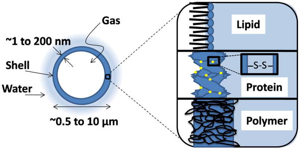
Over the last decade, there has been significant progress towards the development of microbubbles as theranostics for a wide variety of biomedical applications. The unique ability of microbubbles to respond to ultrasound makes them useful agents for contrast ultrasound imaging, molecular imaging, and targeted drug and gene delivery. The general composition of a microbubble is a gas core stabilized by a shell comprised of proteins, lipids or polymers. Each type of microbubble has its own unique advantages and can be tailored for specialized functions. In this review, different microbubbles compositions and physiochemical properties are discussed in the context of current progress towards developing novel constructs for biomedical applications, with specific emphasis on molecular imaging and targeted drug/gene delivery.


Figures

Figure 1
Figure 1
Cartoon showing structure of a typical microbubble with different shell compositions. Microbubbles used for biomedical purposes are typically between 0.5 and 10 μm diameter (the upper limit for passage through the lung capillaries). The gas core is a single chamber and comprises a large majority of the total particle volume. The shell acts as a barrier between the encapsulated gas and the surrounding aqueous medium. Different shell materials may be used, including lipid (~3 nm thick), protein (15–20 nm thick) and polymer (100–200 nm thick). The lipid molecules are held together through physical force fields, such as hydrophobic and van der Waals interactions. The protein is cross-linked by covalent disulfide bonds. The polymer chains are covalently cross-linked and/or entangled to form a bulk-like material.
Figure 2
Figure 2
Microbubble shell morphologies. (A) A lysozyme protein microbubble imaged with SEM taken from Calaveri et al. The microbubble diameter is roughly 1 μm. (B) A diC20:0 phospholipid microbubble imaged with fluorescence microscopy taken from Borden et al. Scale bar denotes 20 μm. (C) A PLA-PFO polymer microbubble imaged with SEM taken from Böhmer et al. All images have been reproduced with permission.
Figure 3
Figure 3
Useful ultrasound-mediated effects of microbubbles. Microbubbles insonified at MHz frequencies produce a variety of effects which may be beneficial for ultrasound imaging or drug delivery. (A) Oscillation of the gas core at moderate pressures produces a detectable backscatter. (B) Streaming of the fluid around the oscillating microbubble creates shear forces that may facilitate drug release and uptake by nearby cells. (D) Insonation at high pressures results in microbubble fragmentation. (D) Insonation at moderate pressures below the fragmentation threshold results in dissolution of the gas core. (E) Insonation at lower frequencies and higher pressures results in inertial cavitation, which can produce shock waves and involuted jets (water hammer). (F) Insonation at low pressures near microbubble resonance results in radiation force, which displaces the microbubble away from the transducer.
Figure 4
Figure 4
Schematic showing technique to enhance targeting selectivity using a microbubble with ultrasound-gated adhesiveness. (A) shows the microbubble traveling to the target site, where the ligand is buried by an overbrush layer of methylated PEG. This protects the microbubble from adhering to non-target tissue and protects the ligand from binding to serum proteins, such as C3b, which would alter the ligand binding specificity and lead to premature clearance by the immune system. (B) shows microbubble activity during insonification, where ultrasound radiation force drives the microbubbles up against the target endothelium and oscillation of the gas core transiently reveals the ligand for binding to promote firm adhesion. (C) optical microscopy images showing selective adhesion of RGD-microbubbles with the buried-ligand architecture to plated HUVEC cells expressing αVβ3 integrin, where adhesion is only observed within the transducer focus. Images taken from Borden et al. All images have been reproduced with permission.
Figure 5
Figure 5
Schematic representation of radiation-fragmentation pulse. A) Microbubbles passing through the vasculature are B) given low intensity ultrasound pulses to effectively force the bubbles against the endothelial layer. C) High intensity pulses are then applied to fragment the microbubble in order to release the drug cargo in proximity to the endothelium.

Similar articles

Cited by

References

    1. Unger EC, Porter T, Culp W, Labell R, Matsunaga T, Zutshi R. Adv Drug Delivery Rev. 2004;56:1291–1314. - PubMed
    1. Ferrara K, Pollard R, Borden M. Annu Rev Biomed Eng. 2007;9:415–447. - PubMed
    1. Lindner JR. Nature Rev Drug Discovery. 2004;3:527–532. - PubMed
    1. Feinstein SB. Am J Physiol. 2004;287:H450–H457. - PubMed
    1. Dayton PA, Rychak JJ. Frontiers in Bioscience. 2007;12:5124–5142. - PubMed

LinkOut - more resources