. 2010 Dec;67(24):4213-32.
doi: 10.1007/s00018-010-0431-6. Epub 2010 Jun 24.

ADAMTS-2 functions as anti-angiogenic and anti-tumoral molecule independently of its catalytic activity

Affiliations

ADAMTS-2 functions as anti-angiogenic and anti-tumoral molecule independently of its catalytic activity

J Dubail et al. Cell Mol Life Sci. 2010 Dec.

Abstract

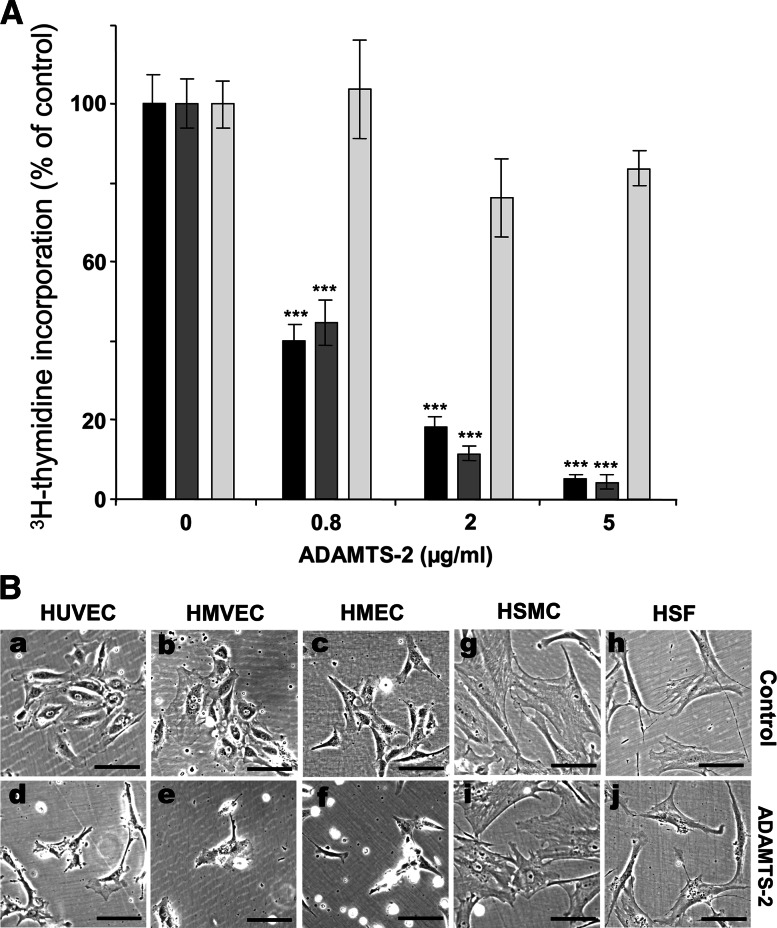
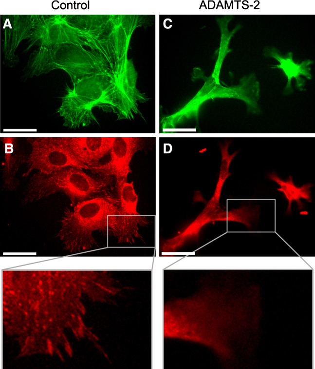
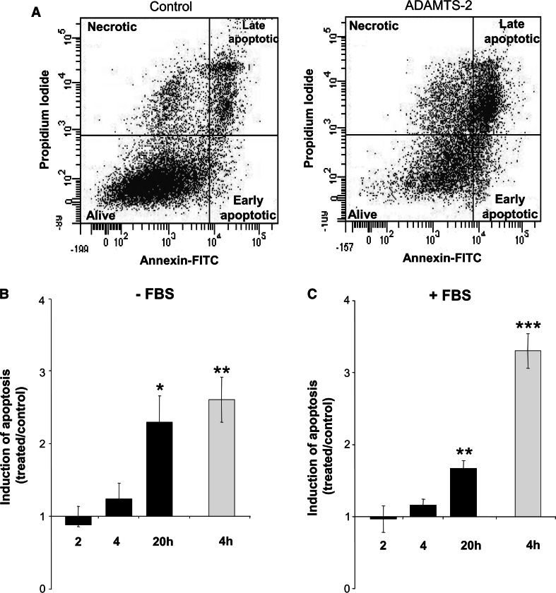
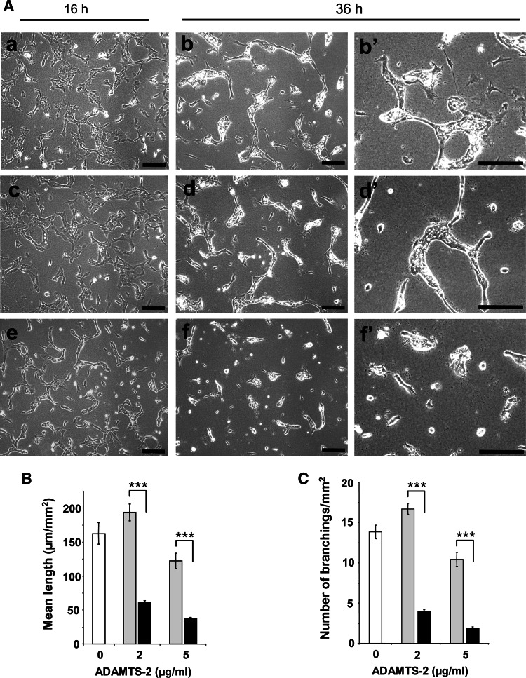
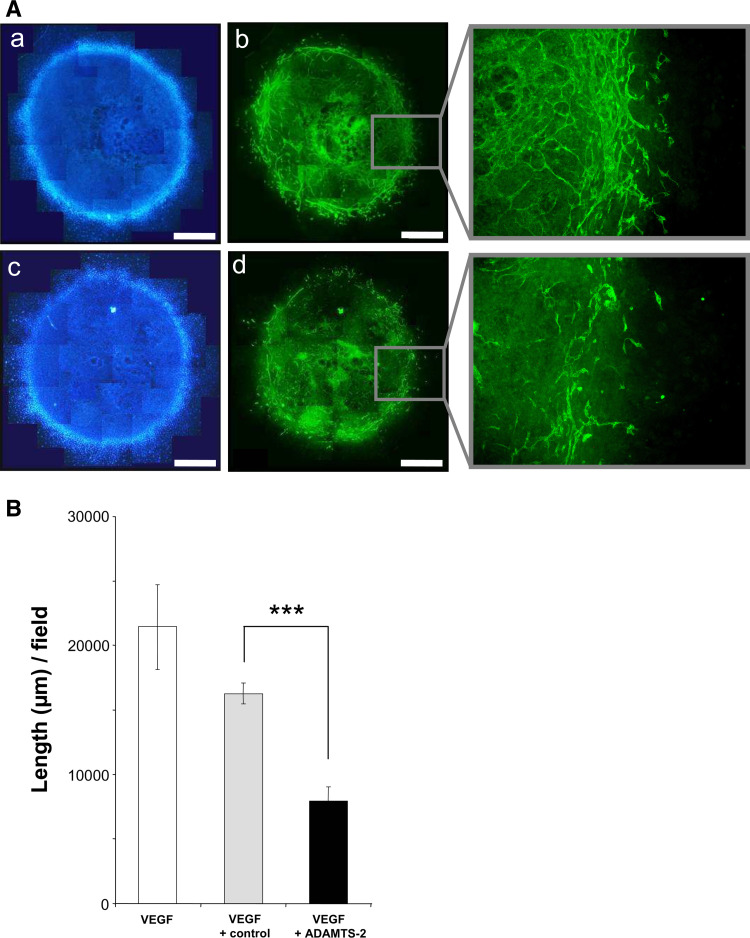
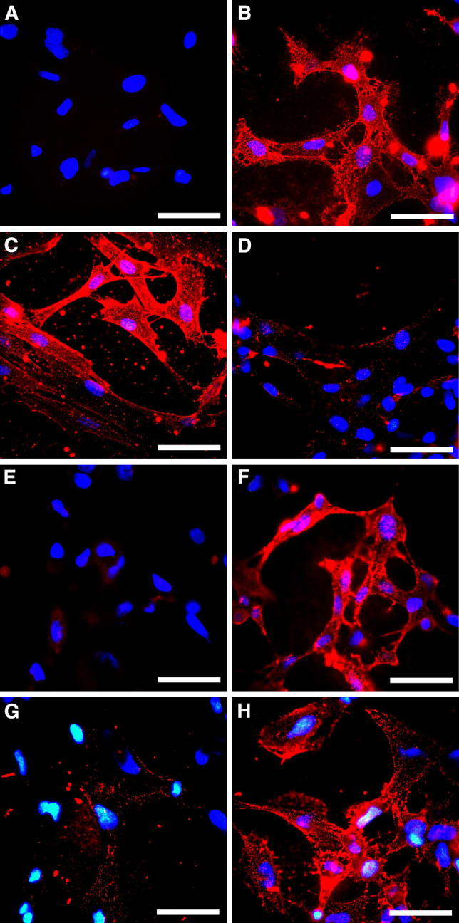
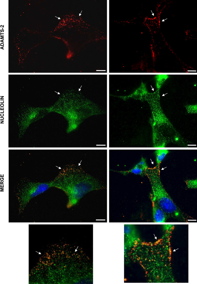
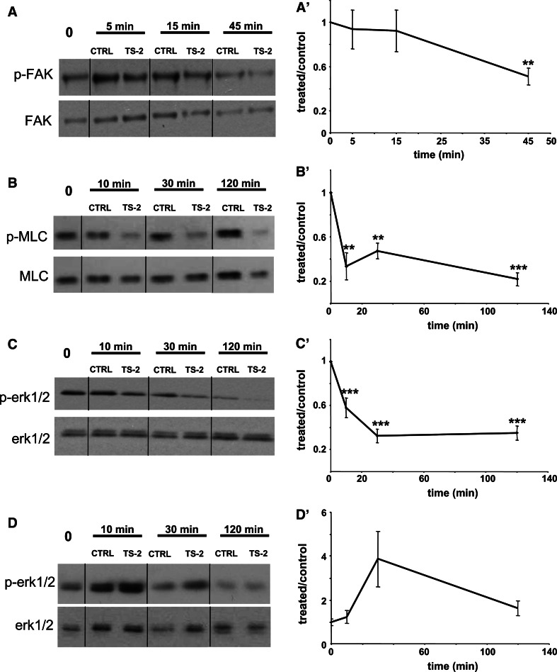
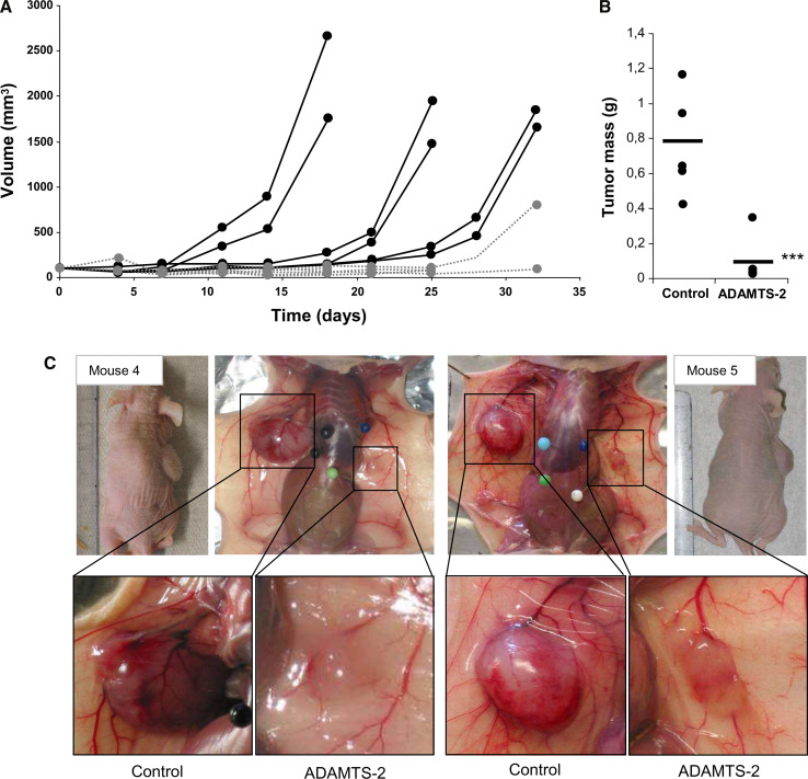
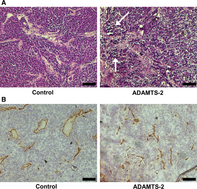
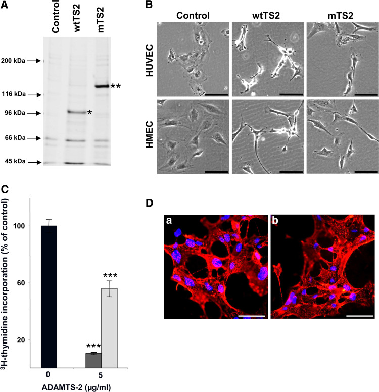
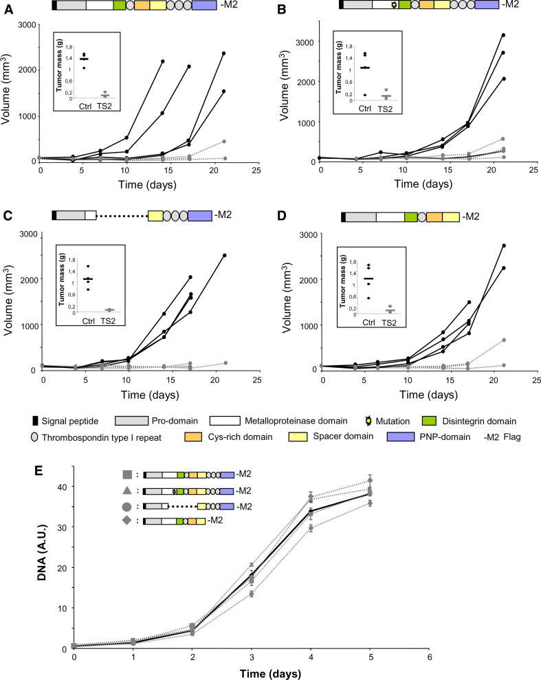
ADAMTS-2 is a metalloproteinase that plays a key role in the processing of fibrillar procollagen precursors into mature collagen molecules by excising the amino-propeptide. We demonstrate that recombinant ADAMTS-2 is also able to reduce proliferation of endothelial cells, and to induce their retraction and detachment from the substrate resulting in apoptosis. Dephosphorylation of Erk1/2 and MLC largely precedes the ADAMTS-2 induced morphological alterations. In 3-D culture models, ADAMTS-2 strongly reduced branching of capillary-like structures formed by endothelial cells and their long-term maintenance and inhibited vessels formation in embryoid bodies (EB). Growth and vascularization of tumors formed in nude mice by HEK 293-EBNA cells expressing ADAMTS-2 were drastically reduced. A similar anti-tumoral activity was observed when using cells expressing recombinant deleted forms of ADAMTS-2, including catalytically inactive enzyme. Nucleolin, a nuclear protein also found to be associated with the cell membrane, was identified as a potential receptor mediating the antiangiogenic properties of ADAMTS-2.


Figures

Fig. 1
Fig. 1
Recombinant ADAMTS-2 inhibits endothelial cells proliferation and alters their morphology. a The proliferation rate of HUVEC (black bars), HMVEC (dark grey bars), or human skin fibroblasts (light grey bars) was measured by 3H-thymidine incorporation in cultures (n = 6) supplemented with 0.5% FBS and increasing concentrations (0.8–5 μg/ml) of ADAMTS-2, or of the same volume of the control preparation (0 μg/ml). The results are expressed in percentage of incorporation as compared to the respective controls. The results are representative of three separate experiments for HMVEC and fibroblasts and ten separate experiments for HUVEC. (***p < 0.001). B Cells were seeded on gelatin coat for 3 h in EBM supplemented with 0.5% FBS and cultured for 16 h in control medium (a, b, c, g, h) or in the presence of ADAMTS-2 at 5 μg/ml (d, e, f, i, j). Bar 100 μm
Fig. 2
Fig. 2
ADAMTS-2 causes disassembly of actin stress fibers and focal adhesions in HUVEC. HUVEC were seeded on fibronectin coat for 3 h in serum-free EBM and cultured for 16 h in control medium (a, b) or in medium supplemented with ADAMTS-2 at 5 μg/ml (c, d). Cells were fixed, permeabilized with 0.1% Triton X-100, and stained to visualize actin stress fibers (FITC-Phalloidin; a, c) and focal adhesions (anti-vinculin mAb and TRITC-conjugated secondary antibody; b, d). Focal adhesion appears as small red spots at the extention of actin stress fibers while the diffuse red staining around the nucleus corresponds to cytoplasmic vinculin. Bar 25 μm
Fig. 3
Fig. 3
ADAMTS-2 induces endothelial cell apoptosis. Three hours after seeding without or with 0.5% FBS, culture medium was supplemented with control preparation, ADAMTS-2 (5 μg/ml) or staurosporine (1 μg/ml) as a positive pro-apoptotic control. Cells were then collected after increasing times in culture, stained with annexin V-FITC and PI, and analyzed by FACS. a An example of cell distribution is provided (20 h of incubation without FBS in control conditions or in the presence of ADAMTS-2). Total number of cells stained by annexin V-FITC only (early apoptotic) and double stained by annexin V-FITC and PI (late apoptotic) was used to monitor induction of apoptosis by ADAMTS-2 in a time-course experiment in the absence (b) or in the presence of (c) FBS. The results are expressed as the ratio between the apoptosis induced by ADAMTS-2 (black bars) or Staurosporine (grey bars) and their respective control (treated/control). b n = 3; c n = 4; ***p < 0.001; **p < 0.01; *p < 0.05 according to the one-way ANOVA statistical test
Fig. 4
Fig. 4
Effect of ADAMTS-2 on capillary-like structures formed by HUVEC. A HUVEC were cultured between two collagen gels in medium containing 10% FBS alone (a, b), or supplemented with the control preparation (c, d) or with ADAMTS-2 at 5 μg/ml (e, f). Pictures were taken after 16 h (a, c, e) or 36 h (b, d, f). Bars 100 μm. Mean length of all visible structures (B) and number of branching of the capillary-like structures (C) were calculated (n = 30 pictures) for cultures in medium alone (open bars) or supplemented with ADAMTS-2 at 2 or 5 μg/ml (black bars) or with the same volume of control sample (grey bars). The results are representative of at least three separate experiments (***p < 0.001)
Fig. 5
Fig. 5
ADAMTS-2 inhibits the formation of vascular structures in embryoid bodies. A Embryoid bodies were formed by culturing ES cells in medium containing VEGF alone (not shown) or further supplemented with the control preparation (a, b) or with ADAMTS-2 at 0.2 μg/ml (c, d). Nuclei were stained with DAPI (a, c) and forming vessels with an anti-CD31 antibody (b, d). Vessels formed by differentiated endothelial cells appear as elongated and branched green structures. Bar 1 mm. B Mean length of vessels in EB cultured in medium containing VEGF alone (open bars, n = 3), or supplemented with control sample (light grey bars, n = 20), or with ADAMTS-2 at 0.2 μg/ml (black bars, n = 18) were calculated (***p < 0.001)
Fig. 6
Fig. 6
Binding of ADAMTS-2 at the cell surface. HMEC (a, b, d–h) and fibroblasts (c) were incubated for 6 h at 8°C in serum-free medium supplemented with control preparation (a) or ADAMTS-2 at 2 μg/ml (b–h) in the absence (a–c, e–h) or presence of 10 μg/ml heparin (d) and then immunostained for the presence of ADAMTS-2 immobilized at the cell surface. For a second set of experiments, HMEC were first pretreated for 16 h with 20 mM sodium chlorate alone (e) or with sodium chlorate in the presence of 10 mM sodium sulfate (f) or for 4 h with 6 mU/ml heparinase III (g) or 0.1 U/ml chondroitinase (h). After pre-treatment, medium was changed and cells were cultured for 6 h at 8°C in the presence of ADAMTS-2 before staining as described above. Bar 50 μm
Fig. 7
Fig. 7
ADAMTS-2 and nucleolin co-localization at the cell-surface HMEC were pretreated for 4 h with 6 mU/ml heparinase III, incubated for 6 h at 8°C in serum-free medium supplemented with ADAMTS-2 at 0.5 μg/ml, and stained to visualize ADAMTS-2 (red) and nucleolin (green) at the cell surface. A merged picture of the two stainings shows co-localization of the two proteins (arrows). Bar 10 μm
Fig. 8
Fig. 8
Effect of ADAMTS-2 on the phosphorylation of Erk1/2, MLC and FAK. HUVEC (a, b, c) or fibroblasts (d) were seeded in serum-free medium and allowed to spread for 3 h at 37°C. ADAMTS-2 (TS-2) and control preparation (CTRL) were then added for various times. Cell lysates were analyzed by Western blotting using antibodies for phosphorylated FAK (p-FAK) and total FAK (a), phosphorylated MLC (p-MLC) and total MLC (b) and phosphorylated Erk1/2 (p-Erk1/2) and total Erk1/2 (c, d). Scanning density of the bands obtained in at least three independent experiments was used to calculate the relative level of p-FAK versus total FAK (n = 6) (a′), p-MLC versus total MLC (n = 3) (b′) and p-Erk1/2 versus total Erk1/2 (n = 8) (c′, d′) in the various cultures conditions (***p < 0.001; **p < 0.01)
Fig. 9
Fig. 9
ADAMTS-2 inhibits growth of HEK 293-EBNA tumors in nude mice. HEK 293-EBNA cells stably transfected with empty vector or expressing ADAMTS-2 were mixed with Matrigel and subcutaneously injected into each flank of the mice, one with control cells (control) and the other with ADAMTS-2 (TS-2) expressing cells (n = 6). a Growth of plugs containing control cells (plain black lines) or ADAMTS-2 expressing cells (dotted grey lines) was evaluated by measuring the tumor's volume every 3–4 days. Mice were killed when the apparent volume of one of the bilateral tumors reached 1,500 mm3. b Mean values of tumor mass for control cells and ADAMTS-2-expressing cells (***p < 0.001). c Aspect of the tumors in toto and before dissection in two representative mice
Fig. 10
Fig. 10
Immunohistological examination of control and ADAMTS-2-expressing tumors. a Paraffin sections of tumors formed by control or by ADAMTS-2-expressing cells were stained with hematoxylin and eosin stain (H&E). Necrotic cells were visible only in ADAMTS-2 tumors (arrows). b Cryostat sections of tumors were stained by an anti-CD31 antibody for visualizing blood vessels (in brown). Bar 100 μm
Fig. 11
Fig. 11
The catalytic activity of ADAMTS-2 is dispensable for antiangiogenic activity. a Sypro Ruby staining of purified control preparation (control), wild-type ADAMTS-2 (wtTS2) or catalytically inactive ADAMTS-2 (mTS-2). Asterisks indicate the most abundant form of ADAMTS-2 (* for wild-type ADAMTS-2 and ** for catalytically inactive ADAMTS-2). b HUVEC or HMEC were seeded on gelatin coats for 3 h in serum-free EBM and cultured for 16 h in medium supplemented with the control preparation (control), the wild-type ADAMTS-2 (wtTS-2) or catalytically inactive ADAMTS-2 (mTS-2), both at 5 μg/ml. Bar 100 μm. c The proliferation rate of HUVEC was measured by 3H-thymidine uptake in cultures (n = 6) in the presence of wtTS2 (dark grey bar), of mTS2 (light grey bar) both at 5 μg/ml, or of the same amount of control preparation (0 μg/ml, black bar). The results are representative of three separate experiments (***p < 0.001). d HMEC were incubated for 6 h at 8°C in serum-free medium supplemented with wild-type ADAMTS-2 (a) or catalytically inactive ADAMTS-2 (b) both at 2 μg/ml and immunostained for the presence of ADAMTS-2 immobilized at the cell surface. Bar 50 μm
Fig. 12
Fig. 12
Effect of different recombinant forms of ADAMTS-2 on tumor growth. The experimental procedure was as detailed in Fig. 9 (n = 4). A schematic domains organization of the four different recombinant ADAMTS-2 expressed by cells forming the tumors is provided at the top of each corresponding graphics. Tumor volume was measured every 3–4 days. Control tumors are represented by bold plain black lines while tumors formed by the different forms of ADAMTS-2 are illustrated by dotted grey lines (a wild-type ADAMTS-2; b mutated catalytically inactive ADAMTS-2; c ADAMTS-2 lacking the central domains including the metalloproteinase domain; d ADAMTS-2 lacking the C-terminal domains). Insets in ad illustrate the mean tumor mass after dissection. e The proliferation rate of control HEK-293 EBNA cells (bold dark line) or of HEK-293 EBNA cells expressing the different forms of ADAMTS-2 (dotted grey lines) was evaluated by DNA quantification

Similar articles

Cited by

References

    1. Bix G, Iozzo RV. Matrix revolutions: “tails” of basement-membrane components with angiostatic functions. Trends Cell Biol. 2005;15:52–60. doi: 10.1016/j.tcb.2004.11.008. - DOI - PubMed
    1. Sottile J. Regulation of angiogenesis by extracellular matrix. Biochim Biophys Acta. 2004;1654:13–22. - PubMed
    1. Folkman J. Anti-angiogenesis: new concept for therapy of solid tumors. Ann Surg. 1972;175:409–416. doi: 10.1097/00000658-197203000-00014. - DOI - PMC - PubMed
    1. Armstrong LC, Bornstein P. Thrombospondins 1 and 2 function as inhibitors of angiogenesis. Matrix Biol. 2003;22:63–71. doi: 10.1016/S0945-053X(03)00005-2. - DOI - PubMed
    1. Lee NV, Sato M, Annis DS, Loo JA, Wu L, Mosher DF, Iruela-Arispe ML. ADAMTS1 mediates the release of antiangiogenic polypeptides from TSP1 and 2. EMBO J. 2006;25:5270–5283. doi: 10.1038/sj.emboj.7601400. - DOI - PMC - PubMed

Publication types

MeSH terms