. 2010 Oct;88(10):1029-40.
doi: 10.1007/s00109-010-0642-1. Epub 2010 Jun 24.

Methylation matters: binding of Ets-1 to the demethylated Foxp3 gene contributes to the stabilization of Foxp3 expression in regulatory T cells

Affiliations

Methylation matters: binding of Ets-1 to the demethylated Foxp3 gene contributes to the stabilization of Foxp3 expression in regulatory T cells

Julia K Polansky et al. J Mol Med (Berl). 2010 Oct.

Abstract

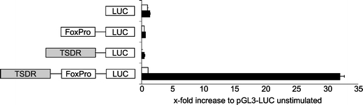
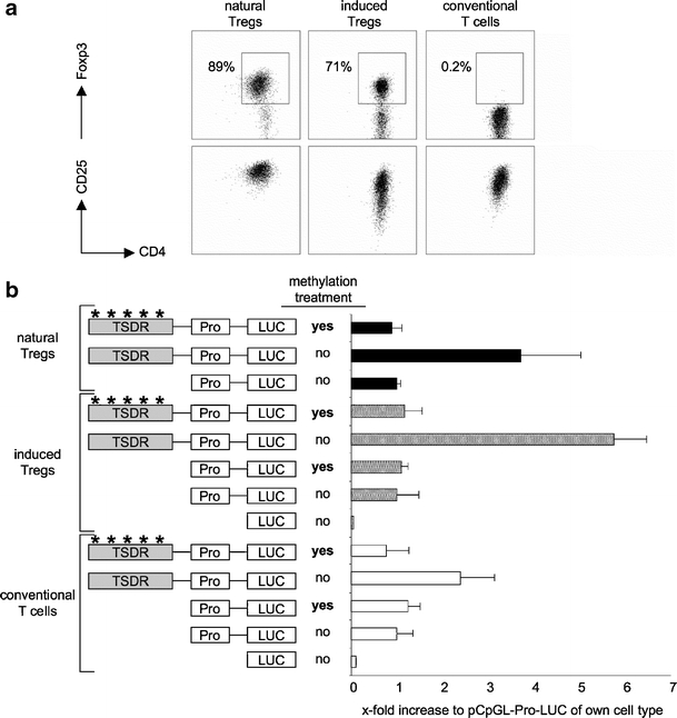
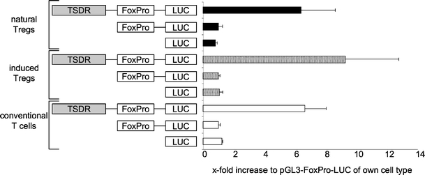
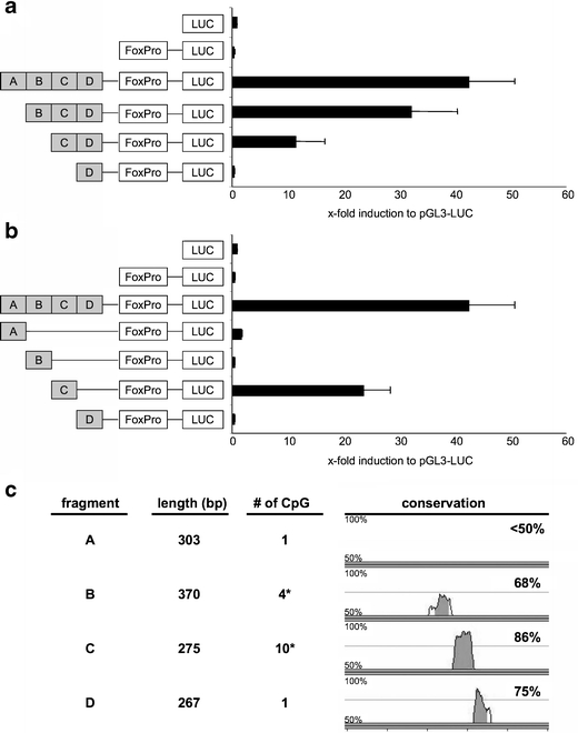
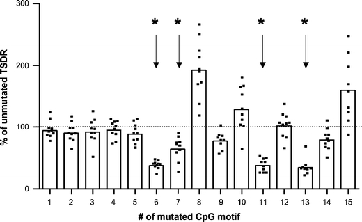
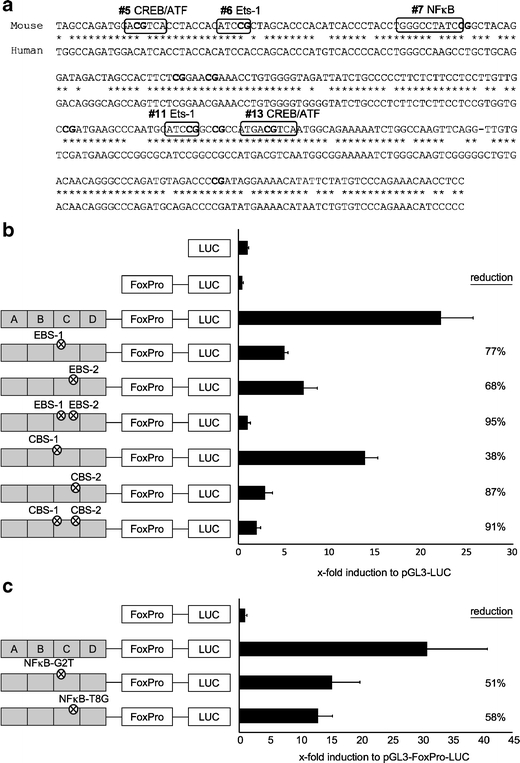
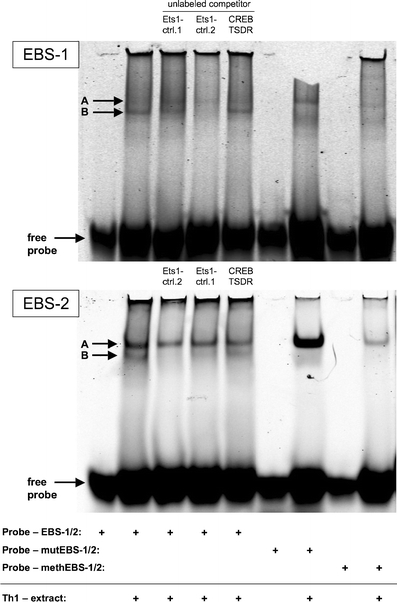
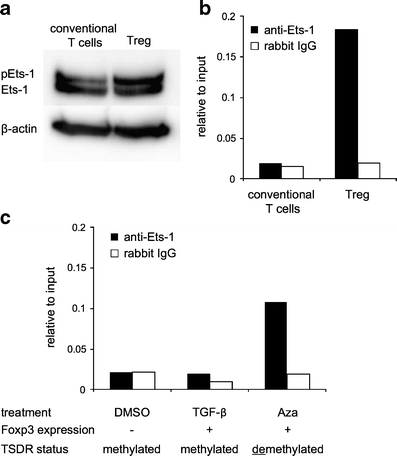
The forkhead-box protein P3 (Foxp3) is a key transcription factor for the development and suppressive activity of regulatory T cells (Tregs), a T cell subset critically involved in the maintenance of self-tolerance and prevention of over-shooting immune responses. However, the transcriptional regulation of Foxp3 expression remains incompletely understood. We have previously shown that epigenetic modifications in the CpG-rich Treg-specific demethylated region (TSDR) in the Foxp3 locus are associated with stable Foxp3 expression. We now demonstrate that the methylation state of the CpG motifs within the TSDR controls its transcriptional activity rather than a Treg-specific transcription factor network. By systematically mutating every CpG motif within the TSDR, we could identify four CpG motifs, which are critically determining the transcriptional activity of the TSDR and which serve as binding sites for essential transcription factors, such as CREB/ATF and NF-κB, which have previously been shown to bind to this element. The transcription factor Ets-1 was here identified as an additional molecular player that specifically binds to the TSDR in a demethylation-dependent manner in vitro. Disruption of the Ets-1 binding sites within the TSDR drastically reduced its transcriptional enhancer activity. In addition, we found Ets-1 bound to the demethylated TSDR in ex vivo isolated Tregs, but not to the methylated TSDR in conventional CD4(+) T cells. We therefore propose that Ets-1 is part of a larger protein complex, which binds to the TSDR only in its demethylated state, thereby restricting stable Foxp3 expression to the Treg lineage.


Figures

Fig. 1
Fig. 1
The TSDR acts as a transcriptional enhancer. The TSDR and the Foxp3 promoter (FoxPro) were cloned into the pGL3-luciferase-reporter vector, and their transcriptional activities were assessed in a dual luciferase assay. The indicated constructs were transfected into RLM-11-1 cells and cells rested for 3 h after transfection. The luciferase activity was measured 20 h after stimulation with PMA and ionomycin (black bars). Unstimulated samples are shown in white bars. All values are depicted in relation to the indicated reference sample, which was set to 1. The graph shows mean values of performed triplicates with standard deviation. LUC coding sequence for the firefly luciferase
Fig. 2
Fig. 2
Functionality of the TSDR is determined by its methylation status. a Primary T cells from Foxp3gfp-reporter mice were sorted and expanded for 5 days in the presence of IL-2 (natural Tregs and conventional T cells) and IL-2 plus TGF-β (induced Tregs). The phenotypes of the expanded cells are shown. b The TSDR was cloned into the CpG-free pCpGL-luciferase reporter plasmid carrying the hEF-promoter (Pro). Subsequently, the plasmids were subjected to in vitro methylation or not. Primary expanded T cell subsets (from a) were transfected with the indicated plasmids and rested over night. On the next day, cells were stimulated for 9 h with PMA and ionomycin and luciferase activity was measured. Asterisks indicate methylated regions of the plasmid. Values are depicted in relation to the indicated reference sample, which was set to 1. The graph shows mean values of performed triplicates with standard deviation. One representative out of two to three independent experiments is depicted. LUC coding sequence for the firefly luciferase
Fig. 3
Fig. 3
The TSDR acts as a transcriptional enhancer even in Foxp3 conventional T cells. Primary murine T cells from BALB/c mice, which had been cultured for 3 days in the presence of IL-2 (natural Tregs and conventional T cells) and IL-2 plus TGF-β (induced Tregs), were transfected with the indicated constructs. Luciferase activity was measured 20 h post-transfection. All values are depicted in relation to the indicated reference sample, which was set to 1. Both graphs show mean values of performed triplicates with standard deviation. One out of three independent experiments is depicted. FoxPro murine Foxp3 promoter; LUC coding sequence for the firefly luciferase
Fig. 4
Fig. 4
The CpG-rich, highly conserved fragment C of the TSDR is of special importance for the transcriptional activity. a and b Dual luciferase reporter assays were performed in RLM-11-1 cells using the indicated constructs. Luciferase activity was measured 20 h after stimulation with PMA and ionomycin. All values are depicted in relation to the indicated reference sample, which was set to 1. Both graphs show mean values of performed triplicates with standard deviation. One out of three independent experiments is depicted. c Characteristics of the tested TSDR fragments. The inter-species conservation was assessed using the mVISTA-tool comparing the murine and the human Foxp3 locus. Due to the primer-design, CpG #5 is present in fragments B and C (indicated by the asterisks)
Fig. 5
Fig. 5
Selective CpG motifs are essential for the transcriptional activity of the TSDR. Point mutations were introduced to destroy single CpG motifs in the TSDR-FoxPro-LUC reporter vector. The resulting 15 mutant-TSDR-plasmids were used in dual luciferase reporter assays in RLM-11-1 cells. After transfection, the cells were rested for 3 h and subsequently stimulated with PMA and ionomycin. The luciferase activity was measured 20 h later. The luciferase activity of the FoxPro-control-plasmid (carrying no TSDR) was subtracted from each value to yield the unmutated TSDR value, which was set to 100%. All other values are depicted in relation to that. Bars represent the mean value of 9–10 replicates (shown as individual dots) taken from three independent experiments. Data sets marked with an asterisk show a statistically significant reduction to 100% according to the Wilcoxon signed-rank test (p ≤ 0.002)
Fig. 6
Fig. 6
Mutation of the Ets-1, the CREB/ATF or the NF-κB binding sites in the TSDR leads to reduction of the transcriptional activity. a Sequence alignment of the murine (upper) and the human (lower) sequence of the TSDR fragment C. Postulated transcription factor binding sites, which span CpG motifs (in bold), are indicated with boxes. b Dual luciferase reporter assays were performed in RLM-11-1 cells using the indicated constructs. Circled multiplication symbol indicates inserted point mutations which destroy the indicated transcription factor binding sites. Luciferase activity was measured 20 h after stimulation with PMA and ionomycin. c Two-point mutations at distinct positions of the NF-κB-binding site were introduced and the resulting TSDR mutants were tested for transcriptional activity as in b. All values are depicted in relation to the indicated reference sample, which was set to 1. Both graphs show mean values of performed triplicates with standard deviation. One out of at least two independent experiments is depicted
Fig. 7
Fig. 7
Two protein complexes bind to the Ets-1 binding sites (EBSs) of the TSDR in vitro. Electromobility shift assays were performed using nuclear protein extracts of in vitro differentiated Th1 cells and oligo-deoxynucleotides (probes) containing the EBS-1 or EBS-2 of the TSDR or their mutated or methylated versions. To compete for Ets-1 binding, unlabeled competitor oligonucleotides containing other known Ets-1 binding sites (Ets-1 contr.1 and 2) were used in 30-fold excess. The CREB/ATF binding site-2 of the TSDR (CREB-TSDR) served as a competition negative control. The gels present one out of two independent experiments
Fig. 8
Fig. 8
In vivo binding of Ets-1 to the demethylated TSDR in stable Foxp3+ Tregs. CD4+CD25+Foxp3gfp+ Tregs and CD4+CD25-Foxp3gfp- conventional T cells were isolated from BALB/c Foxp3gfp reporter mice and analyzed a for Ets-1 protein expression by Western Blotting and b for Ets-1 binding to the TSDR by ChIP. c Isolated conventional T cells were cultured with TCR-stimulation and IL-2 for 5 days. TGF-β (5 ng/ml) or 5-Aza-2′-deoxycytidine (Aza, 1 μM) was added to the culture to induce Foxp3 expression. Control cultures contained DMSO. On day 5, cultures were harvested and sorted for Foxp3gfp+ cells by FACS. Control cultures not containing Foxp3gfp+ cells were used unsorted. All populations were subjected to ChIP using anti-Ets-1 or control antibody. Both graphs show one representative out of three independent experiments

Similar articles

Cited by

References

    1. Brusko TM, Putnam AL, Bluestone JA. Human regulatory T cells: role in autoimmune disease and therapeutic opportunities. Immunol Rev. 2008;223:371–390. doi: 10.1111/j.1600-065X.2008.00637.x. - DOI - PubMed
    1. Riley JL, June CH, Blazar BR. Human T regulatory cell therapy: take a billion or so and call me in the morning. Immunity. 2009;30:656–665. doi: 10.1016/j.immuni.2009.04.006. - DOI - PMC - PubMed
    1. Curiel TJ. Regulatory T cells and treatment of cancer. Curr Opin Immunol. 2008;20:241–246. doi: 10.1016/j.coi.2008.04.008. - DOI - PMC - PubMed
    1. Fontenot JD, Gavin MA, Rudensky AY. Foxp3 programs the development and function of CD4 + CD25+ regulatory T cells. Nat Immunol. 2003;4:330–336. doi: 10.1038/ni904. - DOI - PubMed
    1. Hori S, Nomura T, Sakaguchi S. Control of regulatory T cell development by the transcription factor Foxp3. Science. 2003;299:1057–1061. doi: 10.1126/science.1079490. - DOI - PubMed

Publication types

Substances

LinkOut - more resources