

Projections to the anterodorsal thalamus and lateral mammillary nuclei arise from different cell populations within the postsubiculum: implications for the control of head direction cells
- PMID: 20575008
- PMCID: PMC2975049
- DOI: 10.1002/hipo.20820
Projections to the anterodorsal thalamus and lateral mammillary nuclei arise from different cell populations within the postsubiculum: implications for the control of head direction cells
Abstract
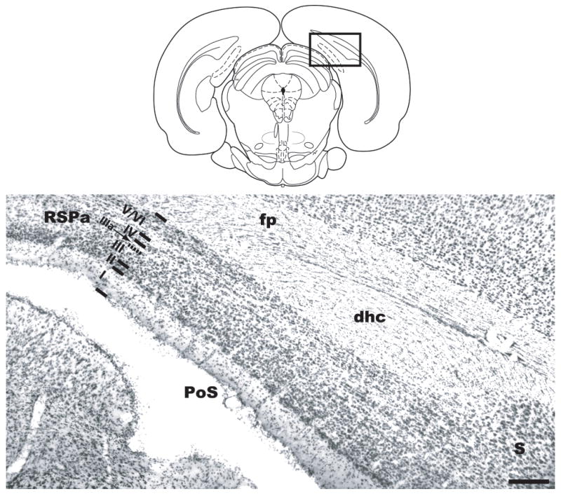
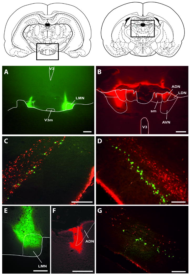
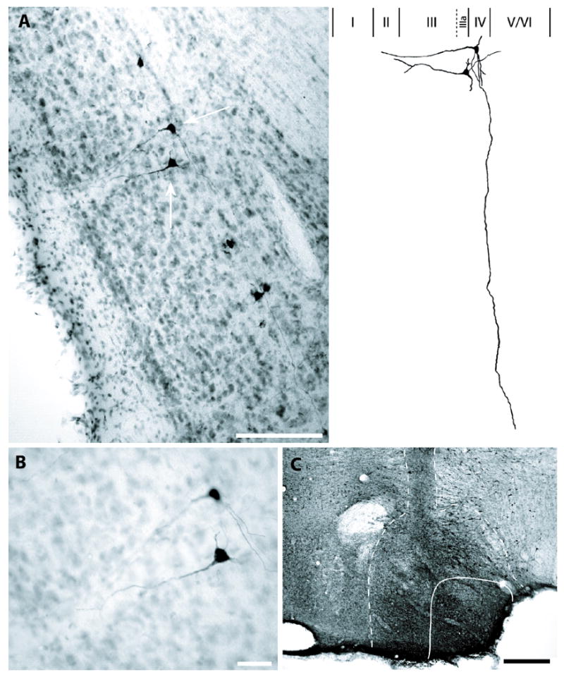
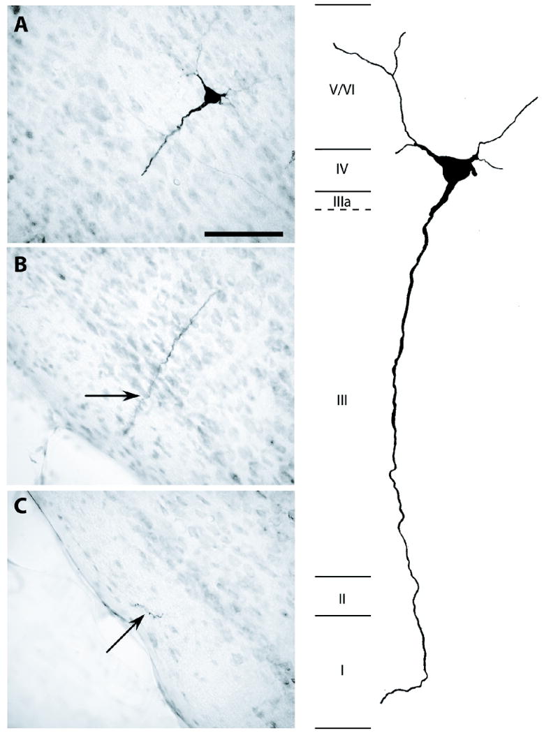
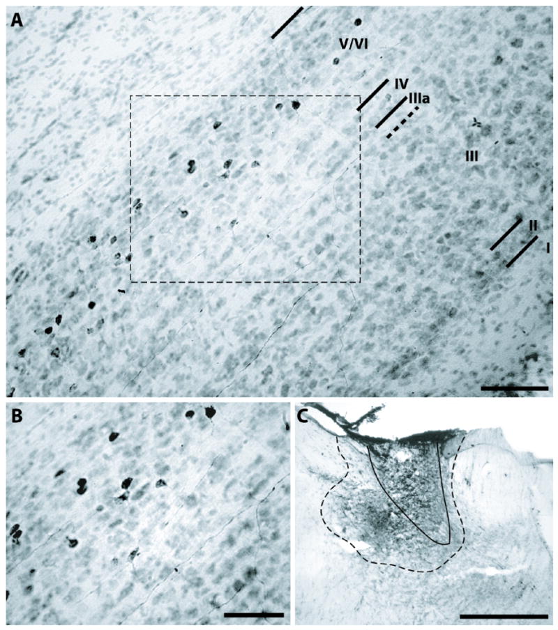
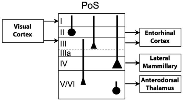
The neural representation of directional heading is encoded by a population of cells located in a circuit that includes the postsubiculum (PoS), anterodorsal thalamus (ADN), and lateral mammillary nuclei (LMN). Throughout this circuit, many cells rely on both movement- and landmark-related information to discharge as a function of the animal's directional heading. The PoS projects to both the ADN and LMN, and these connections may convey critical spatial information about landmarks, because lesions of the PoS disrupt landmark control in head direction (HD) cells and hippocampal place cells [Goodridge and Taube (1997) J Neurosci 17:9315-9330; Calton et al. (2003) J Neurosci 23:9719-9731]. The PoS → ADN projection originates in the deep layers of PoS, but no studies have determined whether the PoS → LMN projection originates from the same cells that project to ADN. To address this issue, two distinct cholera toxin-subunit B (CTB) fluorophore conjugates (Alexa Fluor 488 and Alexa Fluor 594) were injected into the LMN and ADN of the same rats, and PoS sections were examined for cell bodies containing either or both CTB conjugates. Results indicated that the PoS → LMN projection originates exclusively from a thin layer of cells located superficial to the layer(s) of PoS → ADN projection cells, with no overlap. To verify the laminar distribution and morphological characteristics of PoS → LMN and PoS → ADN cells, biotinylated dextran amine was injected into LMN or ADN of different rats, and tissue sections were counterstained with thionin. Results indicated that the PoS → LMN projection arises from large pyramidal cells in layer IV, whereas the PoS → ADN projection arises from a heterogeneous cell population in layers V/VI. This study provides the first evidence that the PoS → ADN and PoS → LMN projections arise from distinct, nonoverlapping cell layers in PoS. Functionally, the PoS may provide landmark information to HD cells in LMN.
Copyright © 2010 Wiley-Liss, Inc.
Figures
References
-
- Allen GV, Hopkins DA. Mammillary body in the rat: topography and synaptology of projections from the subicular complex, prefrontal cortex, and midbrain tegmentum. J Comp Neurol. 1989;286:311–336. - PubMed
-
- Biazoli CE, Jr, Goto M, Campos AM, Canteras NS. The supragenual nucleus: A putative relay station for ascending vestibular signs to head direction cells. Brain Res. 2006;1094:138–148. - PubMed
-
- Blackstad TW. Commissural connections of the hippocampal region in the rat, with special reference to their mode of termination. J Comp Neurol. 1956;105:417–537. - PubMed
-
- Blair HT, Cho J, Sharp PE. Role of the lateral mammillary nucleus in the rat head direction circuit: a combined single unit recording and lesion study. Neuron. 1998;21:1387–1397. - PubMed
Publication types
MeSH terms
Grants and funding
LinkOut - more resources
Full Text Sources
Research Materials
Miscellaneous
