. 2010 Aug 3;49(30):6451-61.
doi: 10.1021/bi100769k.

Conformational transitions in human AP endonuclease 1 and its active site mutant during abasic site repair

Affiliations

Conformational transitions in human AP endonuclease 1 and its active site mutant during abasic site repair

Lyubov Yu Kanazhevskaya et al. Biochemistry. .

Abstract

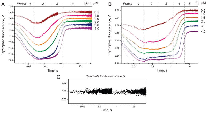
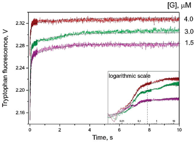
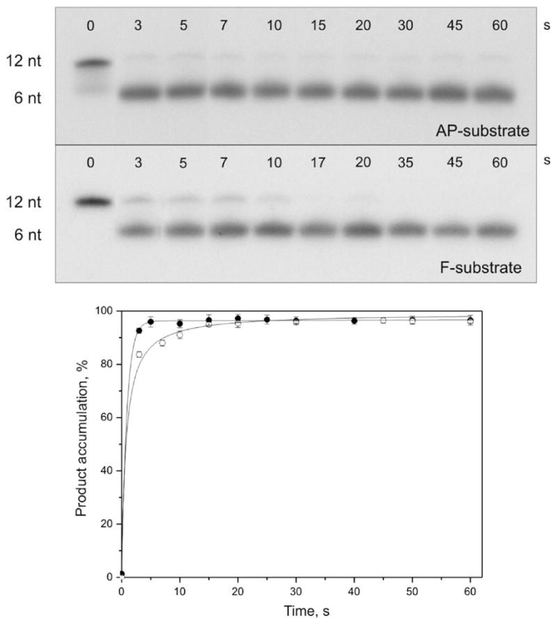
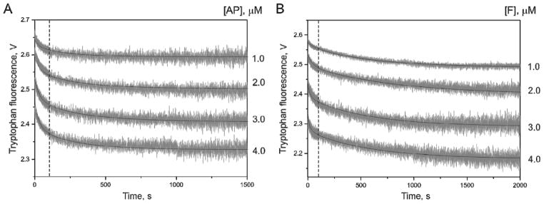
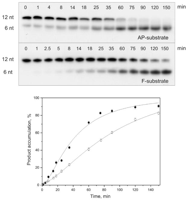
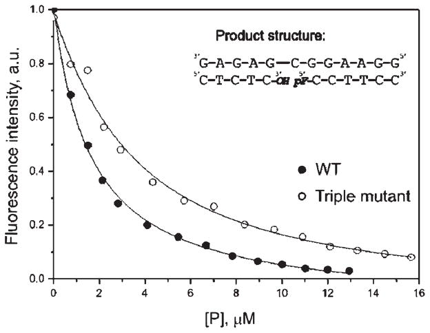
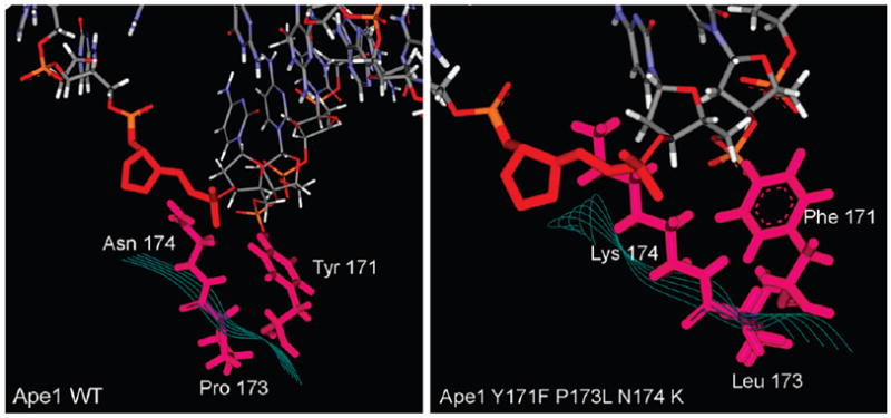
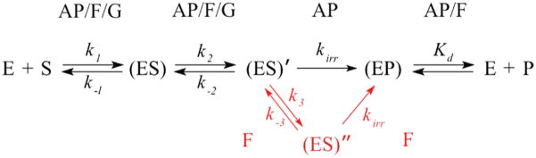
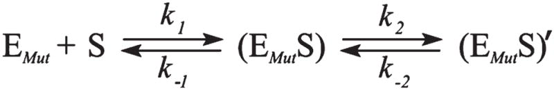
AP endonuclease 1 (APE1) is a crucial enzyme of the base excision repair pathway (BER) in human cells. APE1 recognizes apurinic/apyrimidinic (AP) sites and makes a nick in the phosphodiester backbone 5' to them. The conformational dynamics and presteady-state kinetics of wild-type APE1 and its active site mutant, Y171F-P173L-N174K, have been studied. To observe conformational transitions occurring in the APE1 molecule during the catalytic cycle, we detected intrinsic tryptophan fluorescence of the enzyme under single turnover conditions. DNA duplexes containing a natural AP site, its tetrahydrofuran analogue, or a 2'-deoxyguanosine residue in the same position were used as specific substrates or ligands. The stopped-flow experiments have revealed high flexibility of the APE1 molecule and the complexity of the catalytic process. The fluorescent traces indicate that wild-type APE1 undergoes at least four conformational transitions during the processing of abasic sites in DNA. In contrast, nonspecific interactions of APE1 with undamaged DNA can be described by a two-step kinetic scheme. Rate and equilibrium constants were extracted from the stopped-flow and fluorescence titration data for all substrates, ligands, and products. A replacement of three residues at the enzymatic active site including the replacement of tyrosine 171 with phenylalanine in the enzyme active site resulted in a 2 x 10(4)-fold decrease in the reaction rate and reduced binding affinity. Our data indicate the important role of conformational changes in APE1 for substrate recognition and catalysis.


Figures

Figure 1
Figure 1
Chemical structures of the AP site (AP) and tetrahydrofuran abasic site analogue (F).
Figure 2
Figure 2
Time-dependent fluorescence change associated with the cleavage of AP-substrate (A) and F-substrate (B) by APE1. The fluorescence traces are distributed along the signal axis for better visualization. The final concentrationofAPE1 is1.5 μM. Jagged traces represent experimental data; smooth curves are theoretically fitted. Blue dashed curves (B) represent the results of fitting of the experimental data to Scheme 5 (23). Dashed drop lines approximately correspond to different stages of kinetic Scheme 1. (C) Representative graph of the residuals for fitting of the experimental data (3.0 μM substrate).
Figure 3
Figure 3
Stopped-flow fluorescent traces of APE1 binding the nonspecific G-ligand. The final concentration of APE1 is 1.5 μM. Jagged traces represent experimental data; smooth curves are theoretically fitted. Inset: the same fluorescence traces presented on a log scale.
Figure 4
Figure 4
Cleavage of 32P-labeled AP- and F-substrates by APE1. Top panel, analysis by 20% PAGE; autoradiograms are shown. Reaction mixtures (10 μL) contained 1.5 μM substrate and 1.5 μM enzyme. Bottom panel, time course of the cleaved product accumulation; ●, AP-substrate incision; ○, F-substrate incision. Error bars reflect the standard error of the mean from three independent experiments.
Figure 5
Figure 5
Stopped-flow kinetics of mutant APE1 interactions with specific substrates. Traces of Trp fluorescence observed during the interaction of 3 μM mutant APE1 with the AP-substrate (A) and F-substrate (B). Dashed drop lines approximately correspond to different stages of the kinetics in Scheme 2.
Figure 6
Figure 6
Cleavage of 32P-labeled AP- and F-substrates by the mutant APE1. Top panel, analysis by 20% PAGE; autoradiograms are shown. Reaction mixtures (10 μL) contained 3 μM substrate and 3 μM enzyme. Bottom panel, time course of the cleaved product accumulation; ●, AP-substrate incision; ○, F-substrate incision. Error bars reflect the standard error of the mean from three independent experiments.
Figure 7
Figure 7
Change in Trp fluorescence during titration with oligonucleotides corresponding to the product of F-substrate cleavage. Experimental data for wild-type and mutant APE1’s are shown by filled and open circles, respectively. [P], concentration of the products.
Figure 8
Figure 8
Structure of the wild-type formofAPE1 in comparison with the structure of the active site mutant Y171F-P173L-N174K. Models are based on PDB file 1DEW (8).
Scheme 1
Scheme 1
Kinetic Scheme for the APE1 Interaction with AP-, F-, and G-Containing DNAa aE, APE1; S, free DNA substrate; (ES), bimolecular encounter complex; (ES)′ and (ES)″, subsequent states of the APE1-DNA complex; (EP), complex of APE1 with the DNA product; P, product of the substrate incision. Constants k1, k2, and k3 characterize the forward direction, whereas k−1, k−2, and k−3 are the rate constants for the reverse reactions; kirr corresponds to the irreversible chemical step. Kd is an equilibrium constant calculated as the k4/k−4 ratio. In the case of F-substrate cleavage, an additional stage of the mechanism is shown in red. AP, AP-substrate; F, F-substrate; G, G-ligand.
Scheme 2
Scheme 2
Kinetic Scheme for Interactions of the Triple Mutant with AP- and F-Substratesa aEMut, mutant APE1; S, free DNA substrate; (EMutS), bimolecular encounter complex; (EMutS)′, subsequent state of the mutant APE1-DNA complex.
Scheme 3
Scheme 3
Equilibrium Representing the formation a Complex(EP)between the Protein (E) and the Cleavage Products(P)
Scheme 4
Scheme 4
Minimal Kinetic Scheme Consistent with a Briggs–Haldane Mechanism (from Ref 24)
Scheme 5
Scheme 5
Enzymatic Scheme for DNA Cleavage by APE1 Protein, Derived from the Results Reported and Discussed in Ref

Similar articles

Cited by

References

    1. Lindahl T, Nyberg B. Rate of depurination of native deoxyribonucleic acid. Biochemistry. 1972;11:3610–3618. - PubMed
    1. Lindahl T. Instability and decay of the primary structure of DNA. Nature. 1993;362:709–715. - PubMed
    1. Nakamura J, Swenberg JA. Endogenous apurinic/apyrimidinic sites in genomic DNA of mammalian tissues. Cancer Res. 1999;59:2522–2526. - PubMed
    1. Dianov GL, Sleeth KM, Dianova II, Allinson SL. Repair of abasic sites in DNA. Mutat Res. 2003;531:157–163. - PubMed
    1. Lindahl T. Suppression of spontaneous mutagenesis in human cells by DNA base excision-repair. Mutat Res. 2000;462:129–135. - PubMed

Publication types

Substances