

Adipose-derived mesenchymal stem cells treated with growth differentiation factor-5 express tendon-specific markers
- PMID: 20575691
- PMCID: PMC2928041
- DOI: 10.1089/ten.tea.2009.0710
Adipose-derived mesenchymal stem cells treated with growth differentiation factor-5 express tendon-specific markers
Abstract
Objectives: Adipose-derived mesenchymal stem cells (ADMSCs) are a unique population of stem cells with therapeutic potential in the treatment of connective tissue injuries. Growth differentiation factor-5 (GDF)-5 is known to play a role in tendon repair and maintenance. The aim of this study was to investigate the effects of GDF-5 on proliferation and tendonogenic gene expression of rat ADMSCs.
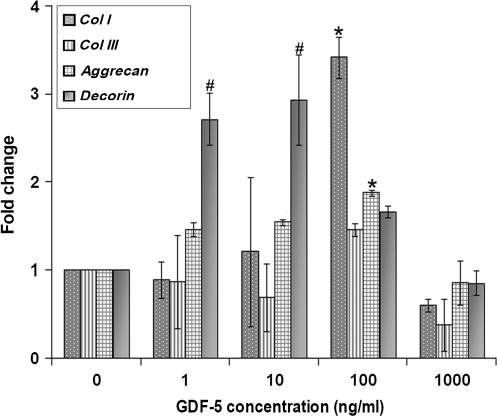
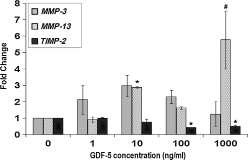
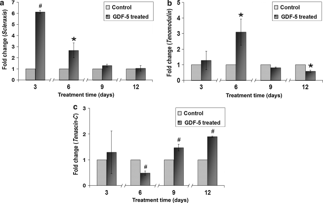
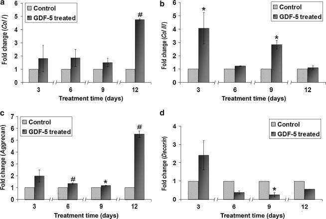
Methods: ADMSCs were treated in culture with different concentrations of GDF-5 (0-1000 ng/mL) for 12 days. Biochemical, temporal, and concentration kinetic studies were done. Extracellular matrix (ECM) synthesis, tendonogenic differentiation, and matrix remodeling gene and protein expression were analyzed.
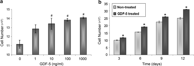
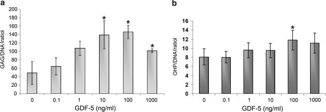
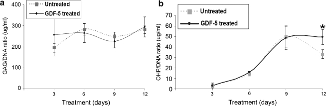
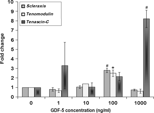
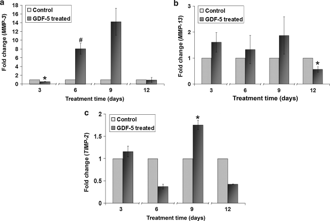
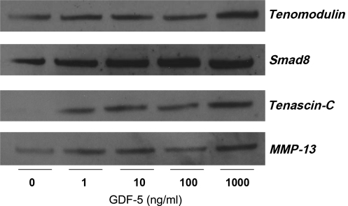
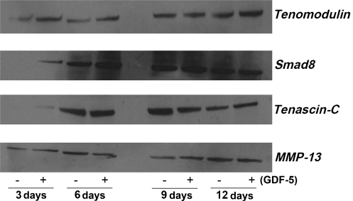
Results: GDF-5 led to increased ADMSC proliferation in a dose- and time-dependent manner. ADMSCs demonstrated enhanced ECM (collagen type I, decorin, and aggrecan) and tendonogenic marker (scleraxis, tenomodulin, and tenascin-C) gene expression with 100 ng/mL of GDF-5 (p < 0.05). ECM and tendon-specific markers showed time-dependent increases at various time points (p < 0.05), although decorin decreased at day 9 (p < 0.05). GDF-5 did alter expression of matrix remodeling genes, with no specific trends observed. Western blot analysis confirmed dose- and time-dependent increases in protein expression of tenomodulin, tenascin-C, Smad-8, and matrix metalloproteinase-13.
Conclusion: In vitro GDF-5 treatment can induce cellular events leading to the tendonogenic differentiation of ADMSCs. The use of combined GDF-5 and ADMSCs tissue-engineered therapies may have a role in the future of tendon repair.
Figures
Similar articles
-
Dose-Response Tendon-Specific Markers Induction by Growth Differentiation Factor-5 in Human Bone Marrow and Umbilical Cord Mesenchymal Stem Cells.Int J Mol Sci. 2020 Aug 17;21(16):5905. doi: 10.3390/ijms21165905. Int J Mol Sci. 2020. PMID: 32824547 Free PMC article.
-
Growth/differentiation factor-5 modulates the synthesis and expression of extracellular matrix and cell-adhesion-related molecules of rat Achilles tendon fibroblasts.Connect Tissue Res. 2011;52(4):353-64. doi: 10.3109/03008207.2010.534208. Epub 2011 Jan 20. Connect Tissue Res. 2011. PMID: 21250863
-
Bone marrow-derived mesenchymal stem cells obtained during arthroscopic rotator cuff repair surgery show potential for tendon cell differentiation after treatment with insulin.Arthroscopy. 2011 Nov;27(11):1459-71. doi: 10.1016/j.arthro.2011.06.029. Epub 2011 Oct 5. Arthroscopy. 2011. PMID: 21978434
-
Growth and differentiation factor-5 contributes to the structural and functional maintenance of the intervertebral disc.Cell Physiol Biochem. 2015;35(1):1-16. doi: 10.1159/000369670. Epub 2015 Jan 2. Cell Physiol Biochem. 2015. PMID: 25547527 Review.
-
Potential of Photobiomodulation to Induce Differentiation of Adipose- Derived Mesenchymal Stem Cells into Neural Cells.Curr Stem Cell Res Ther. 2021;16(3):307-322. doi: 10.2174/1574888X15999200918095834. Curr Stem Cell Res Ther. 2021. PMID: 32957891 Review.
Cited by
-
Tissue-engineering strategies for the tendon/ligament-to-bone insertion.Connect Tissue Res. 2012;53(2):95-105. doi: 10.3109/03008207.2011.650804. Epub 2011 Dec 20. Connect Tissue Res. 2012. PMID: 22185608 Free PMC article. Review.
-
Matrix Nanopatterning Regulates Mesenchymal Differentiation through Focal Adhesion Size and Distribution According to Cell Fate.Biomimetics (Basel). 2019 Jun 25;4(2):43. doi: 10.3390/biomimetics4020043. Biomimetics (Basel). 2019. PMID: 31242712 Free PMC article.
-
Enhancement of tenogenic differentiation of human adipose stem cells by tendon-derived extracellular matrix.Biomaterials. 2013 Dec;34(37):9295-306. doi: 10.1016/j.biomaterials.2013.08.054. Epub 2013 Sep 14. Biomaterials. 2013. PMID: 24044998 Free PMC article.
-
3D Biomimetic Scaffold for Growth Factor Controlled Delivery: An In-Vitro Study of Tenogenic Events on Wharton's Jelly Mesenchymal Stem Cells.Pharmaceutics. 2021 Sep 10;13(9):1448. doi: 10.3390/pharmaceutics13091448. Pharmaceutics. 2021. PMID: 34575523 Free PMC article.
-
Emerging Applications of Stem Cell and Regenerative Medicine to Sports Injuries.Orthop J Sports Med. 2014 Feb 6;2(2):2325967113519935. doi: 10.1177/2325967113519935. eCollection 2014 Feb. Orthop J Sports Med. 2014. PMID: 26535296 Free PMC article. Review.
References
-
- Moller A. Astron M. Westlin N. Increasing incidence of Achilles tendon rupture. Acta Orthop Scand. 1996;67:479. - PubMed
-
- Jozsa L. Kvist M. Balint B.J. Reffy A. Jarvinen M. Lehto M. Barzo M. The role of recreational sport activity in Achilles tendon rupture. A clinical, pathoanatomical, and sociological study of 292 cases. Am J Sports Med. 1989;17:338. - PubMed
-
- Kleinert H.E. Serafin D. Kutz J.E. Atasoy E. Reimplantation of amputated digits and hands. Orthop Clin North Am. 1973;4:957. - PubMed
-
- Morberg P. Jerre R. Sward L. Karlsson J. Long-term results after surgical management of partial Achilles tendon ruptures. Scand J Med Sci Sports. 1997;7:299. - PubMed
-
- Józsa L. Kannus P. Human Tendons: Anatomy, Physiology, and Pathology. Champaign, IL: Human Kinetics; 1997.
Publication types
MeSH terms
Substances
Grants and funding
LinkOut - more resources
Full Text Sources
Other Literature Sources
