. 2010 Jun 24:10:327.
doi: 10.1186/1471-2407-10-327.

Poly(I:C) induces intense expression of c-IAP2 and cooperates with an IAP inhibitor in induction of apoptosis in cancer cells

Affiliations

Poly(I:C) induces intense expression of c-IAP2 and cooperates with an IAP inhibitor in induction of apoptosis in cancer cells

Luc Friboulet et al. BMC Cancer. .

Abstract

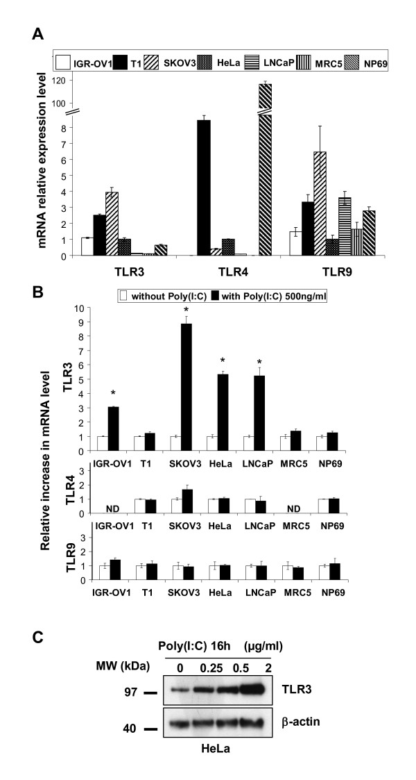
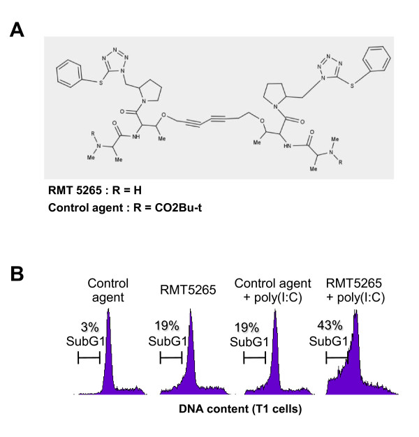
Background: There is increasing evidence that the toll-like receptor 3 (TLR3) is an interesting target for anti-cancer therapy. Unfortunately, most laboratory investigations about the impact of TLR3 stimulation on human malignant cells have been performed with very high concentrations--5 to 100 microg/ml--of the prototype TLR3 ligand, poly(I:C). In a previous study focused on a specific type of human carcinoma - nasopharyngeal carcinoma - we have shown that concentrations of poly(I:C) as low as 100 ng/ml are sufficient to induce apoptosis of malignant cells when combined to a pharmacological antagonist of the IAP family based on Smac mimicry.

Methods: This observation prompted us to investigate the contribution of the IAP family in cell response to poly(I:C) in a variety of human malignant cell types.

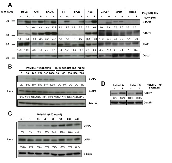
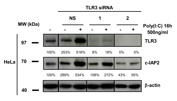
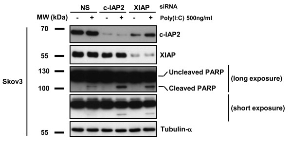
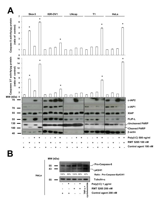
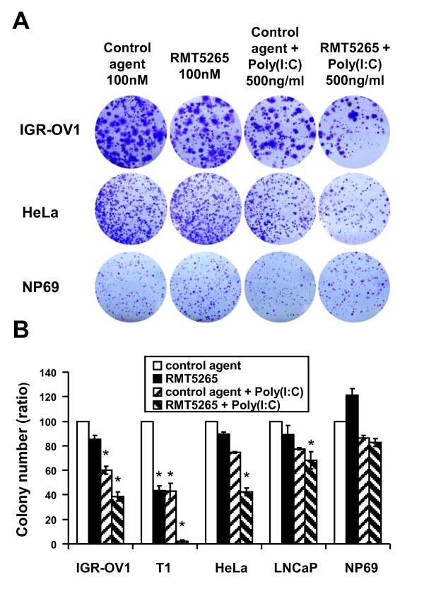
Results: We report a rapid, intense and selective increase in c-IAP2 protein expression observed under stimulation by poly(I:C)(500 ng/ml) in all types of human malignant cells. In most cell types, this change in protein expression is underlain by an increase in c-IAP2 transcripts and dependent on the TLR3/TRIF pathway. When poly(I:C) is combined to the IAP inhibitor RMT 5265, a cooperative effect in apoptosis induction and/or inhibition of clonogenic growth is obtained in a large fraction of carcinoma and melanoma cell lines.

Conclusions: Currently, IAP inhibitors like RMT 5265 and poly(I:C) are the subject of separate therapeutic trials. In light of our observations, combined use of both types of compounds should be considered for treatment of human malignancies including carcinomas and melanomas.


Figures

Figure 1
Figure 1
Assessment of TLR 3, 4 and 9 messenger RNAs and TLR3 protein in a panel of human cell lines in basal conditions and under stimulation by poly(I:C). A) Relative expression levels of TLR3, 4 and 9 transcripts in basal conditions. Levels of mRNAs in HeLa cells were chosen as a reference and arbitrarily set at 1. B) TLR mRNA relative increase in expression under stimulation by poly(I:C) (500 ng/ml) for 16 h. For each cell type, the basal level of expression was taken as a reference and set at 1. C) Western blot detection of the TLR3 protein in HeLa cells treated for 16 h with increasing concentrations of poly(I:C)(from 250 ng/ml to 2 μg/ml). β-actin staining was used to control protein loading. Data presented in A), B) and C) are representative of at least two similar experiments. The stars indicate a statistical difference from respective controls (p < 0.05).
Figure 2
Figure 2
Status of the IAP proteins in a panel of malignant and non-malignant cell types in basal conditions and under stimulation by the poly(I:C). A) Variations in IAP protein concentrations in basal conditions and after 16 h of stimulation by poly(I:C) 500 ng/ml. The c-IAP1, c-IAP2 and XIAP relative concentrations were assessed by iterative staining of blotted membranes. β-actin was used as a loading control for density measurements. B) Exploration of the dose-effects relationships in c-IAP2 induction in HeLa cells treated for 16 h by poly(I:C) (TLR3 agonist) or CpG DNA (TLR9 agonist) at concentrations increasing from 50 to 2000 ng/ml. The same membrane was stained successively with anti-c-IAP2, c-IAP1 and β-actin. C) Western blot analysis of HeLa cells treated with poly(I:C) 500 ng/ml for increasing time intervals from 1 h to 48 h. The same membrane was stained successively with anti-c-IAP2, c-IAP1 and β-actin. D) Increase in c-IAP2 protein expression in malignant cells directly explanted in vitro from fresh human tumors. Cells were prepared for short term primary cultures from tumor biopsy fragments obtained from 2 patients designated A and B. These patients were referred for Head and Neck squamous cell carcinomas (see Methods section). Cells were incubated with poly(I:C) immediately after tumor dispersion for 16 h and collected for protein extraction and western blot analysis of c-IAP2 and β-actin expression. Data presented in A), B) and C) are representative of at least two similar experiments.
Figure 3
Figure 3
Treatment by poly(I:C) increases c-IAP2 gene (BIRC3) transcription in human malignant cells. Inhibition by Bafilomycin A1 and knock-down of TRIF. A) Cell concentrations of BIRC3 messenger RNAs are measured by quantitative RT-PCR in several types of malignant cells mock-treated or treated with poly(I:C) for 16 h at 500 ng/ml. Concentration of BIRC3 transcripts measured in basal conditions were chosen as a reference for each tested cell type and arbitrarily set at 1. B) Kinetics of the increase in the concentration of BIRC3 messenger RNAs in HeLa cells treated with poly(I:C) 500 ng/ml for increasing time intervals from 1 to 48 h. C) The influence of poly(I:C) on BIRC3 transcripts is neutralized by Bafilomycin A1, an inhibitor of endosome acidification and TLR3 signalling. Prior to RNA extraction, HeLa cells are incubated for two hours in the presence of poly(I:C)(500 ng/ml) or recombinant TNF α (20 ng/ml) in combination with Bafilomycin A1 (BFA) 100 nM or 2-aminopurin (2-AP) 5 mM or without additional compound (control condition). D) The induction of c-IAP2 by poly(I:C) in Hela cells is suppressed when the TRIF adaptor protein is knocked-down using 3 distinct specific siRNA whereas a non-specific (NS) RNA used as a negative control has no inhibitory effect. Upper panel: quantitative PCR assessment of the BIRC3 transcripts in the absence or in the presence of specific siRNAs. Lower panel: absence of TRIF protein expression detectable by western blot in Hela cells treated with specific siRNA (1, 2 and 3); in contrast TRIF expression is not altered by a negative control non-specific siRNA (NS). Data are representative of two similar experiments. The stars indicate a statistical difference from respective controls (p < 0.05).
Figure 4
Figure 4
Knocking-down TLR3 suppresses enhancement of c-IAP2 protein expression by poly(I:C). Western blot analysis of protein extracts from HeLa cells treated with poly(I:C) (500 ng/ml for 16 h) with or without pre-treatment with TLR3-specific and control (NS) siRNAs. The same membrane was stained successively with anti-c-IAP2, TLR3 and β-actin. Note that the control siRNA by itself enhanced TLR3 and c-IAP2 expression. This is consistent with previous observations and possibly related to the interaction of these small double-strand RNAs with the TLR3 itself regardless of their sequence [36]. In our transfection protocol siRNAs are at 100 nM for the first 4 h and 50 nM for the next 44 h. TLR3 inhibition was not complete with the siRNA n°1 which was apparently weaker than the siRNA n°2. However the siRNA n°1 was strong enough to prevent enhancement of TLR3 and c-IAP2 expression in the absence of poly(I:C). Data are representative of two similar experiments.
Figure 5
Figure 5
Enhanced apoptosis in Skov3 ovarian carcinoma cells treated with poly(I:C) combined with knocking-down c-IAP2 or XIAP. Skov3 cells were pre-treated with non-specific (NS), c-IAP2 or XIAP siRNAs with or without subsequent addition of poly(I:C). Cell protein extracts were subjected to western blot and stained successively for c-IAP2, XIAP, PARP and tubulin-α. PARP-cleavage was substantially enhanced by combination of poly (I:C) with c-IAP2 and XIAP siRNAs.
Figure 6
Figure 6
Induction of apoptosis in T1 cells treated with poly(I:C) combined with a smac-mimetic (RMT 5265). A) Representation of the RMT 5265 molecule backbone drawn with MARVIN online software: http://atchimiebiologie.free.fr/marvin/doc/dev/oli.html. Substitution of one H on both alanines (R = CO2Bu-t) results in an inactive compound used as a negative control (HS4044). B) Flow cytometry assessment of the sub-G1 cell fraction in the T1 cell line treated with control compound or RMT 5265 (100 nM; 48 h) with or without poly(I:C)(500 ng/ml; last 16 h).
Figure 7
Figure 7
Analysis of biochemical changes in human malignant cell lines treated with poly(I:C) combined to RMT 5265. A) Cell samples representative of five different cell lines were subjected to the following treatments: control compound or RMT 5265 (100 nM for 48 h) with or without poly(I:C) (500 ng/ml; last 16 h). Caspase 3/7 (middle panel) and caspase 8 (upper panel) activities were measured in cell protein extracts using a chemiluminescent assay. c-IAP2, c-IAP1, FLIP-L and β-actin were detected in additional cellular extracts from the same experimental samples by western blotting using specific antibodies applied successively to the same blotted membrane (lower panel). The stars indicate a statistical difference from respective controls (p < 0.05). B) Western blot analysis of caspase-8 cleavage in HeLa cells treated with control compound or RMT 5265 (200 nM for 48 h) with or without poly(I:C) (1 μg/ml; last 16 h). In order to provide a positive control for caspase 8 cleavage, HeLa cells were treated with TNF-α (50 ng/ml) plus cycloheximide (2.5 μg/ml) for 6 h. The same blotted membrane was stained with anti-tubulin-α for protein loading control.
Figure 8
Figure 8
Impact of poly (I:C) and RMT 5265 on the clonogenic growth of human cell lines. Cells from IGR-OV1, T1, HeLa, LNCap and NP69 cell lines were seeded at low densities in 6 well-plates as reported in the Methods section. A) Plates were stained with crystal violet after 2 to 3 weeks of growth. B) Colonies were counted after scanning and digitalization of dried plates (Epson perfection 4990 scanner). For star-marked conditions, the differences in colony numbers with the control condition were statistically significant (p < 0.05).

References

    1. Matsumoto M, Funami K, Oshiumi H, Seya T. Toll-like receptor 3: a link between toll-like receptor, interferon and viruses. Microbiol Immunol. 2004;48:147–154. - PubMed
    1. Chen R, Alvero AB, Silasi DA, Steffensen KD, Mor G. Cancers take their Toll--the function and regulation of Toll-like receptors in cancer cells. Oncogene. 2008;27:225–233. doi: 10.1038/sj.onc.1210907. - DOI - PubMed
    1. Huang B, Zhao J, Unkeless JC, Feng ZH, Xiong H. TLR signaling by tumor and immune cells: a double-edged sword. Oncogene. 2008;27:218–224. doi: 10.1038/sj.onc.1210904. - DOI - PubMed
    1. Laplanche A, Alzieu L, Delozier T, Berlie J, Veyret C, Fargeot P, Luboinski M, Lacour J. Polyadenylic-polyuridylic acid plus locoregional radiotherapy versus chemotherapy with CMF in operable breast cancer: a 14 year follow-up analysis of a randomized trial of the Federation Nationale des Centres de Lutte contre le Cancer (FNCLCC) Breast Cancer Res Treat. 2000;64:189–191. doi: 10.1023/A:1006498121628. - DOI - PubMed
    1. Salaun B, Coste I, Rissoan MC, Lebecque SJ, Renno T. TLR3 can directly trigger apoptosis in human cancer cells. J Immunol. 2006;176:4894–4901. - PubMed

Publication types

MeSH terms