

Transvaginal sonographic cervical length for the prediction of spontaneous preterm birth in twin pregnancies: a systematic review and metaanalysis
- PMID: 20576253
- PMCID: PMC3147231
- DOI: 10.1016/j.ajog.2010.02.064
Transvaginal sonographic cervical length for the prediction of spontaneous preterm birth in twin pregnancies: a systematic review and metaanalysis
Abstract
Objective: To assess the accuracy of transvaginal sonographic cervical length (CL) in predicting spontaneous preterm birth in women with twin pregnancies.
Study design: Systematic review and metaanalysis of predictive test accuracy.
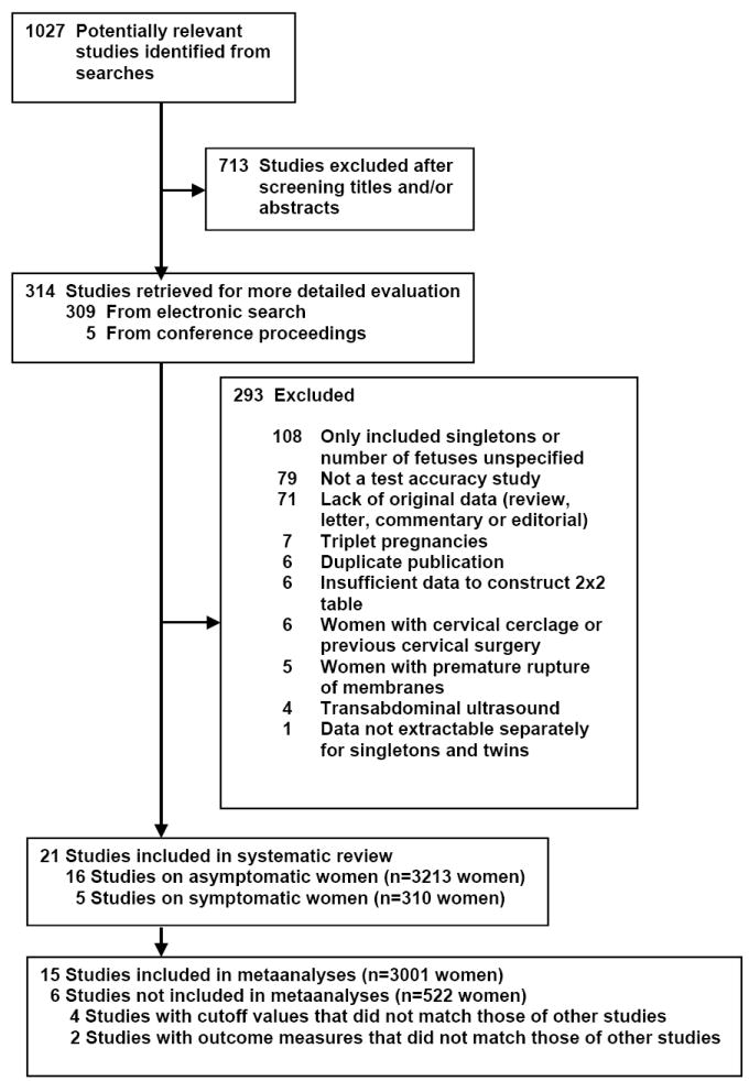
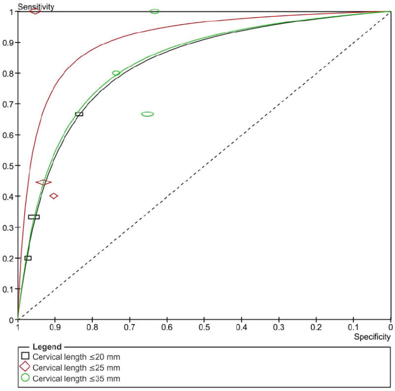
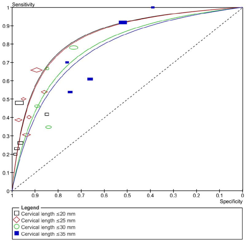
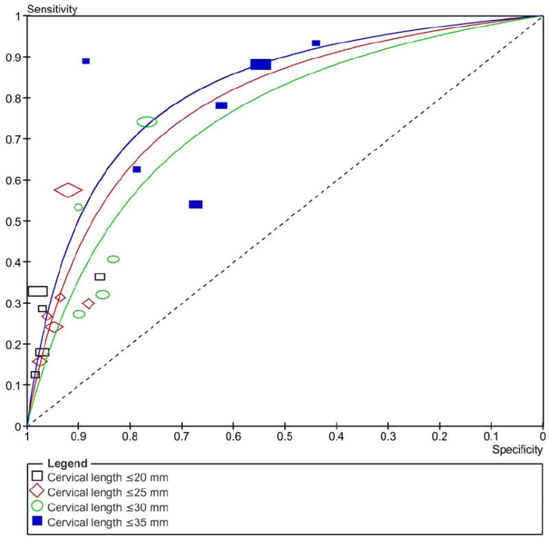
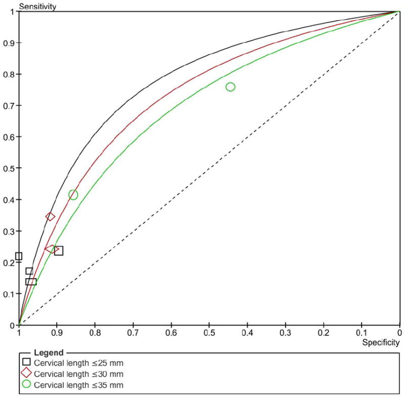
Results: Twenty-one studies (16 in asymptomatic women and 5 in symptomatic women) with a total of 3523 women met the inclusion criteria. Among asymptomatic women, a CL <or=20 mm at 20-24 weeks' gestation was the most accurate in predicting preterm birth <32 and <34 weeks' gestation (pooled sensitivities, specificities, and positive and negative likelihood ratios of 39% and 29%, 96% and 97%, 10.1 and 9.0, and 0.64 and 0.74, respectively). A CL <or=25 mm at 20-24 weeks' gestation had a pooled positive likelihood ratio of 9.6 to predict preterm birth <28 weeks' gestation. The predictive accuracy of CL for preterm birth was low in symptomatic women.
Conclusion: Transvaginal sonographic CL at 20-24 weeks' gestation is a good predictor of spontaneous preterm birth in asymptomatic women with twin pregnancies.
Copyright (c) 2010. Published by Mosby, Inc.
Figures
Similar articles
-
Transvaginal sonographic measurement of cervical length to predict preterm birth in asymptomatic women at increased risk: a systematic review.Ultrasound Obstet Gynecol. 2008 May;31(5):579-87. doi: 10.1002/uog.5323. Ultrasound Obstet Gynecol. 2008. PMID: 18412093
-
Predictive accuracy of changes in transvaginal sonographic cervical length over time for preterm birth: a systematic review and metaanalysis.Am J Obstet Gynecol. 2015 Dec;213(6):789-801. doi: 10.1016/j.ajog.2015.06.015. Epub 2015 Jun 10. Am J Obstet Gynecol. 2015. PMID: 26070703 Free PMC article.
-
Cervical length measurement for the prediction of preterm birth in multiple pregnancies: a systematic review and bivariate meta-analysis.Ultrasound Obstet Gynecol. 2011 Jul;38(1):10-7. doi: 10.1002/uog.9013. Epub 2011 Jun 20. Ultrasound Obstet Gynecol. 2011. PMID: 21465606
-
Vaginal progesterone for preventing preterm birth and adverse perinatal outcomes in twin gestations: a systematic review and meta-analysis.Am J Obstet Gynecol. 2023 Dec;229(6):599-616.e3. doi: 10.1016/j.ajog.2023.05.010. Epub 2023 May 15. Am J Obstet Gynecol. 2023. PMID: 37196896 Free PMC article.
-
Cervicovaginal fetal fibronectin for the prediction of spontaneous preterm birth in multiple pregnancies: a systematic review and meta-analysis.J Matern Fetal Neonatal Med. 2010 Dec;23(12):1365-76. doi: 10.3109/14767058.2010.499484. J Matern Fetal Neonatal Med. 2010. PMID: 21067303 Free PMC article.
Cited by
-
Breastfeeding initiation, duration, and experiences of mothers of late preterm twins: a mixed-methods study.Int Breastfeed J. 2022 Sep 8;17(1):68. doi: 10.1186/s13006-022-00507-3. Int Breastfeed J. 2022. PMID: 36076279 Free PMC article.
-
Prediction of Preterm Delivery by Ultrasound Measurement of Cervical Length and Funneling Changes of the Cervix in Pregnant Women with Preterm Labor at 28-34 weeks of Gestation.J Med Life. 2020 Oct-Dec;13(4):536-542. doi: 10.25122/jml-2020-0069. J Med Life. 2020. PMID: 33456603 Free PMC article.
-
Cervical cerclage versus cervical pessary with or without vaginal progesterone for preterm birth prevention in twin pregnancies and a short cervix: A two-by-two factorial randomised clinical trial.PLoS Med. 2025 Feb 21;22(2):e1004526. doi: 10.1371/journal.pmed.1004526. eCollection 2025 Feb. PLoS Med. 2025. PMID: 39982935 Free PMC article. Clinical Trial.
-
The clinical significance of a positive Amnisure test in women with preterm labor and intact membranes.J Matern Fetal Neonatal Med. 2012 Sep;25(9):1690-8. doi: 10.3109/14767058.2012.657279. Epub 2012 Apr 25. J Matern Fetal Neonatal Med. 2012. PMID: 22280400 Free PMC article.
-
A blueprint for the prevention of preterm birth: vaginal progesterone in women with a short cervix.J Perinat Med. 2013 Jan;41(1):27-44. doi: 10.1515/jpm-2012-0272. J Perinat Med. 2013. PMID: 23314512 Free PMC article. Review.
References
-
- Martin JA, Hamilton BE, Sutton PD, Ventura SJ, Menacker F, Kirmeyer S, Mathews TJ. Births: final data for 2006. Natl Vital Stat Rep. 2009;57:1–102. - PubMed
-
- Leitich H, Brunbauer M, Kaider A, Egarter C, Husslein P. Cervical length and dilatation of the internal cervical os detected by vaginal ultrasonography as markers for preterm delivery: A systematic review. Am J Obstet Gynecol. 1999;181:1465–72. - PubMed
-
- Honest H, Bachmann LM, Coomarasamy A, Gupta JK, Kleijnen J, Khan KS. Accuracy of cervical transvaginal sonography in predicting preterm birth: a systematic review. Ultrasound Obstet Gynecol. 2003;22:305–22. - PubMed
-
- Crane JM, Hutchens D. Transvaginal sonographic measurement of cervical length to predict preterm birth in asymptomatic women at increased risk: a systematic review. Ultrasound Obstet Gynecol. 2008;31:579–87. - PubMed
Publication types
MeSH terms
Grants and funding
LinkOut - more resources
Full Text Sources
Other Literature Sources
Medical
Miscellaneous
