. 2010 Sep 1;169(3):1007-16.
doi: 10.1016/j.neuroscience.2010.05.054. Epub 2010 Jun 1.

Changes in 5-HT2A-mediated behavior and 5-HT2A- and 5-HT1A receptor binding and expression in conditional brain-derived neurotrophic factor knock-out mice

Affiliations

Changes in 5-HT2A-mediated behavior and 5-HT2A- and 5-HT1A receptor binding and expression in conditional brain-derived neurotrophic factor knock-out mice

A B Klein et al. Neuroscience. .

Abstract

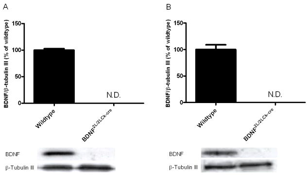
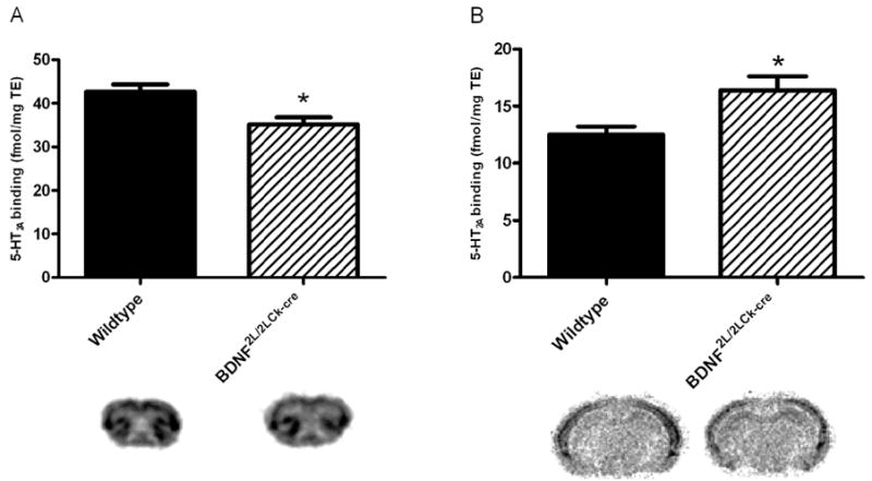
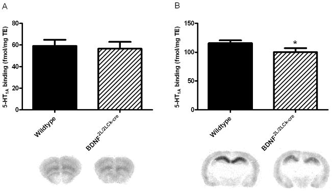
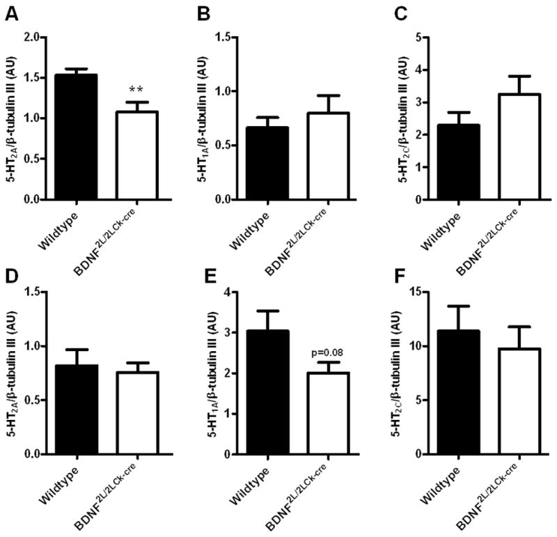
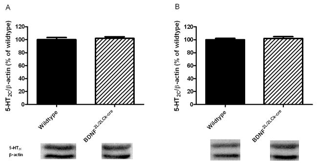
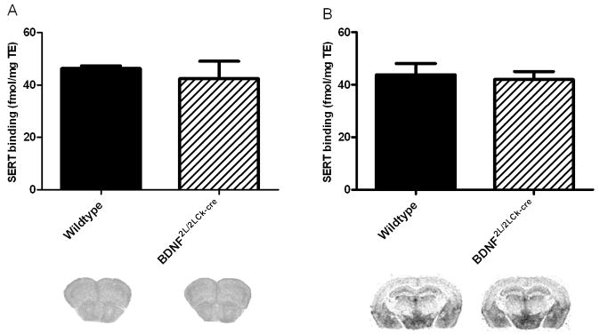
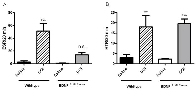
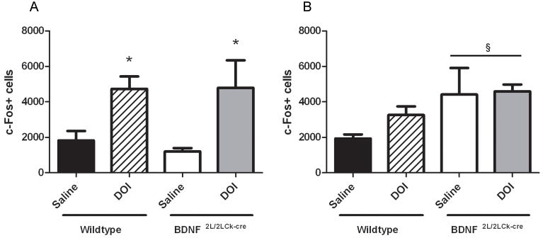
Changes in brain-derived neurotrophic factor (BDNF) expression have been implicated in the etiology of psychiatric disorders. To investigate pathological mechanisms elicited by perturbed BDNF signaling, we examined mutant mice with central depletion of BDNF (BDNF(2L/2LCk-cre)). A severe impairment specific for the serotonin 2A receptor (5-HT(2A)R) in prefrontal cortex was described previously in these mice. This is of much interest, as 5-HT(2A)Rs have been linked to neuropsychiatric disorders and anxiety-related behavior. Here we further characterized the serotonin receptor alterations triggered by BDNF depletion. 5-HT(2A) ([(3)H]-MDL100907) and 5-HT(1A) ([(3)H]-WAY100635) receptor autoradiography revealed site-specific alterations in BDNF mutant mice. They exhibited lower 5-HT(2A) receptor binding in frontal cortex but increased binding in hippocampus. Additionally, 5-HT(1A) receptor binding was decreased in hippocampus of BDNF mutants, but unchanged in frontal cortex. Molecular analysis indicated corresponding changes in 5-HT(2A) and 5-HT(1A) mRNA expression but normal 5-HT(2C) content in these brain regions in BDNF(2L/2LCk-cre) mice. We investigated whether the reduction in frontal 5-HT(2A)R binding was reflected in reduced functional output in two 5-HT(2A)-receptor mediated behavioral tests, the head-twitch response (HTR) and the ear-scratch response (ESR). BDNF(2L/2LCk-cre) mutants treated with the 5-HT(2A) receptor agonist (+/-)-2,5-dimethoxy-4-iodoamphetamine (DOI) showed a clearly diminished ESR but no differences in HTR compared to wildtypes. These findings illustrate the context-dependent effects of deficient BDNF signaling on the 5-HT receptor system and 5-HT(2A)-receptor functional output.


Figures

Figure 1
Figure 1
BDNF protein levels in frontal cortex (A) and hippocampus (B) in wildtype and BDNF2L/2LCk-cre mice. BDNF levels were not detectable in frontal cortex and hippocampus in the BDNF mutant mice using western blot, while wildtype levels corresponded to levels seen in other mice strains. BDNF levels were related to β-tubulin III levels (n=7). Representative bands for BDNF (14 kDa) and β-tubulin III (55 kDa) are shown below the corresponding column. N.D.: not detectable. Error bars indicate SEM.
Figure 2
Figure 2
5-HT2A receptor binding (3H-MDL-100907) in BDNF2L/2LCk-cre mice and wildtype littermates in frontal cortex (A) and dorsal hippocampus (B). There was a significant decrease in binding in frontal cortex in BDNF2L/2LCk-cre mice vs wildtypes, while the opposite was observed for dorsal hippocampus. (n=4, Student’s t-test, *P < 0.05). Representative autoradiograms are shown below the corresponding column. Error bars indicate SEM
Figure 3
Figure 3
5-HT1A receptor binding (3H-WAY100635) in BDNF2L/2LCk-cre mice and wildtype littermates in frontal cortex (A) and dorsal hippocampus (B). There was a significant decrease in binding in dorsal hippocampus in BDNF2L/2LCk-cre mice vs wildtypes, while no change was observed in frontal cortex. (n=4, Student’s t-test, *P < 0.05). Representative autoradiograms are shown below the corresponding column. Error bars indicate SEM
Figure 4
Figure 4
RT-qPCR on 5-HT2A (A, D), 5-HT1A (B, E) and 5-HT2C (C, F) mRNA in frontal cortex (upper panel) and hippocampus (lower panel) of wildtype and BDNF2L/2LCk-cre mice. There was a significant lower 5-HT2A mRNA expression in BDNF2L/2LCk-cre mice in frontal cortex compared to wildtypes (A), while no differences could be observed in hippocampus (D)(n=7–8, Student’s t-test, **P < 0.01). For the 5-HT1A receptor, there was a tendency towards a decrease in expression in BDNF2L/2LCk-cre mice compared to wildtypes (E)(P = 0.08) for hippocampus, while no change was observed in frontal cortex (B). For the 5-HT2C receptor we did not find any differences in expression of mRNA between BDNF2L/2LCk-cre mice and wildtypes. (AU, arbitrary units, Error bars indicate SEM)
Figure 5
Figure 5
5-HT2C receptor protein levels in wildtype and BDNF2L/2LCk-cre mice in frontal cortex (A) and hippocampus (B). No changes in protein levels of this serotonin receptor could be observed (n=7–8, Student’s t-test). Representative bands for 5-HT2C (55 kDa) and β-actin (40 kDa) are shown below the corresponding column. Error bars indicate SEM
Figure 6
Figure 6
SERT binding performed with 3H-escitalopram in frontal cortex (A) and hippocampus (B). We did not detect any differences in SERT binding between wildtypes and BDNF2L/2LCk-cre mice. (n=4, Student’s t-test). Representative autoradiograms are shown below the corresponding column. Error bars indicate SEM
Figure 7
Figure 7
Ear-Scratch Reponse (ESR)(A) and Head-Twitch Reponse (HTR)(B) in BDNF mutant mice and wildtype littermates after administration of DOI (2.5 mg/kg, i.p.) or saline. The registration of ESR and HTR was started 5 min after administration and continued for 20 min. Bars: Black: wildtype, saline; Crossed: wildtype, DOI 2.5 mg/kg, i.p.; White: BDNF2L/2LCk-cre mice, saline; Gray: BDNF2L/2LCk-cre mice (n=7, two-way ANOVA, Bonferroni post test, n.s.=not significant, **P < 0.01, ***P < 0.001. Error bars indicate SEM
Figure 8
Figure 8
c-Fos immunopositive cells in frontal cortex (A) and hippocampus (B). A single DOI (2.5 mg/kg) injection significantly induced c-Fos immunoreactive cells compared to a single saline injection in frontal cortex in both wildtypes and BDNF2L/2LCk-cre mice (A). There was a genotype effect in hippocampus (§) indicating higher cell activation response BDNF2L/2LCk-cre mice compared to wildtypes (B)(n=4, two-way ANOVA, Bonferroni post hoc test, *P < 0.05. §P<0.05. Error bars indicate SEM
Figure 9
Figure 9
Analysis of protein levels using western blotting for synaptophysin (38 kDa), PSD-95 (95 kDa) and MAP2 (250 kDa). Upper panel: frontal cortex, lower panel: hippocampus. BDNF mutants have normal contents of the synaptic markers synaptophysin and PSD-95 and the dendritic marker MAP2 when compared to wildtypes. β-tubulin III (55 kDa) was used as loading control. (n=7–8, Student’s t-test). Error bars indicate SEM

Similar articles

Cited by

References

    1. Akimova E, Lanzenberger R, Kasper S. The serotonin-1A receptor in anxiety disorders. Biol Psychiatry. 2009;66:627–635. - PubMed
    1. Chan JP, Unger TJ, Byrnes J, Rios M. Examination of behavioral deficits triggered by targeting Bdnf in fetal or postnatal brains of mice. Neuroscience. 2006;142:49–58. - PubMed
    1. Cornea-Hebert V, Riad M, Wu C, Singh SK, Descarries L. Cellular and subcellular distribution of the serotonin 5-HT2A receptor in the central nervous system of adult rat. J Comp Neurol. 1999;409:187–209. - PubMed
    1. Darmani NA. Cannabinoids of diverse structure inhibit two DOI-induced 5-HT(2A) receptor-mediated behaviors in mice. Pharmacol Biochem Behav. 2001;68:311–317. - PubMed
    1. Dave KD, Fernando GS, Quinn JL, Harvey JA, Aloyo VJ. Serotonin 5-HT2A receptors in the CA1 field of the hippocampus mediate head movements in the rabbit. Psychopharmacology (Berl) 2004;176:287–295. - PubMed

Publication types

MeSH terms

Substances