

Shear stress-induced volume decrease in C11-MDCK cells by BK-alpha/beta4
- PMID: 20576683
- PMCID: PMC2944292
- DOI: 10.1152/ajprenal.00222.2010
Shear stress-induced volume decrease in C11-MDCK cells by BK-alpha/beta4
Abstract
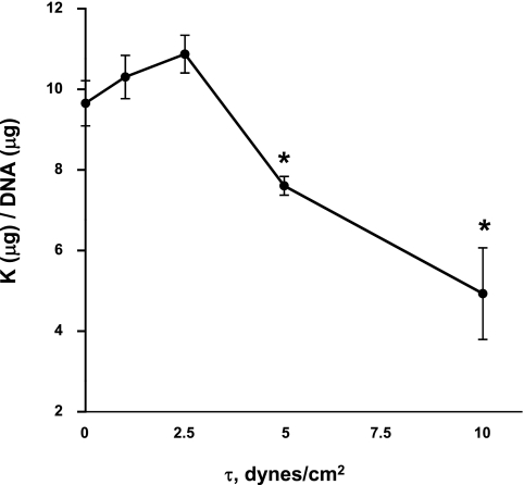
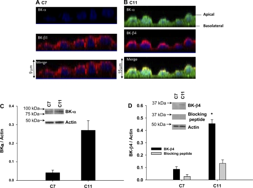
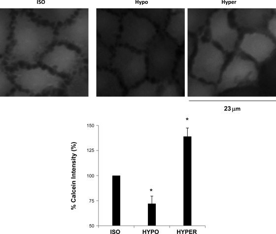
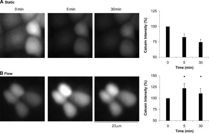
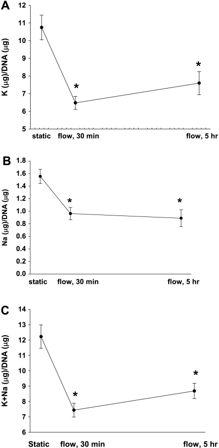
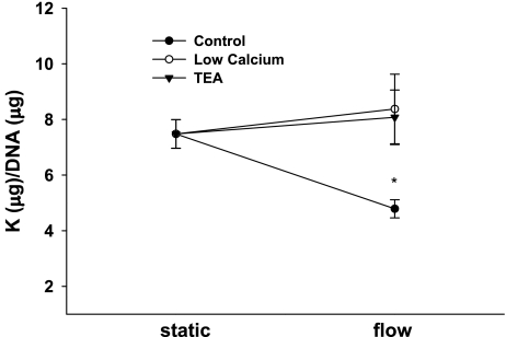
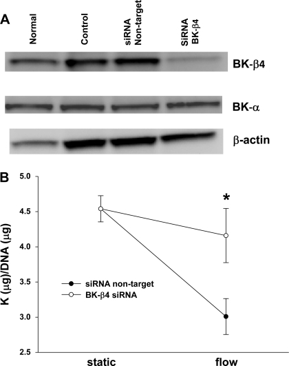
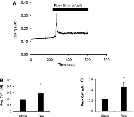
Large-conductance, calcium-activated potassium channels (BK) are expressed in principal cells (PC) and intercalated cells (IC) in mammalian nephrons as BK-alpha/beta1 and BK-alpha/beta4, respectively. IC, which protrude into the lumens of tubules, express substantially more BK than PC despite lacking sufficient Na-K-ATPase to support K secretion. We previously showed in mice that IC exhibit size reduction when experiencing high distal flows induced by a high-K diet. We therefore tested the hypothesis that BK-alpha/beta4 are regulators of IC volume via a shear stress (tau)-induced, calcium-dependent mechanism, resulting in a reduction in intracellular K content. We determined by Western blot and immunocytochemical analysis that C11-Madin-Darby canine kidney cells contained a predominance of BK-alpha/beta4. To determine the role of BK-alpha/beta4 in tau-induced volume reduction, we exposed C11 cells to tau and measured K efflux by flame photometry and cell volume by calcein staining, which changes inversely to cell volume. With 10 dynes/cm(2), calcein intensity significantly increased 39% and monovalent cationic content decreased significantly by 37% compared with static conditions. Furthermore, the shear-induced K loss from C11 was abolished by the reduction of extracellular calcium, addition of 5 mM TEA, or BK-beta4 small interfering (si) RNA, but not by addition of nontarget siRNA. These results show that BK-alpha/beta4 plays a role in shear-induced K loss from IC, suggesting that BK-alpha/beta4 regulate IC volume during high-flow conditions. Furthermore, these results support the use of C11 cells as in vitro models for studying BK-related functions in IC of the kidney.
Figures
References
-
- Bailey MA, Cantone A, Yan Q, MacGregor GG, Leng Q, Amorim JB, Wang T, Hebert SC, Giebisch G, Malnic G. Maxi-K channels contribute to urinary potassium excretion in the ROMK-deficient mouse model of type II Bartter's syndrome and in adaptation to a high-K diet. Kidney Int 70: 51–59, 2006 - PubMed
-
- Bolivar JJ, Cereijido M. Voltage and Ca2+-activated K+ channel in cultured epithelial cells (MDCK). J Membr Biol 97: 43–51, 1987 - PubMed
-
- Brenner R, Chen QH, Vilaythong A, Toney GM, Noebels JL, Aldrich RW. BK channel [beta]4 subunit reduces dentate gyrus excitability and protects against temporal lobe seizures. Nat Neurosci 8: 1752–1759, 2005 - PubMed
-
- Cai Z, Xin J, Pollock DM, Pollock JS. Shear stress-mediated NO production in inner medullary collecting duct cells. Am J Physiol Renal Physiol 279: F270–F274, 2000 - PubMed
-
- Carmines PK, Fowler BC, Bell PD. Segmentally distinct effects of depolarization on intracellular [Ca2+] in renal arterioles. Am J Physiol Renal Fluid Electrolyte Physiol 265: F677–F685, 1993 - PubMed
Publication types
MeSH terms
Substances
Grants and funding
LinkOut - more resources
Full Text Sources