. 2010 Aug;21(8):1326-33.
doi: 10.1681/ASN.2009091001. Epub 2010 Jun 24.

Intraflagellar transport proteins are essential for cilia formation and for planar cell polarity

Affiliations

Intraflagellar transport proteins are essential for cilia formation and for planar cell polarity

Ying Cao et al. J Am Soc Nephrol. 2010 Aug.

Abstract

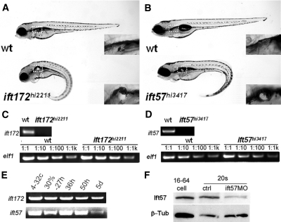
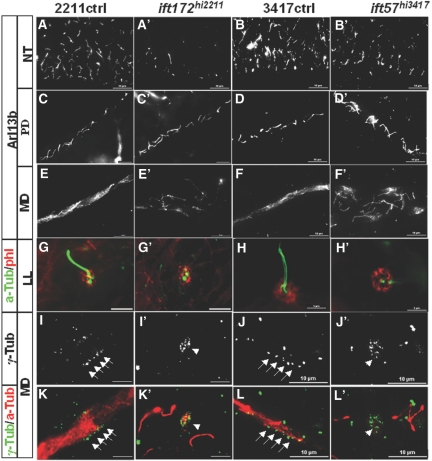
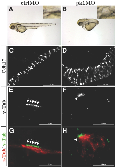
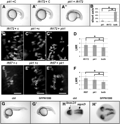
The highly conserved intraflagellar transport (IFT) proteins are essential for cilia formation in multiple organisms, but surprisingly, cilia form in multiple zebrafish ift mutants. Here, we detected maternal deposition of ift gene products in zebrafish and found that ciliary assembly occurs only during early developmental stages, supporting the idea that maternal contribution of ift gene products masks the function of IFT proteins during initial development. In addition, the basal bodies in multiciliated cells of the pronephric duct in ift mutants were disorganized, with a pattern suggestive of defective planar cell polarity (PCP). Depletion of pk1, a core PCP component, similarly led to kidney cyst formation and basal body disorganization. Furthermore, we found that multiple ift genes genetically interact with pk1. Taken together, these data suggest that IFT proteins play a conserved role in cilia formation and planar cell polarity in zebrafish.


Figures

Figure 1.
Figure 1.
hi3417 and hi2211 are zygotic loss-of-function mutants of ift57 and ift172, respectively. (A and B) A wild-type sibling and a mutant at 4 dpf in side views. Box with dashed line: cyst (in mutant) and the lack of cyst (in wild-type sibling) enlarged in insets. (C and D) Loss of wild-type ift172 and ift57 transcripts in hi2211 and hi3417 mutants at 33 hpf, respectively. Upper panels: RT-PCR with a pair of primers spanning the proviral insertion site. Lower panels: loading controls with a pair of elf1-specific primers using cDNAs serially diluted 1:1, 1:10, 1:100, and 1:1000. (E) RT-PCR time course for ift172 and ift57 using wild-type samples. (F) Western blot against Ift57 (Ift57). β-Tub is used as loading control. β-Tub, β-Tubulin; ctrl, uninjected embryo control; ift57MO, ift57 morphants; wt, wild type; 4-32c, 4- to 32-cell stage; 30%–27h, mixture of samples from 30% epiboly to 27 hpf; 36h, 36 hpf; 50h, 50 hfp; 5d, 5dpf; 16–64 cell, sample from embryos at the 16- to 64-cell stage; 20s, samples from embryos at the 20-somite stage.
Figure 2.
Figure 2.
Cilia formation is disrupted in ift172hi2211 and ift57hi3417 mutants in a time-dependent manner. (A through B') Cilia in the neural tube at 34 hpf as shown by anti-Arl13b labeling. (C through D') Cilia in the posterior pronephric duct at 2 dpf as shown by anti-Arl13b labeling. (E through F') Cilia in the MD at 2 dpf as shown by anti-Arl13b labeling. (G through H') Cilia in the lateral line organ as shown by anti–a-Tub in green at 2 dpf. phl labeling in red shows the presence of the LL organ. (I through L') Basal bodies in the MD at 3 pdf as shown by γ-Tub labeling. Anti–a-Tub in (K through L') labeled cilia. Arrows point to arrays of basal bodies, whereas arrowheads point to disorganized clusters of basal bodies. a-Tub, anti-acetylated Tubulin; γ-Tub, anti-γ-Tubulin; LL, lateral line; MD, mid region pronephric duct; NT, neural tube; PD, posterior pronephric duct; phl, Phalloidin; 2211ctrl, wild-type sibling of ift172hi2211; 3417ctrl, wild-type sibling of ift57hi347.
Figure 3.
Figure 3.
Inactivation of pk1 leads to kidney cyst formation and disorganization of basal bodies. (A and B) A control embryo and a pk1 morphant raised with 1-phenyl-2-thiourea at 3 dpf in side views. Insets: enlarged areas showing a cyst (B) and the lack of cyst (A). (C and D) Pronephric ducts labeled with anti-Cdh17 in side views. (E through H) Basal bodies as shown by anti–γ-Tub labeling and cilia by anti-a-Tub labeling. Arrows point to arrayed basal bodies, whereas arrowheads point to clusters of disorganized basal bodies. a-Tub, acetylated Tubulin; ctrlMO, embryos injected with a standard control morpholino; γ-Tub, γ-Tubulin; pk1MO, embryos injected with pk1 morpholino.
Figure 4.
Figure 4.
Interactions between ift57 or ift172 with the PCP pathway. (A through A”) Embryos injected with different combinations of morpholino oligos at the 20-somite stage in side views. (B) Tabulation of embryos with shortened body axis. *P < 0.05, from two independent experiments. (C through C”) and (E through E”) Paraxial cells labeled with a membrane-targeting GFP in dorsal views with the anterior to the top and the middle line to the right in embryos injected with different combinations of morpholinos. (D and F) Tabulation of LWR of paraxial cells along the lateral-medial axis. P < 0.0005. (G and G') A control embryo and an embryo injected with GFP-N1550 mRNA in side views at 1 dpf. (H and H') A control embryo and an embryo injected with GFP-N1550 mRNA labeled with krox20 and myoD in situ probes in dorsal views at the 10-somite stage. Scale bars: 50 μm. c, a standard control morpholino; GFPN1550, embryo injected with GFP-N1550 mRNA; pk1, pk1 morpholino; ift172, ift172 morpholino; ift57, ift57 morpholino.

Comment in

References

    1. DiBella LM, Park A, Sun Z: Zebrafish Tsc1 reveals functional interactions between the cilium and the TOR pathway. Hum Mol Genet 18: 595–606, 2009 - PMC - PubMed
    1. Haycraft CJ, Banizs B, Aydin-Son Y, Zhang Q, Michaud EJ, Yoder BK: Gli2 and Gli3 localize to cilia and require the intraflagellar transport protein polaris for processing and function. PLoS Genet 1: e53, 2005 - PMC - PubMed
    1. Huangfu D, Liu A, Rakeman AS, Murcia NS, Niswander L, Anderson KV: Hedgehog signalling in the mouse requires intraflagellar transport proteins. Nature 426: 83–87, 2003 - PubMed
    1. Kishimoto N, Cao Y, Park A, Sun Z: Cystic kidney gene seahorse regulates cilia-mediated processes and Wnt pathways. Dev Cell 14: 954–961, 2008 - PubMed
    1. Shillingford JM, Murcia NS, Larson CH, Low SH, Hedgepeth R, Brown N, Flask CA, Novick AC, Goldfarb DA, Kramer-Zucker A, Walz G, Piontek KB, Germino GG, Weimbs T: From the Cover: The mTOR pathway is regulated by polycystin-1, and its inhibition reverses renal cystogenesis in polycystic kidney disease. Proc Natl Acad Sci U S A 103: 5466–5471, 2006 - PMC - PubMed

Publication types

Substances

LinkOut - more resources