. 2010 Jul 30;329(5991):538-41.
doi: 10.1126/science.1189345. Epub 2010 Jun 24.

Tissue-engineered lungs for in vivo implantation

Affiliations

Tissue-engineered lungs for in vivo implantation

Thomas H Petersen et al. Science. .

Abstract

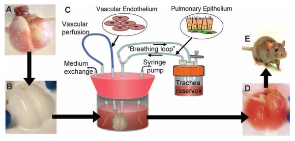
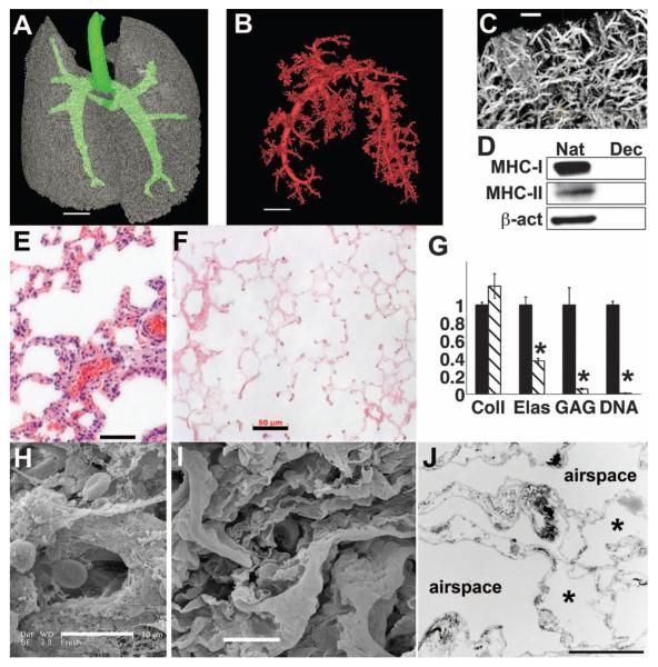
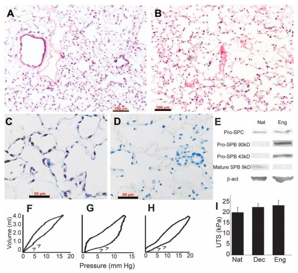
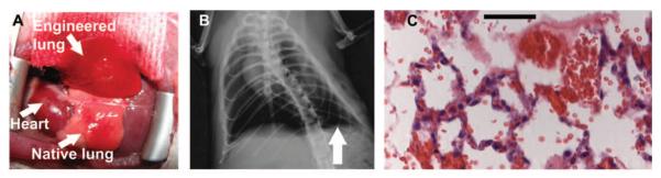
Because adult lung tissue has limited regeneration capacity, lung transplantation is the primary therapy for severely damaged lungs. To explore whether lung tissue can be regenerated in vitro, we treated lungs from adult rats using a procedure that removes cellular components but leaves behind a scaffold of extracellular matrix that retains the hierarchical branching structures of airways and vasculature. We then used a bioreactor to culture pulmonary epithelium and vascular endothelium on the acellular lung matrix. The seeded epithelium displayed remarkable hierarchical organization within the matrix, and the seeded endothelial cells efficiently repopulated the vascular compartment. In vitro, the mechanical characteristics of the engineered lungs were similar to those of native lung tissue, and when implanted into rats in vivo for short time intervals (45 to 120 minutes) the engineered lungs participated in gas exchange. Although representing only an initial step toward the ultimate goal of generating fully functional lungs in vitro, these results suggest that repopulation of lung matrix is a viable strategy for lung regeneration.


Figures

Fig. 1
Fig. 1
Schema for lung tissue engineering. (A) Native adult rat lung is cannulated in the pulmonary artery and trachea for infusion of decellularization solutions. (B) Acellular lung matrix is devoid of cells after 2 to 3 hours of treatment. (C) Acellular matrix is mounted inside a biomimetic bioreactor that allows seeding of vascular endothelium into the pulmonary artery and pulmonary epithelium into the trachea. (D) After 4 to 8 days of culture, the engineered lung is removed from the bioreactor and is suitable for implantation into (E) the syngeneic rat recipient.
Fig. 2
Fig. 2
Characterization of acellular lung matrix. (A) 3D micro-CT of the acellular matrix airway compartment. Large airways are in green. (B) Micro-CT angiography of vascular compartment, thresholded to visualize only large vessels. In (A) and (B), voxel size is 58 μm; scale bar, 4 mm. (C) Micro-CT angiography of smaller vessels in acellular lung. Voxel size, 6.5 μm; scale bar, 500 μm. (D) Immunoblot for MHC-1, MHC-II, and β-actin in native (Nat) and decellularized (Dec) lungs, showing removal of cellular proteins. (E) Hematoxylin and eosin (H&E) stain of native rat lung. (F) H&E stain of acellular lung matrix. Scale bar, 50 μm in (E) and (F). (G) Collagen (Coll), elastin (Elas), glycosaminoglycan (GAG), and DNA contents of native lung (black bars) and acellular matrices (hatched bars). Values are mean ± SD per lung (n ≥ 4 lungs for all measures), scaled to 1 for native, with asterisk indicating P < 0.05 for difference between native and acellular matrices. (H) SEM of native rat lung. (I) SEM of acellular matrix. Scale bar, 10 μm in (H) and (I). (J) TEM of acellular lung matrix. Asterisk indicates capillaries in alveolar septa. Scale bar, 5 μm.
Fig. 3
Fig. 3
Repopulation of the matrix with lung epithelial and endothelial cells and mechanical assessment of the engineered lungs. (A) H&E stain of mixed neonatal pulmonary epithelium seeded into acellular matrix and cultured for 8 days. (B) H&E stain of lung microvascular endothelium seeded into vascular compartment and cultured for 8 days. Scale bars, 100 μm in (A) and (B). (C) Proliferating cell nuclear antigen stain of epithelial culture after 8 days; brown nuclei are dividing. (D) Terminal deoxynucleotidyl transferase–mediated deoxyuridine triphosphate nick end labeling stain of epithelium after 4 days of culture detects no apoptotic cells (positive cells stain brown; green indicates nuclear counterstain). Scale bars, 50 μm in (C) and (D). (E) Immunoblots for pro-SPC indicates similar expression for native (Nat) and engineered (Eng) lung. Engineered lung expresses SPB precursor proteins at 43 and 90 kD, but no mature SPB. β-actin confirms similar protein loading. (F to H) Quasi-static compliance curves for (F) typical native, (G) acellular, and (H) engineered lungs; arrows indicate inflation arm of loop. (I) Mean ultimate tensile strengths (UTS) of native (n = 4), acellular (n = 10), and engineered (n = 5) lungs. Error bars are SD; there were no significant differences between any groups.
Fig. 4
Fig. 4
Implantation of engineered lungs into rats. (A) Tissue-engineered left lung was implanted into adult Fischer 344 rat recipient and photographed ~30 min later. (B) X-ray image of rat showing the implanted engineered left lung (white arrow) and the right native lung. (C) H&E stain of explanted lung. Red blood cells perfusing septa are evident, and some red blood cells are present in airspaces. Scale bar, 50 μm.

Comment in

  • Reconstructing the lung.
    Wagner WR, Griffith BP. Wagner WR, et al. Science. 2010 Jul 30;329(5991):520-2. doi: 10.1126/science.1194087. Science. 2010. PMID: 20671176 No abstract available.

References

    1. American Lung Association [accessed 4 March 2010];Lung Disease Data 2008. www.lungusa.org.
    1. Orens JB, Garrity ER., Jr. Proc. Am. Thorac. Soc. 2009;6:13. - PubMed
    1. Ott HC, et al. Nat. Med. 2008;14:213. - PubMed
    1. Gilbert TW, Sellaro TL, Badylak SF. Biomaterials. 2006;27:3675. - PubMed
    1. Baptista PM, et al. IEEE Eng. Med. Biol. Soc. 2009;31:6526. - PubMed

Publication types

MeSH terms