. 2010 Jun 25;328(5986):1662-8.
doi: 10.1126/science.1188302.

Reconstituting organ-level lung functions on a chip

Affiliations

Reconstituting organ-level lung functions on a chip

Dongeun Huh et al. Science. .

Abstract

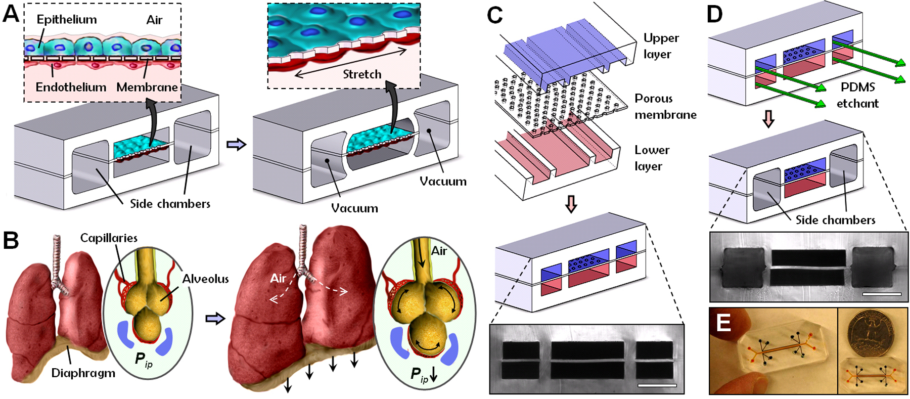
Here, we describe a biomimetic microsystem that reconstitutes the critical functional alveolar-capillary interface of the human lung. This bioinspired microdevice reproduces complex integrated organ-level responses to bacteria and inflammatory cytokines introduced into the alveolar space. In nanotoxicology studies, this lung mimic revealed that cyclic mechanical strain accentuates toxic and inflammatory responses of the lung to silica nanoparticles. Mechanical strain also enhances epithelial and endothelial uptake of nanoparticulates and stimulates their transport into the underlying microvascular channel. Similar effects of physiological breathing on nanoparticle absorption are observed in whole mouse lung. Mechanically active "organ-on-a-chip" microdevices that reconstitute tissue-tissue interfaces critical to organ function may therefore expand the capabilities of cell culture models and provide low-cost alternatives to animal and clinical studies for drug screening and toxicology applications.


Figures

Fig. 1.
Fig. 1.
Biologically inspired design of a human breathing lung-on-a-chip microdevice. (A) The microfabricated lung mimic device utilizes compartmentalized PDMS microchannels to form an alveolar-capillary barrier on a thin porous flexible PDMS membrane coated with ECM. The device recreates physiological breathing movements by applying vacuum to the side chambers and causing mechanical stretching of the PDMS membrane forming the alveolar-capillary barrier. (B) During inhalation in the living lung, contraction of the diaphragm causes a reduction in intrapleural pressure (Pip), leading to distension of the alveoli and physical stretching of the alveolar-capillary interface. (C) Three PDMS layers are aligned and irreversibly bonded to form two sets of three parallel microchannels separated by a 10 μm-thick PDMS membrane containing an array of through-holes with an effective diameter of 10 μm (scale bar, 200 μm). (D) Following permanent bonding, PDMS etchant is flowed through the side channels. Selective etching of the membrane layers in these channels produces two large side chambers to which vacuum is applied to cause mechanical stretching (scale bar, 200 μm). (E) Images of an actual lung-on-a-chip microfluidic device viewed from above.
Fig. 2.
Fig. 2.
On-chip formation and mechanical stretching of an alveolar-capillary interface. (A) Long-term microfluidic co-culture produces a tissue-tissue interface consisting of a single layer of the alveolar epithelium (Epithelium; stained with CellTracker Green) closely apposed to a monolayer of the microvascular endothelium (Endothelium; stained with CellTracker Red), both of which express intercellular junctional structures stained with antibodies to Occludin or VE-cadherin, separated by a flexible ECM-coated PDMS membrane (bar, 50 μm). (B) Surfactant production by the alveolar epithelium during air-liquid interface culture in our device detected by cellular uptake of the fluorescent dye, quinacrine that labels lamellar bodies (white dots; bar, 25 μm). (C) Air-liquid interface (ALI) culture leads to a greater increase in trans-bilayer electrical resistance (TER) and produces tighter alveolar-capillary barriers with higher TER (over 800 Ω·cm2), as compared to the tissue layers formed under submerged liquid culture conditions. (D) Alveolar barrier permeability measured by quantitating the rate of fluorescent albumin transport is significantly reduced in air-liquid interface (ALI) cultures compared to liquid cultures (*p < 0.001). Data in (C) and (D) represent the mean ± SEM from three separate experiments. (E) Membrane stretching-induced mechanical strain visualized by the displacements of individual fluorescent quantum dots that were immobilized on the membrane in hexagonal and rectangular patterns before (red) and after (green) stretching (bar, 100 μm). (F) Membrane stretching exerts tension on the cells and causes them to distort in the direction of the applied force, as illustrated by the overlaid outlines of a single cell before (blue) and after (red) application of 15% strain. The pentagons in the micrographs represent microfabricated pores in the membrane. Endothelial cells were used for visualization of cell stretching.
Fig. 3.
Fig. 3.
Reconstitution and direct visualization of complex organ-level responses involved in pulmonary inflammation and infection in the lung-on-a-chip device. (A) Epithelial stimulation with TNF-α (50 ng/ml) upregulates ICAM-1 expression (red) on the endothelium; control shows lack of ICAM-1 expression in the absence of TNF-α treatment. Cells were stretched with 10% strain at 0.2 Hz in both cases. (B) Fluorescently-labeled human neutrophils (white dots) avidly to the activated endothelium within one minute after introduction into the vascular channel. (C) Time-lapse microscopic images showing a captured neutrophil (white arrow) that spreads via firm adhesion and then crawls over the apical surface of the activated endothelium (not visible in this view; direction indicated by yellow arrows) until it forces itself through the cell-cell boundary within about 2 minutes after adhesion (times indicated in seconds). During the following 3 to 4 minutes, the neutrophil transmigrates through the alveolar-capillary barrier by passing through a pentagonal pore in the PDMS membrane, and then it moves away from the focal plane, causing it to appear blurry in the micrographs. (D) Phase contrast microscopic images show a neutrophil (arrow) emerging from the apical surface of the alveolar epithelium at the end of its transmigration over a period of approximately 3 minutes; thus, complete passage takes approximately 6 minutes in total. (E) Time-lapse fluorescence microscopic images showing phagocytosis of two GFP-expressing E. coli (green) bacteria on the epithelial surface by a neutrophil (red) that transmigrated from the vascular microchannel to the alveolar compartment. (bar, 50 μm in (A), (B) and 20 μm in (C)-(E)).
Fig. 4.
Fig. 4.
Microengineered model of pulmonary nanotoxicology. (A) Ultrafine silica nanoparticles introduced through an air-liquid interface overlying the alveolar epithelium induce ICAM-1 expression (red) in the underlying endothelium and adhesion of circulating neutrophils (white dots) in the lower channel (bar, 50 μm). Graph shows that physiological mechanical strain and silica nanoparticles synergistically upregulate ICAM-1 expression (*p < 0.005; **p < 0.001). (B) Alveolar epithelial cells increase ROS production when exposed to silica nanoparticles (100 μg/ml) in conjunction with 10% cyclic strain (square) (p < 0.0005), whereas nanoparticles (triangle) or strain (diamond) alone had no effect on intracellular ROS levels relative to control cells (circle); ROS generation was normalized to the mean ROS value at time 0. (C) The alveolar epithelium responds to silica nanoparticles in a strain-dependent manner (*p < 0.001). (D) Addition of 50 nm superparamagnetic nanoparticles produced only a transient elevation of ROS in the epithelial cells subjected to 10% cyclic strain (p < 0.0005). (E) Application of physiological mechanical strain (10%) promotes increased cellular uptake of 100 nm polystyrene nanoparticles (magenta) relative to static cells, as illustrated by representative sections (a-d) through fluorescent confocal images. Internalized nanoparticles are indicated with arrows; green and blue show cytoplasmic and nuclear staining, respectively. (F) Transport of nanomaterials across the alveolar-capillary interface of the lung is simulated by nanoparticle transport from the alveolar chamber to the vascular channel of the lung mimic device. (G) Application of 10% mechanical strain (closed square) significantly increased the rate of nanoparticle translocation across the alveolar-capillary interface compared to static controls in this device (closed triangle) or in a Transwell culture system (open triangle) (p < 0.0005). (H) Fluorescence micrographs of a histological section of the whole lung showing 20 nm fluorescent nanoparticles (white dots, indicated with arrows in the inset at upper right that shows the region enclosed by the dashed square at higher magnification) present in the lung after intratracheal injection of nebulized nanoparticles and ex vivo ventilation in the mouse lung model. Nanoparticles cross the alveolar-capillary interface and are found on the surface of the alveolar epithelium, in the interstitial space, and on the capillary endothelium (PC, pulmonary capillary; AS, alveolar space; blue, epithelial nucleus; bar, 20 μm). (I) Physiological cyclic breathing generated by mechanical ventilation in whole mouse lung produces more than a 5-fold increase in nanoparticle absorption into the blood perfusate when compared to lungs without lung ventilation (p < 0.0005). The graph indicates the number of nanoparticles detected in the pulmonary blood perfusate over time, as measured by drying the blood (1 μl) on glass and quantitating the number of particles per unit area (0.5 mm2). (J) The rate of nanoparticle translocation was significantly reduced by adding NAC to scavenge free radicals (*p < 0.001).

Similar articles

Cited by

  • A gravity-driven tissue chip to study the efficacy and toxicity of cancer therapeutics.
    Rafsanjani Nejad P, Lamichhane A, Guragain P, Luker G, Tavana H. Rafsanjani Nejad P, et al. Lab Chip. 2024 Nov 19;24(23):5251-5263. doi: 10.1039/d4lc00404c. Lab Chip. 2024. PMID: 39485368 Free PMC article.
  • Genomic responses in mouse models poorly mimic human inflammatory diseases.
    Seok J, Warren HS, Cuenca AG, Mindrinos MN, Baker HV, Xu W, Richards DR, McDonald-Smith GP, Gao H, Hennessy L, Finnerty CC, López CM, Honari S, Moore EE, Minei JP, Cuschieri J, Bankey PE, Johnson JL, Sperry J, Nathens AB, Billiar TR, West MA, Jeschke MG, Klein MB, Gamelli RL, Gibran NS, Brownstein BH, Miller-Graziano C, Calvano SE, Mason PH, Cobb JP, Rahme LG, Lowry SF, Maier RV, Moldawer LL, Herndon DN, Davis RW, Xiao W, Tompkins RG; Inflammation and Host Response to Injury, Large Scale Collaborative Research Program. Seok J, et al. Proc Natl Acad Sci U S A. 2013 Feb 26;110(9):3507-12. doi: 10.1073/pnas.1222878110. Epub 2013 Feb 11. Proc Natl Acad Sci U S A. 2013. PMID: 23401516 Free PMC article. Clinical Trial.
  • Navigating ethical challenges in the development and translation of biomaterials research.
    Hunckler MD, Levine AD. Hunckler MD, et al. Front Bioeng Biotechnol. 2022 Sep 20;10:949280. doi: 10.3389/fbioe.2022.949280. eCollection 2022. Front Bioeng Biotechnol. 2022. PMID: 36204464 Free PMC article.
  • Self-assembling short immunostimulatory duplex RNAs with broad-spectrum antiviral activity.
    Si L, Bai H, Oh CY, Jiang A, Hong F, Zhang T, Ye Y, Jordan TX, Logue J, McGrath M, Belgur C, Calderon K, Nurani A, Cao W, Carlson KE, Prantil-Baun R, Gygi SP, Yang D, Jonsson CB, tenOever BR, Frieman M, Ingber DE. Si L, et al. Mol Ther Nucleic Acids. 2022 Sep 13;29:923-940. doi: 10.1016/j.omtn.2022.08.031. Epub 2022 Aug 24. Mol Ther Nucleic Acids. 2022. PMID: 36032397 Free PMC article.
  • Harnessing organs-on-a-chip to model tissue regeneration.
    Tavakol DN, Fleischer S, Vunjak-Novakovic G. Tavakol DN, et al. Cell Stem Cell. 2021 Jun 3;28(6):993-1015. doi: 10.1016/j.stem.2021.05.008. Cell Stem Cell. 2021. PMID: 34087161 Free PMC article. Review.

References

    1. Davila JC, Rodriguez RJ, Melchert RB, Acosta D., Annu. Rev. Pharmacol. Toxicol 38, 63 (1998). - PubMed
    1. Pampaloni F, Reynaud EG, Stelzer EHK, Nat. Rev. Mol. Cell Biol 8, 839 (2007). - PubMed
    1. Ingber DE, FASEB J 20, 811 (2006). - PubMed
    1. Whitesides GM, Ostuni E, Takayama S, Jiang XY, Ingber DE, Annu. Rev. Biomed. Eng 3, 335 (2001). - PubMed
    1. Khademhosseini A, Langer R, Borenstein J, Vacanti JP, Proc. Natl. Acad. Sci. U. S. A 103, 2480 (2006). - PMC - PubMed

Publication types

MeSH terms

Substances