

From coarse to fine? Spatial and temporal dynamics of cortical face processing
- PMID: 20576927
- PMCID: PMC3020585
- DOI: 10.1093/cercor/bhq112
From coarse to fine? Spatial and temporal dynamics of cortical face processing
Abstract
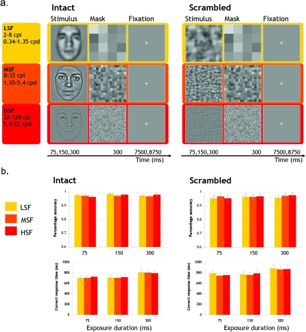
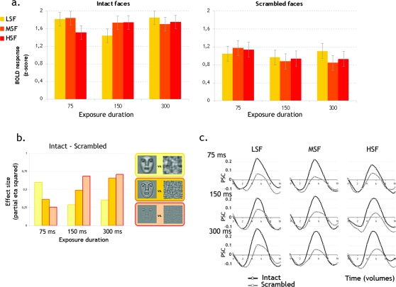
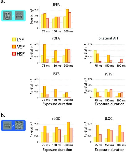
Primary vision segregates information along 2 main dimensions: orientation and spatial frequency (SF). An important question is how this primary visual information is integrated to support high-level representations. It is generally assumed that the information carried by different SF is combined following a coarse-to-fine sequence. We directly addressed this assumption by investigating how the network of face-preferring cortical regions processes distinct SF over time. Face stimuli were flashed during 75, 150, or 300 ms and masked. They were filtered to preserve low SF (LSF), middle SF (MSF), or high SF (HSF). Most face-preferring regions robustly responded to coarse LSF, face information in early stages of visual processing (i.e., until 75 ms of exposure duration). LSF processing decayed as a function of exposure duration (mostly until 150 ms). In contrast, the processing of fine HSF, face information became more robust over time in the bilateral fusiform face regions and in the right occipital face area. The present evidence suggests the coarse-to-fine strategy as a plausible modus operandi in high-level visual cortex.
Figures
Similar articles
-
The effects of face spatial frequencies on cortical processing revealed by magnetoencephalography.Neurosci Lett. 2005 May 20-27;380(1-2):54-9. doi: 10.1016/j.neulet.2005.01.016. Epub 2005 Jan 24. Neurosci Lett. 2005. PMID: 15854750
-
Face inversion disrupts the perception of vertical relations between features in the right human occipito-temporal cortex.J Neuropsychol. 2009 Mar;3(Pt 1):45-67. doi: 10.1348/174866408X292670. J Neuropsychol. 2009. PMID: 19338716
-
Faster, stronger, lateralized: low spatial frequency information supports face processing.Neuropsychologia. 2011 Nov;49(13):3583-90. doi: 10.1016/j.neuropsychologia.2011.08.027. Epub 2011 Sep 16. Neuropsychologia. 2011. PMID: 21939676
-
Retinotopic and lateralized processing of spatial frequencies in human visual cortex during scene categorization.J Cogn Neurosci. 2013 Aug;25(8):1315-31. doi: 10.1162/jocn_a_00397. Epub 2013 Apr 11. J Cogn Neurosci. 2013. PMID: 23574583
-
The neural bases of spatial frequency processing during scene perception.Front Integr Neurosci. 2014 May 7;8:37. doi: 10.3389/fnint.2014.00037. eCollection 2014. Front Integr Neurosci. 2014. PMID: 24847226 Free PMC article. Review.
Cited by
-
Natural Contrast Statistics Facilitate Human Face Categorization.eNeuro. 2022 Oct 6;9(5):ENEURO.0420-21.2022. doi: 10.1523/ENEURO.0420-21.2022. Print 2022 Sep-Oct. eNeuro. 2022. PMID: 36096649 Free PMC article.
-
Target specificity improves search, but how universal is the benefit?Atten Percept Psychophys. 2020 Nov;82(8):3878-3894. doi: 10.3758/s13414-020-02111-1. Atten Percept Psychophys. 2020. PMID: 32901340 Free PMC article.
-
Face and object discrimination in autism, and relationship to IQ and age.J Autism Dev Disord. 2014 May;44(5):1039-54. doi: 10.1007/s10803-013-1955-z. J Autism Dev Disord. 2014. PMID: 24150884
-
Evidence for Separate Contributions of High and Low Spatial Frequencies during Visual Word Recognition.Front Hum Neurosci. 2017 Jun 22;11:324. doi: 10.3389/fnhum.2017.00324. eCollection 2017. Front Hum Neurosci. 2017. PMID: 28690505 Free PMC article.
-
Ultra rapid object categorization: effects of level, animacy and context.PLoS One. 2013 Jun 28;8(6):e68051. doi: 10.1371/journal.pone.0068051. Print 2013. PLoS One. 2013. PMID: 23840810 Free PMC article.
References
-
- Baker CI, Hutchison TL, Kanwisher N. Does the fusiform face area contain subregions highly selective for nonfaces? Nat Neurosci. 2007;10:3–4. - PubMed
-
- Bar M. The proactive brain: using analogies and associations to generate predictions. Trends Cogn Sci. 2007;11:280–289. - PubMed
-
- Barton JJ, Press DZ, Keenan JP, O'Connor M. Lesions of the fusiform face area impair perception of facial configuration in prosopagnosia. Neurology. 2002;58:71–78. - PubMed
MeSH terms
Substances
LinkOut - more resources
Full Text Sources
Miscellaneous
