

Inhibition of bone morphogenetic proteins protects against atherosclerosis and vascular calcification
- PMID: 20576934
- PMCID: PMC2994650
- DOI: 10.1161/CIRCRESAHA.110.219071
Inhibition of bone morphogenetic proteins protects against atherosclerosis and vascular calcification
Abstract
Rationale: The bone morphogenetic proteins (BMPs), a family of morphogens, have been implicated as mediators of calcification and inflammation in the vascular wall.
Objective: To investigate the effect of altered expression of matrix Gla protein (MGP), an inhibitor of BMP, on vascular disease.
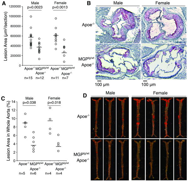
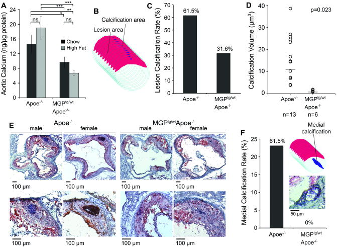
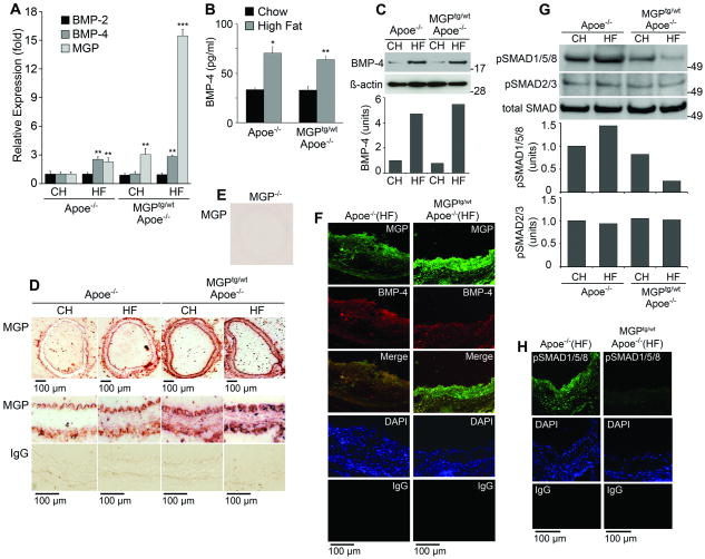
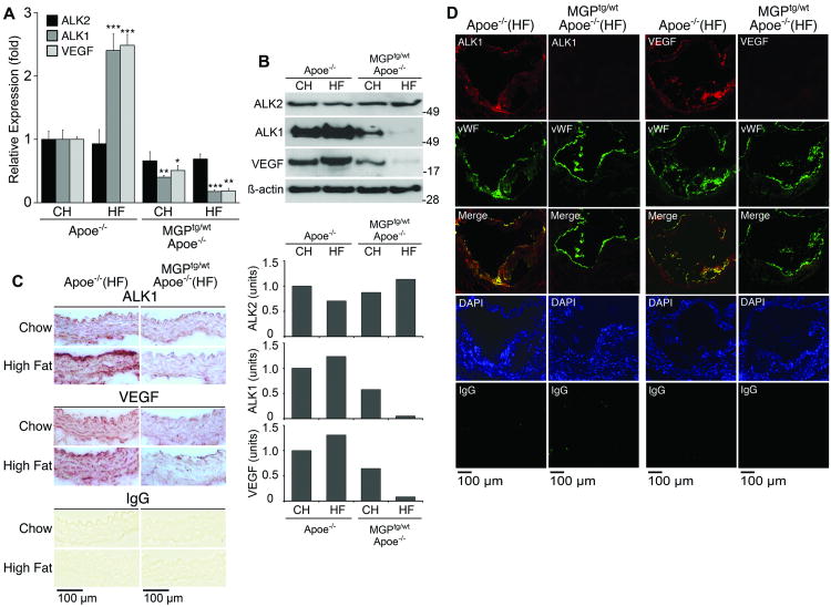
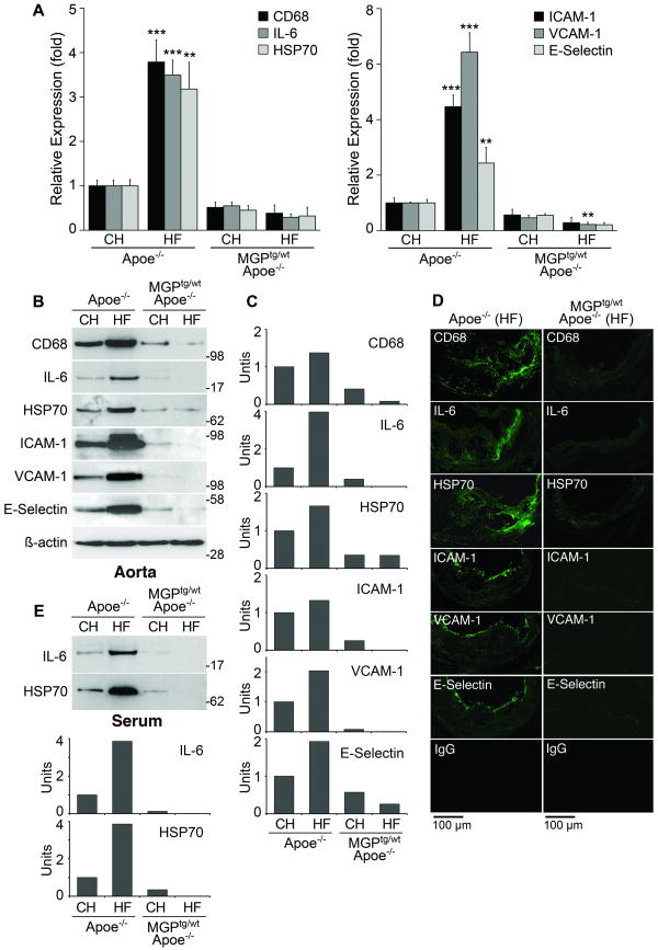
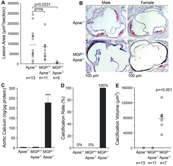
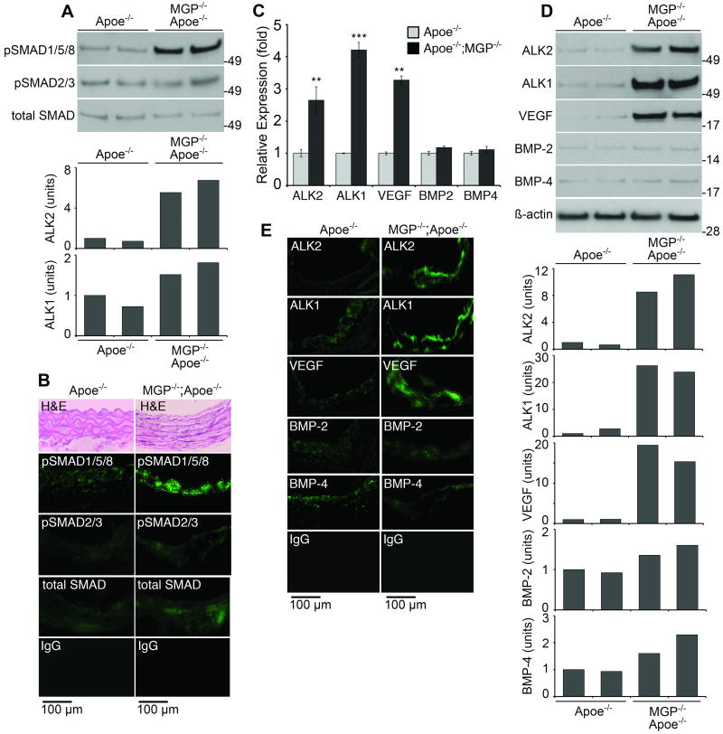
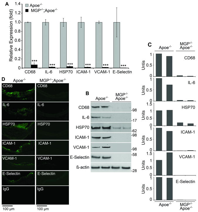
Methods and results: We used MGP transgenic or MGP-deficient mice bred to apolipoprotein E mice, a model of atherosclerosis. MGP overexpression reduced vascular BMP activity, atherosclerotic lesion size, intimal and medial calcification, and inflammation. It also reduced expression of the activin-like kinase receptor 1 and the vascular endothelial growth factor, part of a BMP-activated pathway that regulates angiogenesis and may enhance lesion formation and calcification. Conversely, MGP deficiency increased BMP activity, which may explain the diffuse calcification of vascular medial cells in MGP deficient aortas and the increase in expression of activin-like kinase receptor 1 and vascular endothelial growth factor. Unexpectedly, atherosclerotic lesion formation was decreased in MGP-deficient mice, which may be explained by a dramatic reduction in expression of endothelial adhesion molecules limiting monocyte infiltration of the artery wall.
Conclusions: Our results indicate that BMP signaling is a key regulator of vascular disease, requiring careful control to maintain normal vascular homeostasis.
Figures
Similar articles
-
Heat shock protein 70 enhances vascular bone morphogenetic protein-4 signaling by binding matrix Gla protein.Circ Res. 2009 Sep 11;105(6):575-84. doi: 10.1161/CIRCRESAHA.109.202333. Epub 2009 Aug 6. Circ Res. 2009. PMID: 19661459 Free PMC article.
-
Activation of vascular bone morphogenetic protein signaling in diabetes mellitus.Circ Res. 2011 Feb 18;108(4):446-57. doi: 10.1161/CIRCRESAHA.110.236596. Epub 2010 Dec 30. Circ Res. 2011. PMID: 21193740 Free PMC article.
-
Proline and gamma-carboxylated glutamate residues in matrix Gla protein are critical for binding of bone morphogenetic protein-4.Circ Res. 2008 May 9;102(9):1065-74. doi: 10.1161/CIRCRESAHA.107.166124. Epub 2008 Mar 27. Circ Res. 2008. PMID: 18369157
-
[Matrix Gla-protein and its role in vascular wall calcification].Fiziol Zh (1994). 2011;57(4):96-112. Fiziol Zh (1994). 2011. PMID: 22167840 Review. Ukrainian.
-
BMP signaling in vascular diseases.FEBS Lett. 2012 Jul 4;586(14):1993-2002. doi: 10.1016/j.febslet.2012.04.030. Epub 2012 May 3. FEBS Lett. 2012. PMID: 22710160 Review.
Cited by
-
Crossveinless 2 regulates bone morphogenetic protein 9 in human and mouse vascular endothelium.Blood. 2012 May 24;119(21):5037-47. doi: 10.1182/blood-2011-10-385906. Epub 2012 Apr 3. Blood. 2012. PMID: 22474252 Free PMC article.
-
Serum anti-DIDO1, anti-CPSF2, and anti-FOXJ2 antibodies as predictive risk markers for acute ischemic stroke.BMC Med. 2021 Jun 9;19(1):131. doi: 10.1186/s12916-021-02001-9. BMC Med. 2021. PMID: 34103026 Free PMC article.
-
Histologic identification of brown adipose and peripheral nerve involvement in human atherosclerotic vessels.Hum Pathol. 2012 Dec;43(12):2213-22. doi: 10.1016/j.humpath.2012.03.013. Epub 2012 Jun 27. Hum Pathol. 2012. PMID: 22748303 Free PMC article.
-
Warfarin calcifies human aortic valve interstitial cells at high-phosphate conditions via pregnane X receptor.J Bone Miner Metab. 2019 Nov;37(6):944-956. doi: 10.1007/s00774-019-01001-3. Epub 2019 Apr 8. J Bone Miner Metab. 2019. PMID: 30963258
-
Molecular Interactions Between Vascular Smooth Muscle Cells and Macrophages in Atherosclerosis.Front Cardiovasc Med. 2021 Oct 15;8:737934. doi: 10.3389/fcvm.2021.737934. eCollection 2021. Front Cardiovasc Med. 2021. PMID: 34722670 Free PMC article.
References
-
- Csiszar A, Smith KE, Koller A, Kaley G, Edwards JG, Ungvari Z. Regulation of bone morphogenetic protein-2 expression in endothelial cells: role of nuclear factor-kappaB activation by tumor necrosis factor-alpha, H2O2, and high intravascular pressure. Circulation. 2005;111:2364–2372. - PubMed
-
- Sorescu GP, Song H, Tressel SL, Hwang J, Dikalov S, Smith DA, Boyd NL, Platt MO, Lassegue B, Griendling KK, Jo H. Bone morphogenic protein 4 produced in endothelial cells by oscillatory shear stress induces monocyte adhesion by stimulating reactive oxygen species production from a nox1-based NADPH oxidase. Circ Res. 2004;95:773–779. - PubMed
-
- Dhore CR, Cleutjens JP, Lutgens E, Cleutjens KB, Geusens PP, Kitslaar PJ, Tordoir JH, Spronk HM, Vermeer C, Daemen MJ. Differential expression of bone matrix regulatory proteins in human atherosclerotic plaques. Arterioscler Thromb Vasc Biol. 2001;21:1998–2003. - PubMed
-
- Schluesener HJ, Meyermann R. Immunolocalization of BMP-6, a novel TGF-beta-related cytokine, in normal and atherosclerotic smooth muscle cells. Atherosclerosis. 1995;113:153–156. - PubMed
-
- Al-Aly Z, Shao JS, Lai CF, Huang E, Cai J, Behrmann A, Cheng SL, Towler DA. Aortic Msx2-Wnt calcification cascade is regulated by TNF-alpha-dependent signals in diabetic Ldlr-/- mice. Arterioscler Thromb Vasc Biol. 2007;27:2589–2596. - PubMed
Publication types
MeSH terms
Substances
Grants and funding
LinkOut - more resources
Full Text Sources
Medical
Molecular Biology Databases
Miscellaneous
