

Rapid progress for non-nuclear estrogen receptor signaling
- PMID: 20577045
- PMCID: PMC2898619
- DOI: 10.1172/JCI43756
Rapid progress for non-nuclear estrogen receptor signaling
Abstract
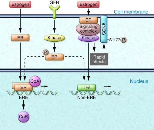
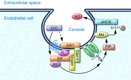
Estrogen receptors are best known as ligand-activated transcription factors that regulate vascular cell gene expression. For many years now, a rapid signaling pathway mediated by cell membrane-associated estrogen receptors also has been recognized, but the physiological relevance of this pathway has remained unclear. In this issue of the JCI, Chambliss et al. provide new data to indicate that activation of non-nuclear estrogen receptor signaling regulates processes central to cardiovascular health and disease. These investigators show that an estrogen-dendrimer conjugate (EDC), which activates estrogen receptors but remains non-nuclear, stimulates vascular EC migration in vitro and protects against vascular injury in vivo. They show further that the vascular benefits of EDC in vivo occur selectively in the vasculature, without stimulating the uterus or enhancing growth of breast cancer xenografts. Taken together, these findings indicate that activation of non-nuclear estrogen receptor signaling regulates vascular events of physiological relevance and suggest that translation of these findings into clinically relevant therapeutic interventions is a logical next goal.
Figures
Comment on
-
Non-nuclear estrogen receptor alpha signaling promotes cardiovascular protection but not uterine or breast cancer growth in mice.J Clin Invest. 2010 Jul;120(7):2319-30. doi: 10.1172/JCI38291. Epub 2010 Jun 23. J Clin Invest. 2010. PMID: 20577047 Free PMC article.
Similar articles
-
Non-nuclear estrogen receptor alpha signaling promotes cardiovascular protection but not uterine or breast cancer growth in mice.J Clin Invest. 2010 Jul;120(7):2319-30. doi: 10.1172/JCI38291. Epub 2010 Jun 23. J Clin Invest. 2010. PMID: 20577047 Free PMC article.
-
Estrogen and growth factor receptor interactions in human breast and non-small cell lung cancer cells.Steroids. 2005 May-Jun;70(5-7):372-81. doi: 10.1016/j.steroids.2005.02.017. Epub 2005 Mar 25. Steroids. 2005. PMID: 15862820
-
Unconventional Estrogen Signaling in Health and Disease.Endocrinology. 2020 Apr 1;161(4):bqaa030. doi: 10.1210/endocr/bqaa030. Endocrinology. 2020. PMID: 32128594 Free PMC article. Review.
-
Nuclear and extranuclear pathway inputs in the regulation of global gene expression by estrogen receptors.Mol Endocrinol. 2008 Sep;22(9):2116-27. doi: 10.1210/me.2008-0059. Epub 2008 Jul 10. Mol Endocrinol. 2008. PMID: 18617595 Free PMC article.
-
To ERR in the estrogen pathway.Trends Endocrinol Metab. 2002 Jul;13(5):220-5. doi: 10.1016/s1043-2760(02)00592-1. Trends Endocrinol Metab. 2002. PMID: 12185669 Review.
Cited by
-
Estrogen receptor subcellular localization and cardiometabolism.Mol Metab. 2018 Sep;15:56-69. doi: 10.1016/j.molmet.2018.05.009. Epub 2018 May 16. Mol Metab. 2018. PMID: 29807870 Free PMC article. Review.
-
Pathophysiology of Aortic Valve Stenosis: Is It Both Fibrocalcific and Sex Specific?Physiology (Bethesda). 2017 May;32(3):182-196. doi: 10.1152/physiol.00025.2016. Physiology (Bethesda). 2017. PMID: 28404735 Free PMC article. Review.
-
Estrogen Receptors and Estrogen-Induced Uterine Vasodilation in Pregnancy.Int J Mol Sci. 2020 Jun 18;21(12):4349. doi: 10.3390/ijms21124349. Int J Mol Sci. 2020. PMID: 32570961 Free PMC article. Review.
-
Sex differences in the physiology of eating.Am J Physiol Regul Integr Comp Physiol. 2013 Dec;305(11):R1215-67. doi: 10.1152/ajpregu.00446.2012. Epub 2013 Jul 31. Am J Physiol Regul Integr Comp Physiol. 2013. PMID: 23904103 Free PMC article. Review.
-
Rapid estrogen receptor signaling mediates estrogen-induced inhibition of vascular smooth muscle cell proliferation.Arterioscler Thromb Vasc Biol. 2013 Aug;33(8):1837-43. doi: 10.1161/ATVBAHA.112.300752. Epub 2013 Jun 6. Arterioscler Thromb Vasc Biol. 2013. PMID: 23744991 Free PMC article.
References
-
- Hsia J, et al. Conjugated equine estrogens and coronary heart disease. Arch Intern Med. 2006;166(3):356–365. - PubMed
