. 2010 Jul;120(7):2528-36.
doi: 10.1172/JCI41402. Epub 2010 Jun 23.

Mitigation of hematologic radiation toxicity in mice through pharmacological quiescence induced by CDK4/6 inhibition

Affiliations

Mitigation of hematologic radiation toxicity in mice through pharmacological quiescence induced by CDK4/6 inhibition

Søren M Johnson et al. J Clin Invest. 2010 Jul.

Abstract

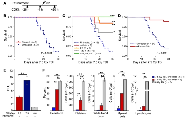
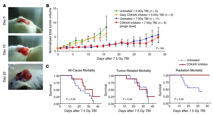
Total body irradiation (TBI) can induce lethal myelosuppression, due to the sensitivity of proliferating hematopoietic stem/progenitor cells (HSPCs) to ionizing radiation (IR). No effective therapy exists to mitigate the hematologic toxicities of TBI. Here, using selective and structurally distinct small molecule inhibitors of cyclin-dependent kinase 4 (CDK4) and CDK6, we have demonstrated that selective cellular quiescence increases radioresistance of human cell lines in vitro and mice in vivo. Cell lines dependent on CDK4/6 were resistant to IR and other DNA-damaging agents when treated with CDK4/6 inhibitors. In contrast, CDK4/6 inhibitors did not protect cell lines that proliferated independently of CDK4/6 activity. Treatment of wild-type mice with CDK4/6 inhibitors induced reversible pharmacological quiescence (PQ) of early HSPCs but not most other cycling cells in the bone marrow or other tissues. Selective PQ of HSPCs decreased the hematopoietic toxicity of TBI, even when the CDK4/6 inhibitor was administered several hours after TBI. Moreover, PQ at the time of administration of therapeutic IR to mice harboring autochthonous cancers reduced treatment toxicity without compromising the therapeutic tumor response. These results demonstrate an effective method to mitigate the hematopoietic toxicity of IR in mammals, which may be potentially useful after radiological disaster or as an adjuvant to anticancer therapy.


Figures

Figure 1
Figure 1. PQ enhances radioresistance in vitro.
(A) Images of phospho–γ-H2AX foci (green) and phalloidin staining (red) of tHDFs, with or without 6 Gy IR and 24 hours of 100 nM PD0332991 exposure. Original magnification, ×40. Scale bar: 50 μm. (B) Western blots showing phospho-p53 (p-p53; arrow) induction in tHDF lysates after 6 Gy IR, with and without PD0332991 treatment. (C) Comet tail area, after 24 hours of exposure to 100 nM PD0332991, prior to the indicated IR dose. Forty cells in each condition were imaged at an original magnification of ×20. *P < 0.01, **P < 0.0001, for pair-wise comparisons.
Figure 2
Figure 2. PQ enhances radioresistance in vitro.
(A) An in vitro schedule of CDK inhibitor (CDKi) and doxorubicin (Dox) exposure using tHDFs. Assays were performed where indicated on the timeline with arrows. WB, Western blot. (B) Flow cytometry analysis of phospho–γ-H2AX formation after PD0332991 and doxorubicin treatment of tHDFs. (C) Western blot analysis of DNA damage response markers in tHDFs, at indicated times after PD0332991 treatment, with or without doxorubicin treatment. The black bar indicates lysates harvested immediately after IR exposure (t = 0 hours), and the gray bar indicates lysates harvested 12 hours after IR exposure (t = 12 hours). (D) WST assay for cell proliferation at 7 days after doxorubicin exposure. All absorbances are normalized to DMSO. #P < 0.01 vs. DMSO, ##P < 0.001 vs. DMSO, *P < 0.01 vs. doxorubicin, **P < 0.001 vs. doxorubicin, ***P < 0.0001, for the indicated pair-wise comparisons. “Wash” indicates time in media without CDK inhibitor or doxorubicin, as shown in A.
Figure 3
Figure 3. CDK4/6 inhibition decreases HSPC proliferation.
(A) Flow cytometry gating scheme for HSCs and MPPs (top) and myeloid progenitors (bottom) using cell surface antigens. Gray regions represent cell populations used for further analysis as indicated. FSc, forward scatter. (B) Representative contour plots of proliferation in indicated HSPC populations, as measured by BrdU incorporation and Ki67 expression, after 48 hours of no treatment (n = 6) or PD0332991 treatment (n = 6) in the presence of BrdU for 24 hours. Contours represent 5% density. (C) Quantification of BrdU and Ki67 data in all HSPC subpopulations. (D) Relative frequency of Lin, HSC, MPP, or LincKit+ Sca1 populations after 48 hours of PD0332991 treatment and 24 hours of BrdU exposure. *P < 0.05, **P < 0.01, ***P < 0.001.
Figure 4
Figure 4. PQ at the time of TBI increases survival.
(A) The PD0332991 treatment schedule in initial radioprotection experiments. The arrows indicate doses of PD0332991 before or after TBI (shown as lightning bolt). (B) Kaplan-Meier analysis of survival after 7.5 Gy of TBI, with or without PD0332991 treatment. (C) Survival after 7.5 Gy TBI, with the indicated dosing schedules of PD0332991. (D) Aggregate results of animals gavaged 4 hours prior to TBI, representing results from 3 murine strains with animals of both sexes. Results of strains considered independently were comparable in all groups (see Figure 6C and Supplemental Figure 6C). All survival curve P values were calculated using the log-rank test as a pair-wise comparison with the untreated group. (E) Caspase activation in BM-MNCs after exposure to 7.5 Gy TBI (x-rays; see Methods). (F) Complete blood counts with differential at 21 days after 7.5 Gy TBI, with and without PD0332991 treatment. Treated animals received PD0332991 by oral gavage at 28 and 4 hours prior to (–28 and –4, respectively) and 20 hours after (+20) IR dose on day 0. Myeloid cells include granulocytes and monocytes, and # indicates that the maximum value of the cohort is shown in lieu of error bars where cells numbers were too small to reliably quantify. *P < 0.05, **P < 0.01, ***P < 0.001.
Figure 5
Figure 5. Cell cycle arrest after lethal TBI improves survival and requires both p21CIP and CDK4/6 inhibition for optimal benefit.
Kaplan-Meier survival curves for wild-type, heterozygous, and p21CIP-null mice, exposed to lethal TBI 4 hours after oral gavage with (A) vehicle or (B) PD0332991. Mice were irradiated using an x-ray source (see Methods). The log-rank test for trend is shown.
Figure 6
Figure 6. CDK4/6 inhibition improves survival without compromising tumor sensitivity to radiation therapy.
(A) Representative images of the progression of an autochthonous TyrRas Ink4a/Arf–/– melanoma, despite daily oral therapy with PD0332991. (B) Tumor growth with or without continuous daily PD0332991 treatment compared with tumor growth with or without PD0332991 administered as a single dose 4 hours prior to 7.5 Gy TBI. “NS” is nonsignificant for the comparison between groups receiving 7.5 Gy TBI. (C) Kaplan-Meier survival curves showing overall mortality and mortality subdivided by cause. P values were calculated using the log-rank test.

Comment in

  • Radioprotection: smart games with death.
    Gudkov AV, Komarova EA. Gudkov AV, et al. J Clin Invest. 2010 Jul;120(7):2270-3. doi: 10.1172/JCI43794. Epub 2010 Jun 23. J Clin Invest. 2010. PMID: 20577043 Free PMC article. Review.

Similar articles

Cited by

References

    1. Sinclair WK, Morton RA. X-ray sensitivity during cell generation cycle of cultured chinese hamster cells. Radiat Res. 1966;29(3):450–474. doi: 10.2307/3572025. - DOI - PubMed
    1. Terasima T, Tolmach LJ. X-ray sensitivity and DNA synthesis in synchronous populations of HeLa cells. Science. 1963;140:490–492. doi: 10.1126/science.140.3566.490. - DOI - PubMed
    1. Little JB. Repair of sub-lethal and potentially lethal radiation damage in plateau phase cultures of human cells. Nature. 1969;224(5221):804–806. doi: 10.1038/224804a0. - DOI - PubMed
    1. Elkind MM, Sutton H. X-ray damage and recovery in mammalian cells in culture. Nature. 1959;184:1293–1295. - PubMed
    1. Elkind MM, Sutton H. Radiation response of mammalian cells grown in culture. 1. Repair of x-ray damage in surviving chinese hamster cells. Radiat Res. 1960;13:556–593. doi: 10.2307/3570945. - DOI - PubMed

Publication types

Substances