

Distinct dacryoadenitides autoadoptively transferred to rabbits by different subpopulations of lymphocytes activated ex vivo
- PMID: 20577087
- PMCID: PMC2945426
- DOI: 10.1097/ICO.0b013e3181d0090e
Distinct dacryoadenitides autoadoptively transferred to rabbits by different subpopulations of lymphocytes activated ex vivo
Abstract
Purpose: To test whether CD4+ T cells proliferate in mixed cell reactions with autologous lacrimal gland (LG) acinar cells and whether these cells can autoadoptively transfer disease.
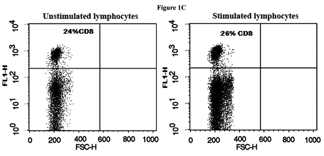
Methods: Purified acinar cells were gamma irradiated and cocultured with peripheral blood lymphocytes. Activated CD4+ T cells were sorted by fluorescence-activated cell sorting (FACS). Unfractionated activated peripheral blood lymphocytes (UF), CD4+-enriched and CD4+-depleted T cells from an autologous mixed cell reaction were injected into the donor rabbit's remaining LG. After 4 weeks, ocular examinations were performed, and the rabbits were euthanized; LGs were removed for histopathology, immunohistochemistry, and real-time reverse transcription-polymerase chain reaction studies.
Results: CD4 T cells increased in the autologous mixed cell reaction from 20% to 80%. Tear production decreased in the induced disease/UF (ID/UF) group and declined even more in the ID/CD4+-enriched group. Tear breakup times decreased and rose bengal staining increased in all groups. All LGs exhibited significant histopathology and increased messenger RNAs for tumor necrosis factor α. The ID/UF group exhibited the largest increases of CD4+ and rabbit T-lymphocyte antigen-positive cells. The ID/CD4+-enriched group contained fewer infiltrating CD4 cells but more eosinophils, severely altered acinar morphology, and increased fibrosis. LG of the ID/CD4+-depleted group exhibited large increases of CD18, major histocompatibility complex II, and CD4+ cells. Messenger RNAs for interleukin 2, interleukin 4, and CD4+ increased in the ID/CD4+-enriched group compared with the CD4+-depleted group.
Conclusions: Autoreactive CD4+ effector cells activated ex vivo and autoadoptively transferred, caused what seems to be a distinct dacryoadenitis. The CD4+-depleted cell fraction also contained pathogenic effector cells capable of inducing disease.
Figures


Similar articles
-
Autoimmune dacryoadenitis and sialadenitis induced in rabbits by intravenous injection of autologous lymphocytes activated ex vivo against lacrimal antigens.Cornea. 2012 Jun;31(6):693-701. doi: 10.1097/ICO.0b013e31823f8e47. Cornea. 2012. PMID: 22333667
-
Lacrimal histopathology and ocular surface disease in a rabbit model of autoimmune dacryoadenitis.Cornea. 2003 Jan;22(1):25-32. doi: 10.1097/00003226-200301000-00007. Cornea. 2003. PMID: 12502944
-
Tumor necrosis factor inhibitor gene expression suppresses lacrimal gland immunopathology in a rabbit model of autoimmune dacryoadenitis.Cornea. 2003 May;22(4):343-51. doi: 10.1097/00003226-200305000-00012. Cornea. 2003. PMID: 12792478
-
Adeno-associated virus-mediated IL-10 gene transfer suppresses lacrimal gland immunopathology in a rabbit model of autoimmune dacryoadenitis.Invest Ophthalmol Vis Sci. 2010 Oct;51(10):5137-44. doi: 10.1167/iovs.10-5423. Epub 2010 May 26. Invest Ophthalmol Vis Sci. 2010. PMID: 20505195 Free PMC article.
-
Dry eye as a mucosal autoimmune disease.Int Rev Immunol. 2013 Feb;32(1):19-41. doi: 10.3109/08830185.2012.748052. Int Rev Immunol. 2013. PMID: 23360156 Free PMC article. Review.
Cited by
-
Multiple Natural and Experimental Inflammatory Rabbit Lacrimal Gland Phenotypes.Ocul Surf. 2016 Oct;14(4):460-483.e3. doi: 10.1016/j.jtos.2016.07.001. Epub 2016 Jul 15. Ocul Surf. 2016. PMID: 27423911 Free PMC article.
-
Adipose-Derived Mesenchymal Stem Cells Reduce Lymphocytic Infiltration in a Rabbit Model of Induced Autoimmune Dacryoadenitis.Invest Ophthalmol Vis Sci. 2016 Oct 1;57(13):5161-5170. doi: 10.1167/iovs.15-17824. Invest Ophthalmol Vis Sci. 2016. PMID: 27699412 Free PMC article.
References
-
- Bardos T, Mikecz K, Finnegan A, Zhang J, Glant TT. T and B cell recovery in arthritis adoptively transferred to SCID mice: antigen-specific activation is required for restoration of autopathogenic CD4+ Th1 cells in a syngeneic system. J Immunol. 2002;168:6013–6021. - PubMed
-
- Berlo SE, van Kooten PJ, Ten Brink CB, Hauet-Broere F, Oosterwegel MA, Glant TT, Van Eden W, Broeren CP. Naive transgenic T cells expressing cartilage proteoglycan-specific TCR induce arthritis upon in vivo activation. J Autoimmun. 2005;25:172–180. - PubMed
-
- Maffia P, Brewer JM, Gracie JA, Ianaro A, Leung BP, Mitchell PJ, Smith KM, McInnes IB, Garside P. Inducing experimental arthritis and breaking self-tolerance to joint-specific antigens with trackable, ovalbumin-specific T cells. J Immunol. 2004;173:151–156. - PubMed
-
- Pepose JS, Akata RF, Pflugfelder SC, Voigt W. Mononuclear cell phenotypes and immunoglobulin gene rearrangements in lacrimal gland biopsies from patients with Sjogren's syndrome. Ophthalmology. 1990;97:1599–1605. - PubMed
-
- Pflugfelder SC, Wilhelmus KR, Osato MS, Matoba AY, Font RL. The autoimmune nature of aqueous tear deficiency. Ophthalmology. 1986;93:1513–1517. - PubMed
Publication types
MeSH terms
Substances
Grants and funding
LinkOut - more resources
Full Text Sources
Medical
Research Materials
