. 2011 Feb;35(2):117-25.
doi: 10.1097/SHK.0b013e3181ecb57c.

Sepsis-induced alterations in protein-protein interactions within mTOR complex 1 and the modulating effect of leucine on muscle protein synthesis

Affiliations

Sepsis-induced alterations in protein-protein interactions within mTOR complex 1 and the modulating effect of leucine on muscle protein synthesis

Abid A Kazi et al. Shock. 2011 Feb.

Abstract

Sepsis-induced muscle atrophy is produced in part by decreased protein synthesis mediated by inhibition of mTOR (mammalian target of rapamycin). The present study tests the hypothesis that alteration of specific protein-protein interactions within the mTORC1 (mTOR complex 1) contributes to the decreased mTOR activity observed after cecal ligation and puncture in rats. Sepsis decreased in vivo translational efficiency in gastrocnemius and reduced the phosphorylation of eukaryotic initiation factor (eIF) 4E-binding protein (BP) 1, S6 kinase (S6K) 1, and mTOR, compared with time-matched pair-fed controls. Sepsis decreased T246-phosphorylated PRAS40 (proline-rich Akt substrate 40) and reciprocally increased S792-phosphorylated raptor (regulatory associated protein of mTOR). Despite these phosphorylation changes, sepsis did not alter PRAS40 binding to raptor. The amount of the mTOR-raptor complex did not differ between groups. In contrast, the binding and retention of both 4E-BP1 and S6K1 to raptor were increased, and, conversely, the binding of raptor with eIF3 was decreased in sepsis. These changes in mTORC1 in the basal state were associated with enhanced 5'-AMP activated kinase activity. Acute in vivo leucine stimulation increased muscle protein synthesis in control, but not septic rats. This muscle leucine resistance was associated with coordinated changes in raptor-eIF3 binding and 4E-BP1 phosphorylation. Overall, our data suggest the sepsis-induced decrease in muscle protein synthesis may be mediated by the inability of 4E-BP1 and S6K1 to be phosphorylated and released from mTORC1 as well as the decreased recruitment of eIF3 necessary for a functional 48S complex. These data provide additional mechanistic insight into the molecular mechanisms by which sepsis impairs both basal protein synthesis and the anabolic response to the nutrient signal leucine in skeletal muscle.


Figures

Fig. 1
Fig. 1
Regulation of muscle protein synthesis. Simplified schematic of the integrating role of mTOR in controlling diverse cellular signals, such as hormones, nutrients, and stress, regulating protein synthesis. The mTORC1 consisting of mTOR, raptor (regulatory associated protein of mTOR), GβL (also known as mLST8), PRAS40, and DEPTOR stimulates translation initiation and protein synthesis by increasing phosphorylation of both 4E-BP1 (eIF4E BP-1) and S6K1 (ribosomal protein S6K1). In contrast, mTORC2, consisting of mTOR, rictor (rapamycin-insensitive companion of mTOR), GβL, DEPTOR, mSIN1 (mammalian stress-activated protein kinase-interacting protein), and PRR5 (proline-rich protein 5), seems to have minimal impact on muscle protein synthesis. The phosphorylation of 4E-BP1 decreases the inactive 4E-BP1 00B7 eIF4E complex and increases the eIF4E 00B7 eIF4G complex, thereby enhancing cap-dependent translation. Activation of S6K1 phosphorylates a host of proteins, which can differentially regulate protein synthesis. Growth factors, such as insulin and IGF-1 (insulinlike growth factor 1), signal through binding to their cognate receptors and regulate mTORC1 via a PI3K-Akt–dependent pathway. Akt destabilizes the TSC1/TSC2 protein-protein complex, thereby activating mTOR via the small GTPase Rheb (Ras homolog enriched in brain). In addition, nutrients, such as the branched-chain amino acid leucine, increase translation via a mechanism affecting mTOR probably distal to or at the level of Rheb and possibly mediated by the family of Rag G-proteins or Vps34 (vacuolar protein sorting 34). The cellular energy status (i.e., AMP/ATP ratio) is transduced by LKB1 modulation of AMPK (52032-AMP activated protein kinase) activity.
Fig. 2
Fig. 2
Effect of sepsis on the relative content of total and phosphorylated proteins regulating translation initiation in skeletal muscle. Gastrocnemius was sampled 24 h after CLP or from time-matched pair-fed sham control rats. A, Muscle homogenates were processed, and representative Western blots for phosphorylated mTOR, 4E-BP1, S6, and eIF4G are presented. B, eIF4E was immunoprecipitated (IP) from muscle homogenates and immunoblotted for either 4E-BP1, eIF4G, or eIF4E, and representative blots shown. Arrow refers to the most heavily phosphorylated form of 4E-BP1 and hence most active form of the protein. Sample size was 7 to 10 rats per group for each protein, and representative immunoblots are presented.
Fig. 3
Fig. 3
Effect of sepsis on the total amount and phosphorylation of AMPK, ACC, and LKB1 as well as total REDD1 protein in gastrocnemius. A and B, Representative Western blots for total and phosphorylated proteins. Bar graphs, quantitation of all Western blot data for Thr172-phosphorylated AMPK (C), Ser428-phosphorylated LKB1 (D), Ser79-phosphorylated ACC (E), and total REDD1 (F) normalized to the total amount of the respective protein or loading control, and the control value set at 100 arbitrary units (AUs). Values are means ± SEM; n = 7–10 rats each. *P < 0.05, compared with time-matched pair-fed control values.
Fig. 4
Fig. 4
Effect of sepsis on phosphorylation of Akt and downstream target proteins PRAS40 and GSK in gastrocnemius. A and B are representative Western blots of phosphorylated and total Akt, PRAS40, and GSK. C and E, Quantitation of all Western blot data for Thr308- and Ser473-phosphorylated Akt, respectively, normalized to total Akt. D and F, Quantitation of all Western blot data for Thr246-phosphorylated PRAS40 and Ser21/9-phosphorylated GSKα/β, respectively, normalized to total protein. Values for control animals were set at 100 AU. Values are means ± SEM; n = 7–10 rats each. *P < 0.05, compared with time-matched pair-fed control values.
Fig. 5
Fig. 5
Effect of sepsis on TSC in gastrocnemius. Top three panels, Representative Western blots from Thr1462-phosphorylated TSC2 and total TSC2, and total TSC1, respectively. Bottom two panels, Representative immunoblots of TSC1 and TSC2 performed after immunoprecipitation (IP) of TSC2 from muscle homogenates. Statistical analysis of data from 7 to 10 rats per group indicate no statistical significance for any end point between control and septic rats (data not shown).
Fig. 6
Fig. 6
Effect of sepsis on raptor phosphorylation and total DEPTOR. A, Representative Western blots for Ser792-phosphorylated raptor as well as total raptor, GβL, and DEPTOR , respectively. Statistical analysis of all data indicated that there was no significant difference in the amount of total raptor or GβL between muscle from control and septic rats (data not shown). B and C, Quantitation of Western blot data for phosphorylated raptor and total DEPTOR, respectively, normalized to loading protein and control value set at 100 AU. Values are means ± SEM; n = 5–10 rats each. *P < 0.05, compared with time-matched pair-fed control values.
Fig. 7
Fig. 7
Effect of sepsis on binding of raptor to various partner proteins in gastrocnemius. A, Raptor was immunoprecipitated (IP) and then immunoblotted for PRAS40, 4E-BP1, S6K1, GβL, mTOR, or raptor. B and C, Quantitation of Western blot data normalized to amount of raptor in the IP, with control value set at 100 AU. Values are means ± SEM; n = 6 rats each. *P < 0.05, compared with time-matched pair-fed control values.
Fig. 8
Fig. 8
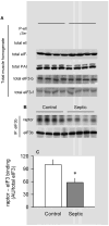
Effect of sepsis on total eIF3 and eIF3 00B7 raptor in muscle. A, Representative Western blots of total and Ser422-phosphorylated eIF4B, eIF4A1, PABP, eIF3-b, and eIF3-f. For each of these proteins, there was no statistical difference in the relative amount of the protein in muscle between control and septic rats (quantitative data not shown). B, eIF3b was immunoprecipitated (IP) from muscle and immunoblotting performed for both raptor and eIF3b. C, Bar graph, quantitation of Western blot data of eIF3 00B7 raptor binding normalized to the amount of eIF3b in the IP, where control value was set at 100 AU. Values are means ± SEM; n = 5 rats each. *P < 0.05, compared with time-matched pair-fed control values.
Fig. 9
Fig. 9
Leucine-induced changes in protein synthesis and mTORC1 in skeletal muscle from control and septic rats. Gastrocnemius was sampled 30 min after oral administration of a maximally stimulating dose of the branched-chain amino acid leucine. In vivo muscle protein synthesis was determined as described in Materials and Methods (A). Bar graphs are quantitation of immunoblots after immunoprecipitation (IP) of either raptor (B) or eIF3b (C). Western blot data from whole muscle tissue homogenates have also been quantitated (D, E, and F). There was no sepsis or leucine effect on the total amount of raptor, mTOR, eIF3b, 4E-BP1, or PRAS40 (data not shown). Saline-treated control values were arbitrarily set to 1.0 AU. Values are means ± SEM; n = 7–9 rats per group. Values with different letters are statistically different (P < 0.05).
Fig. 10
Fig. 10
Schematic for sepsis-induced changes in mTORC1. Under basal conditions, S6K1 is associated with the eIF3 at the PIC (preinitiation complex). In addition, 4E-BP1 is bound to the eIF4E, which in turn is bound to the 7m-GTP cap of the mRNA at the 52032 end. Under inhibitory conditions (as in rapamycin-treated), mTOR/raptor complex is not part of this mRNA PIC. Upon stimulation (as in after anabolic stimulation), mTOR/raptor complex is recruited to the eIF3 complex. Activated mTOR then phosphorylates both S6K1 (at T389) and 4E-BP1 (at several sites in a hierarchy manner). Upon phosphorylation, these mTOR substrates (S6K1 and 4E-BP1) dissociate from the eIF3. The unbound dissociated (phosphorylated) S6K1 can be further phosphorylated by PDK1 (3-phosphoinositide–dependent kinase 1) at T229; this latter secondary phosphorylation is believed to be required for the full activation of the S6K which is capable of phosphorylating more than 10 known substrates. All abbreviations are defined in the text and in Figure 1.

Similar articles

Cited by

References

    1. Long CL, Jeevanandam M, Kim BM, Kinney JM. Whole body protein synthesis and catabolism in septic man. Am J Clin Nutr. 1977;30:1340–1344. - PubMed
    1. Lang CH, Frost RA. Sepsis-induced suppression of skeletal muscle translation initiation mediated by tumor necrosis factor alpha. Metabolism. 2007;56:49–57. - PubMed
    1. Lang CH, Frost RA, Vary TC. Regulation of muscle protein synthesis during sepsis and inflammation. Am J Physiol Endocrinol Metab. 2007;293:E453–E459. - PubMed
    1. Hinton TM, Coldwell MJ, Carpenter GA, Morley SJ, Pain VM. Functional analysis of individual binding activities of the scaffold protein eIF4G. J Biol Chem. 2007;282:1695–1708. - PubMed
    1. Lang CH, Frost RA. Glucocorticoids and TNFalpha interact cooperatively to mediate sepsis-induced leucine resistance in skeletal muscle. Mol Med. 2006;12:291–299. - PMC - PubMed

Publication types