. 2010 Jun 24;465(7301):1044-8.
doi: 10.1038/nature09196.

Structure and mechanism of human DNA polymerase eta

Affiliations

Structure and mechanism of human DNA polymerase eta

Christian Biertümpfel et al. Nature. .

Erratum in

  • Nature. 2011 Aug 18;476(7360):360

Abstract

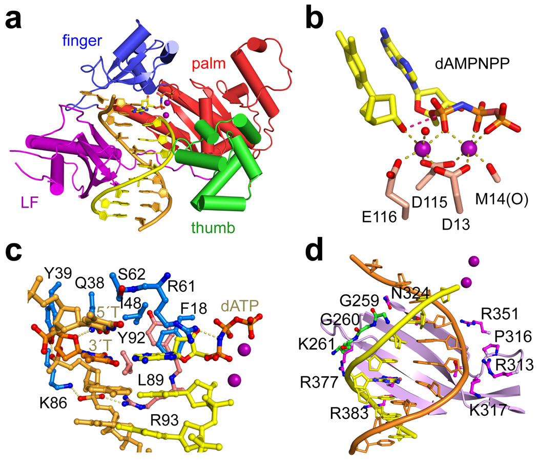
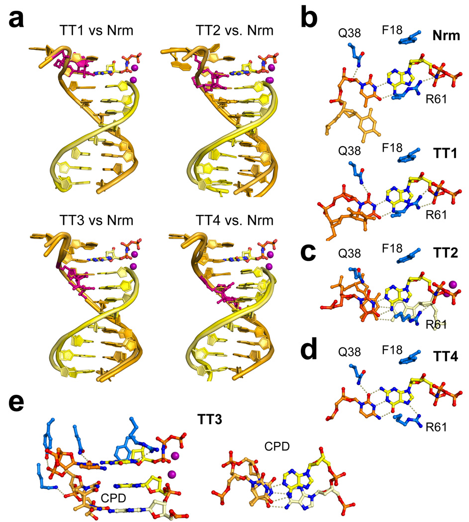
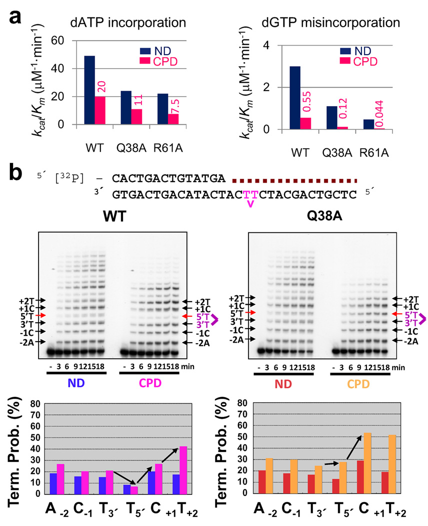
The variant form of the human syndrome xeroderma pigmentosum (XPV) is caused by a deficiency in DNA polymerase eta (Poleta), a DNA polymerase that enables replication through ultraviolet-induced pyrimidine dimers. Here we report high-resolution crystal structures of human Poleta at four consecutive steps during DNA synthesis through cis-syn cyclobutane thymine dimers. Poleta acts like a 'molecular splint' to stabilize damaged DNA in a normal B-form conformation. An enlarged active site accommodates the thymine dimer with excellent stereochemistry for two-metal ion catalysis. Two residues conserved among Poleta orthologues form specific hydrogen bonds with the lesion and the incoming nucleotide to assist translesion synthesis. On the basis of the structures, eight Poleta missense mutations causing XPV can be rationalized as undermining the molecular splint or perturbing the active-site alignment. The structures also provide an insight into the role of Poleta in replicating through D loop and DNA fragile sites.


Figures

Figure 1
Figure 1
Structure of hPol η. a. The ternary complex of hPol η with a normal DNA. Protein domains are shown in distinct colors and labeled. The DNA template is colored orange and the primer yellow. Oxygen and nitrogen atoms are colored red and blue, respectively. dAMPNPP is shown as stick-and-balls, and Mg2+ as purple spheres. All structural figures were made using PyMOL (www.pymol.org). b. The active site. Mg2+ coordination is indicated by pale yellow dashed lines. The 3´-OH of the primer strand is 3.2Å from the α-phosphate as indicated by the red dashed line. c. hPol η-DNA interactions around the active site. Protein side chains from the finger and palm domain are shown as light blue and pink sticks, respectively. d. Interactions with the upstream DNA. LF (shown as light purple ribbon diagram) contacts both template and primer, and the thumb domain (shown in green) only makes 3–4 hydrogen bonds with the primer strand. Side chains that make DNA contacts are highlighted in sticks and labeled.
Figure 2
Figure 2
Structures of lesion DNAs. a. Overlay of TT1, TT2, TT3 and TT4 DNA with the DNA in Nrm (undamaged). CPDs are shown as red sticks. DNA and Mg2+ are colored as in Fig. 1. bd. The replicating base pairs in four ternary complexes (highly similar in TT3 and TT4) are each shown with hPol η residues that contact the bases and deoxyribose. Hydrogen bonds are shown as dashed lines. e. CPD in TT3 and TT4 forms WC-like hydrogen bonds but cannot base stack with neighboring nucleotides.
Figure 3
Figure 3
Functional analyses of Q38A and R61A Pol η. a. Efficiencies (kcat/KM) of WT and two mutants in correct and incorrect nucleotide incorporation on normal (ND) and CPD DNA. These values are derived from Supplementary Table 3. b. CPD-bypass by WT (left) and Q38A mutant Pol η (right). The DNA sequence for primer extension assays is shown. Magenta colored TT represents the CPD. The same DNA template with a 2 nt longer primer was used for the kinetic assay shown in Fig. 3a. Reactions were carried out at a substrate:enzyme molar ratio of 100:1 for the indicated incubation time. Termination probabilities ([In] /Σ n-end[I], where In is band intensity at the nth position) on normal (ND) and CPD DNA are plotted below.
Figure 4
Figure 4
hPol η is a molecular splint. a. The DNA binding surfaces of various Y-family polymerases are shown with electrostatic potentials. Blue and red represent the positive and negative charge potential at the + and − 10 kT/e scale, respectively. The thumb domain in all cases and the N-clasp of Polκ are removed for clarity. Five phosphorus atoms at position -1 to -5 in the hPol η Nrm (yellow) and TT4 (orange) structures are also shown. b. The extensive interactions between hPolη and the template strand in TT4 (as an example) are shown. Hydrogen bonds are indicated by dashed lines. The finger and LF domains are colored in light purple and light green. Two residues forming hydrophobic interactions with the CPD bases are shown in teal. c. A view approximately 90° from b. Detailed hPol η-DNA interactions are shown in Supplementary Fig. 9.
Figure 5
Figure 5
XPV mutations. a. R111 contacts DNA template and stacks with P316 of LF. b. Mapping of 8 missense XPV mutations. The protein is represented by the Cα trace, DNA as tube-and-ladders, and the altered residues are shown as cyan ball-and-sticks. c. Local interactions of A117 and T122 and their relation to the active site. Most side chains are removed for clarity. d. R361 supports P316 and the β strand that forms an extensive hydrogen-bond network with the DNA template. e. G263 and A264 are adjacent to the thumb-DNA primer interface. G263V and A264P substitutions would clash with L258 and K261. Electron densities are shown as meshes in panels c and e.

Comment in

Similar articles

Cited by

References

    1. Brash DE. Sunlight and the onset of skin cancer. Trends Genet. 1997;13:410–414. - PubMed
    1. Masutani C, et al. The XPV (xeroderma pigmentosum variant) gene encodes human DNA polymerase η. Nature. 1999;399:700–704. - PubMed
    1. Johnson RE, Kondratick CM, Prakash S, Prakash L. hRAD30 mutations in the variant form of xeroderma pigmentosum. Science. 1999;285:263–265. - PubMed
    1. Hishida T, Kubota Y, Carr AM, Iwasaki H. RAD6-RAD18-RAD5-pathway-dependent tolerance to chronic low-dose ultraviolet light. Nature. 2009;457:612–615. - PubMed
    1. Chaney SG, Campbell SL, Bassett E, Wu Y. Recognition and processing of cisplatin- and oxaliplatin-DNA adducts. Crit Rev Oncol Hematol. 2005;53:3–11. - PubMed

Publication types

MeSH terms