. 2010 Jun 24;465(7301):1093-6.
doi: 10.1038/nature09127.

Activation of autophagy during cell death requires the engulfment receptor Draper

Affiliations

Activation of autophagy during cell death requires the engulfment receptor Draper

Christina K McPhee et al. Nature. .

Abstract

Autophagy degrades cytoplasmic components that are required for cell survival in response to starvation. Autophagy has also been associated with cell death, but it is unclear how this is distinguished from autophagy during cell survival. Drosophila salivary glands undergo programmed cell death that requires autophagy genes, and engulfment of salivary gland cells by phagocytes does not appear to occur. Here we show that Draper (Drpr), the Drosophila melanogaster orthologue of the Caenorhabditis elegans engulfment receptor CED-1, is required for autophagy during cell death. Null mutations in, and salivary gland-specific knockdown of, drpr inhibit salivary gland degradation. Knockdown of drpr prevents the induction of autophagy in dying salivary glands, and expression of the Atg1 autophagy regulator in drpr mutants suppresses the failure in degradation of salivary glands. Surprisingly, drpr is required in the same dying salivary gland cells in which it regulates autophagy induction, but drpr knockdown does not prevent starvation-induced autophagy in the fat body, which is associated with survival. In addition, components of the conserved engulfment pathway are required for clearance of dying salivary glands. To our knowledge, this is the first example of an engulfment factor that is required for self-clearance of cells. Further, Drpr is the first factor that distinguishes autophagy that is associated with cell death from autophagy associated with cell survival.


Figures
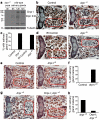
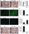
Figure 1
Figure 1. Draper is required for salivary gland cell degradation
a, Protein extracts from drpr null (w; drprΔ5/drprΔ5) pupae at puparium formation (0h) and wild type (Canton-S) salivary glands 6h, 12h, and 14h after puparium formation, were analyzed by Western Blotting with anti-Drpr antibody. b, Control animals (+/w; +/drprΔ5), n=12, and drpr null mutants (w; drprΔ5/drprΔ5), n=47, were analyzed by histology for the presence of salivary gland material (red circles) 24h after puparium formation. c, quantification of data from b and d. d, Control animals (+/w; +/UAS-drprIR), n=11, and those with salivary gland-specific knockdown of drpr (fkh-GAL4/w; UAS-drprIR/+), n=19, were analyzed by histology for the presence of salivary gland material (red circles) 24h after puparium formation. e, Control animals (+/w; +/UAS-drpr-IIR), n=9, and those with salivary gland-specific knockdown of drpr-I (fkh-GAL4/w; UAS-drpr-IIR/+), n=20, were analyzed by histology for the presence of salivary gland material (red circles) 24h after puparium formation. f, quantification of data from e. g, drpr null animals (+/w; +/UAS-Drpr-I; drprΔ5/drprΔ5), n=9, and those with salivary gland-specific expression of Drpr-I (fkh-GAL4/w; UAS-Drpr-I/+; drprΔ5/drprΔ5), n=20, were analyzed by histology for the presence of salivary gland material (red circles) 24h after puparium formation. h, quantification of data from g.
Figure 2
Figure 2. Draper functions downstream or in parallel to caspases during salivary gland cell death
a, Animals with salivary gland-specific expression of p35 (fkh-GAL4/+; UAS-p35/+), n=18, drpr null animals (+/w; +/UAS-p35; drprΔ5/drprΔ5), n=10, and drpr null animals with salivary gland-specific expression of p35 (fkh-GAL4/w; UAS-p35/+; drprΔ5/drprΔ5), n=16, were analyzed by histology for the presence of salivary gland material 24h after puparium formation. Cell fragments are in red circles, and gland fragments are in the yellow circle. b, quantification of data from a. c, Salivary glands were dissected from animals expressing drprIR specifically in GFP-marked clone cells (hsflp/w; UAS-drprIR/+; act<FRT,cd2, FRT>Gal4, UAS-GFP/+) 6h and 14h after puparium formation. Salivary glands were stained with GFP antibody (green) to label cells expressing drprIR, and Lamin antibody (red).
Figure 3
Figure 3. Draper is required for the induction of autophagy in dying salivary gland cells
a, Animals with salivary gland-specific knockdown of Atg12 (fkh-GAL4/w; UAS-Atg12IR/+), n=21, and those with salivary gland-specific knockdown of both Atg12 and drpr-I (fkh-GAL4/w; UAS-Atg12IR/+; UAS-drpr-IIR/+), n=19, were analyzed by histology for the presence of salivary gland material (red circles) 24h after puparium formation. b, quantification of data from a. c, GFP-LC3 was expressed in salivary glands of control animals (+/w; UAS-GFP-LC3/+; fkh-GAL4/+) and those with salivary gland-specific knockdown of drpr (w; UAS-drprIR/UAS-GFP-LC3; fkh-GAL4/+). Salivary glands were dissected 6h and 14h after puparium formation, imaged for GFP-LC3, and LC3 puncta were quantified using Zeiss Automeasure software. d, quantification of data from c. Error bars represent s.e.m.; n ≥ 10; p < 0.0000001. e, LAMP-GFP was expressed in control animals (tub-LAMP-GFP/w; +/fkh-GAL4) and those with salivary gland-specific knockdown of drpr (tub-LAMP-GFP/w; UAS-drprIR/+; fkh-GAL4/+). Salivary glands were dissected 14h after puparium formation, imaged for LAMP-GFP, and LAMP puncta were quantified using Zeiss Automeasure software. f, quantification of data from e. Error bars represent s.e.m.; n ≥ 10; p < 0.05. g, drpr mutant animals (+/w; +/UAS-Atg16A; drprΔ5/drprΔ5), n=10, and those with salivary gland-specific expression of Atg16A (fkh-GAL4/w; UAS-Atg16A/+; drprΔ5/drprΔ5), n=16, were analyzed by histology for the presence of salivary gland material (red circles) 24h after puparium formation. h, quantification of data from g.
Figure 4
Figure 4. Drpr is cell-autonomously required for autophagy in dying salivary glands, but not in response to starvation in the fatbody
a, Protein extracts from drpr null (w; drprΔ5/drprΔ5) pupae at puparium formation (0h) and from the fatbodies of wild type (Canton-S) third instar larvae were analyzed by Western Blotting with anti-Drpr antibody. Third instar larvae were either fed or starved 4h. b, Wild-type (Canton-S) third instar larvae were either fed or starved 4h, and fatbodies were dissected, stained with anti-Drpr antibody, and imaged for Drpr (green). Nuclei were stained with DAPI (blue). c, Third instar larvae expressing GFP-Atg8 in all cells, and drprIR specifically in dsRed-marked clone cells (hsflp/w; UAS-drprIR/+; hsGFPAtg8b, act<FRT,cd2, FRT>Gal4, UAS-dsRed/+), were starved 4h. Larval fatbodies were dissected and imaged for GFPAtg8 (green) and dsRed (red). d, Salivary glands of animals expressing GFP-Atg8 in all cells, and drprIR specifically in dsRed-marked clone cells (hsflp/w; UAS-drprIR/+; hsGFPAtg8b, act<FRT,cd2, FRT>Gal4, UAS-dsRed/+) were dissected 14h after puparium formation. Salivary glands were imaged for GFPAtg8 (green) and dsRed (red). Nuclei were stained with Höescht (blue).

Similar articles

Cited by

References

    1. Kurant E, Axelrod S, Leaman D, Gaul U. Six-microns-under acts upstream of Draper in the glial phagocytosis of apoptotic neurons. Cell. 2008;133:498. - PMC - PubMed
    1. Kuraishi T, et al. Pretaporter, a Drosophila protein serving as a ligand for Draper in the phagocytosis of apoptotic cells. EMBO J. 2009;28:3868. - PMC - PubMed
    1. Pandey UB, et al. HDAC6 rescues neurodegeneration and provides an essential link between autophagy and the UPS. Nature. 2007;447:859. - PubMed
    1. Hay BA, Wolff T, Rubin GM. Expression of baculovirus P35 prevents cell death in Drosophila. Development. 1994;120:2121. - PubMed
    1. Rusten TE, et al. Programmed autophagy in the Drosophila fat body is induced by ecdysone through regulation of the PI3K pathway. Dev. Cell. 2004;7:179. - PubMed

Publication types

MeSH terms