. 2011 Jan;18(1):99-108.
doi: 10.1038/cdd.2010.79. Epub 2010 Jun 25.

Sustained calcium signalling and caspase-3 activation involve NMDA receptors in thymocytes in contact with dendritic cells

Affiliations

Sustained calcium signalling and caspase-3 activation involve NMDA receptors in thymocytes in contact with dendritic cells

P Affaticati et al. Cell Death Differ. 2011 Jan.

Abstract

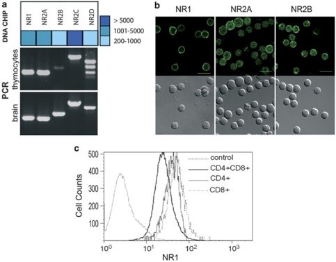
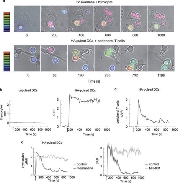
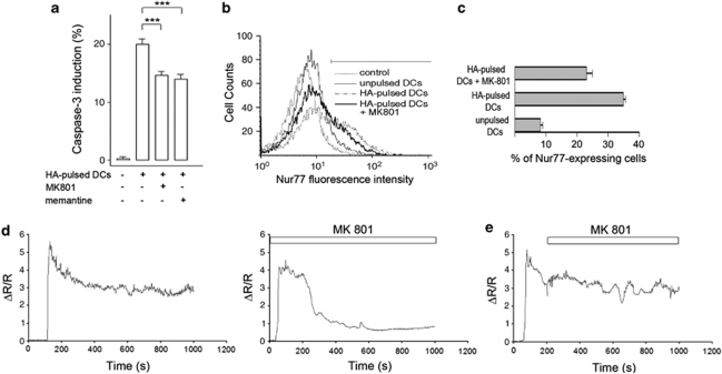
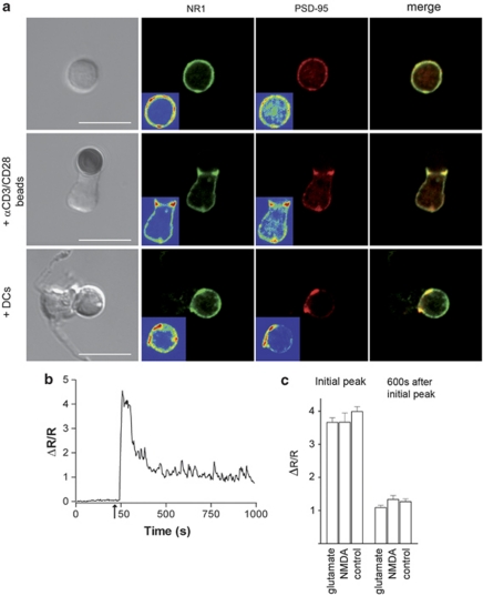
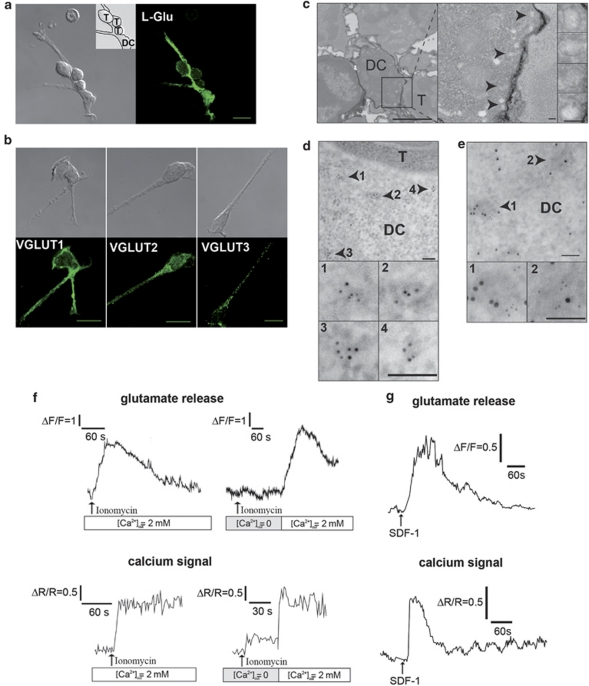
L-glutamate, the major excitatory neurotransmitter, also has a role in non-neuronal tissues and modulates immune responses. Whether NMDA receptor (NMDAR) signalling is involved in T-cell development is unknown. In this study, we show that mouse thymocytes expressed an array of glutamate receptors, including NMDARs subunits. Sustained calcium (Ca(2+)) signals and caspase-3 activation in thymocytes were induced by interaction with antigen-pulsed dendritic cells (DCs) and were inhibited by NMDAR antagonists MK801 and memantine. NMDARs were transiently activated, triggered the sustained Ca(2+) signal and were corecruited with the PDZ-domain adaptor postsynaptic density (PSD)-95 to thymocyte-DC contact zones. Although T-cell receptor (TCR) activation was sufficient for relocalization of NMDAR and PSD-95 at the contact zone, NMDAR could be activated only in a synaptic context. In these T-DC contacts, thymocyte activation occurred in the absence of exogenous glutamate, indicating that DCs could be a physiological source of glutamate. DCs expressed glutamate, glutamate-specific vesicular glutamate transporters and were capable of fast glutamate release through a Ca(2+)-dependent mechanism. We suggest that glutamate released by DCs could elicit focal responses through NMDAR-signalling in T cells undergoing apoptosis. Thus, synapses between T and DCs could provide a functional platform for coupling TCR activation and NMDAR signalling, which might reflect on T-cell development and modulation of the immune response.


Figures

Figure 1
Figure 1
T cells express a large array of i- and m-GluRs. (a) Oligoarray (Neurotrans) analysis of NMDAR mRNA expression on thymocytes compared with brain (upper panel), and validated by RT-PCR compared with brain (lower panel). Oligoarray data are shown as a heat map. Data for all GluRs are shown in Supplementary Figure S1 using primers shown in Supplementary Table S1. (b) Immunofluorescence analysis of NMDAR subunit expression on thymocytes. For each indicated NMDAR subunit (NR1, NR2A, NR2B), a Nomarski image (bottom row) and a confocal fluorescence image is shown in an equatorial plane (top row). Images for other ionotropic glutamate receptors (iGluRs) are shown in Supplementary Figure S1c. Control staining consisting in IgG2b isotype control (for anti-NR1 antibody) or purified rabbit IgG (for anti-NR2A, NR2B, GluR2/3, KA2 antibodies) were negative using the same settings as the specific staining (Supplementary Figure S1c). (c) Flow cytometry analysis of NR1 expression in thymocytes. Antibodies that detect NR1 as a band at the expected molecular weight by western blotting in brain microsomal preparations were used. Relative fluorescence intensity is shown for CD4+, CD8+ and DP subpopulations. Data are representative of three independent experiments. IgG2b control isotype is shown as control for NR1 staining
Figure 2
Figure 2
NMDAR modulates Ca2+ signalling in thymocytes in contact with DCs, in the absence of exogenous glutamate. (a) Representative time-lapse microscopy images (DIC/Fura-2 overlay) of thymocytes (upper set) and peripheral T cells (lower set) making contact with HA-pulsed DCs. The scale indicates Ca2+ level, expressed as ΔR/R values, ranging from 0 (blue) to 3 (red). In thymocytes, steep [Ca2+]i increases were recorded 44±3 s (n=101) after initial contact with the DC, and was, in most synapses, rapidly followed by the establishment of a stable contact (50±2 s, n=93) associated with a sustained high-level plateau lasting at least 10 min. (b) Representative Ca2+ signal in Fura-2 loaded thymocytes in contact with unpulsed DCs (left panel) or with HA-pulsed DCs (right panel) Arrows: contact. (c) Representative Ca2+ signal in Fura-2 loaded peripheral T cells in contact with HA-pulsed DCs. Arrow: contact. (d) Representative traces of the Ca2+ response in thymocytes in contact with HA-pulsed DCs in the presence (black curves) or absence (grey curves) of the NMDAR antagonists memantine (100 μM) (left panel) and MK801 (100 μM) (right panel). Arrows: contact
Figure 3
Figure 3
NMDAR triggers a sustained Ca2+ signal, is transiently activated and is involved in caspase-3 activation and Nur77 induction, in thymocyte-DC contacts. (a) Inhibition of caspase-3 (t=6 h) expression in DP thymocytes co-cultured with unpulsed or HA-pulsed DCs with or without MK801 (100 μM) and memantine (100 μM). Data are means of six replicates for five independent cultures (*** P<0.001). The effect of memantine or MK801 on apoptosis was dose-dependent and was significant at 10 μM, with the maximal effect observed at 100 μM. A dose of 10 μM MK801 resulted in 11.4±1% inhibition of apoptosis (n=6) and a transient Ca2+ signal in 50% thymocytes (n=10) (data not shown). (b) Flow cytometry analysis of Nur77 expression in DP thymocytes, before and 4 h after contact with antigen-loaded DCs, in the presence or absence of MK801 (100 μM). The graphs are representative of three experiments. (c) Quantification of Nur77-expressing DP thymocytes before and 4 h after contact with HA-loaded DCs, in the presence or absence of MK801 (100 μM). (d) Representative traces of the Ca2+ response in thymocytes in contact with HA-pulsed DCs. Thymocytes were preincubated with MK801 (100 μM), NMDA (300 μM) and -serine (20 μM) to block any NMDARs that would be present at the membrane before synapse formation. Thymocytes were then washed and added to HA-pulsed DCs in the absence or presence of the NMDAR antagonists MK801 (100 μM). (e) Representative traces of the Ca2+ response in thymocytes in contact with HA-pulsed DCs MK801 (100 μM) was applied after the beginning of Ca2+ response (t=208 s)
Figure 4
Figure 4
TCR triggering is sufficient to induce NMDAR relocalization at the thymocyte-DC contact zone, whereas NMDAR can be activated only in a synaptic context. (a) Double labelling of PSD-95 (red) and NR1 (green) on isolated thymocytes, thymocytes in contact with anti-CD3/CD28-coated beads and thymocytes in contact with a DC. Merged pictures are shown in each case (yellow), Scale bars, 10 μm. Profiles of distribution of fluorescence intensities and colocalization analysis of PSD-95 and NR1 are shown in Supplementary Figure S4. Controls consisted in mouse IgG2b isotype control for NR1 and purified rabbit IgG for PSD-95 (not shown). (b) Representative trace of Ca2+ response in thymocytes in contact with anti-CD3/CD28-coated beads. Arrow: contact. (c) Histogram of the mean Ca2+ amplitudes from 6 to 11 representative traces at the initial peak and at the plateau phase (600 s after the initial peak), in the absence or presence of glutamate (300 μM) and NMDA (100 μM)
Figure 5
Figure 5
DCs may be the physiological source of glutamate in thymocyte-DC synapses. (a) Confocal immunofluorescence image of glutamate labelling in a DC in contact with three thymocytes (right). Corresponding Nomarski image (left), with an inset indicating the cell contours. Scale bar, 10 μm. (b) Immunofluorescence analysis of VGLUT1, VGLUT2 and VGLUT3 in DCs (bottom row), with the corresponding Nomarski images (top row). Scale bar, 10 μm. Typical punctate fluorescent staining of VGLUT2 and VGLUT3, was observed in DCs (Supplementary Figure S5b). (c) Morphological electron micrograph of thymocyte-DC conjugates showing a group of vesicle structures at the cell–cell interface. Original image (left, scale bar, 2 μm). Magnification of the synapse (middle, scale bar, 100 nm) and of the vesicles (right, scale bar, 100 nm). Arrows: individual vesicles in the DC. (d) Immunogold staining of VGLUT2 in the T cell-DC contact zone. Top: low magnification with vesicles in the DC indicated by arrows. Bottom: higher magnification. Scale bar, 100 nm. (e) Immunogold colocalization of VGLUT2 (5 nm) and synaptotagmin I (10 nm) on the same vesicular structures (arrows). Top: individual vesicles indicated by arrows (scale bar, 2 μm). Bottom: magnifications (scale bars, 100 nm). Control staining is shown in Supplementary Figure S5d (f) Typical traces of glutamate release (upper panels) from DCs stimulated with ionomycin (1 μM), monitored in continuous culture, by recording NADH fluorescence emission. Horizontal scale: time in seconds. Vertical scale: arbitrary units. The amounts of glutamate released represented 40% of the fluorescence increase induced by 10 μM glutamate. Lower panels: Ca2+ signals induced by ionomycin in the presence or absence of extracellular Ca2+ in Fura-2-loaded DCs. (g) Typical traces of glutamate release (uper panel) from DCs stimulated with SDF-1α (100 nM), monitored as in (f). Vertical scale: arbitrary units. Lower panel: Ca2+ signal induced by SDF-1α in Fura-2-loaded DCs, in the presence of extracellular Ca2+

References

    1. Nedergaard M, Takano T, Hansen AJ. Beyond the role of glutamate as a neurotransmitter. Nat Rev Neurosci. 2002;3:748–755. - PubMed
    1. Skerry TM, Genever PG. Glutamate signalling in non-neuronal tissues. Trends Pharmacol Sci. 2001;22:174–181. - PubMed
    1. Pacheco R, Gallart T, Lluis C, Franco R. Role of glutamate on T-cell mediated immunity. J Neuroimmunol. 2007;185:9–19. - PubMed
    1. Pacheco R, Ciruela F, Casado V, Mallol J, Gallart T, Lluis C, et al. Group I metabotropic glutamate receptors mediate a dual role of glutamate in T cell activation. J Biol Chem. 2004;279:33352–33358. - PubMed
    1. Chiocchetti A, Miglio G, Mesturini R, Varsaldi F, Mocellin M, Orilieri E, et al. Group I mGlu receptor stimulation inhibits activation-induced cell death of human T lymphocytes. Br J Pharmacol. 2006;148:760–768. - PMC - PubMed

Publication types

MeSH terms