. 2010 May 28:4:42.
doi: 10.3389/fnhum.2010.00042. eCollection 2010.

Direct visualization of the perforant pathway in the human brain with ex vivo diffusion tensor imaging

Affiliations

Direct visualization of the perforant pathway in the human brain with ex vivo diffusion tensor imaging

Jean C Augustinack et al. Front Hum Neurosci. .

Abstract

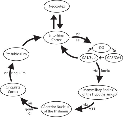
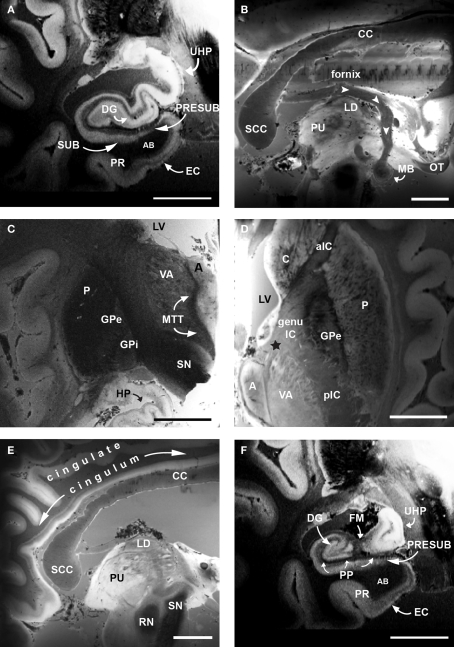
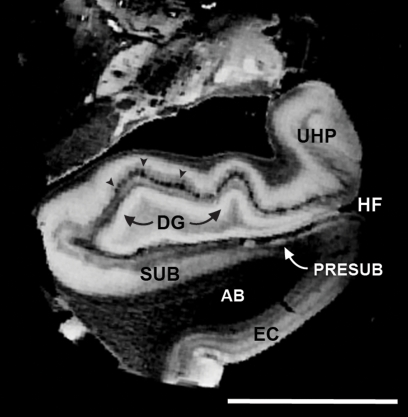
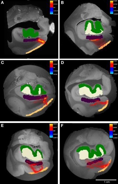
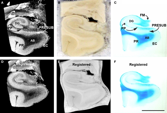
Ex vivo magnetic resonance imaging yields high resolution images that reveal detailed cerebral anatomy and explicit cytoarchitecture in the cerebral cortex, subcortical structures, and white matter in the human brain. Our data illustrate neuroanatomical correlates of limbic circuitry with high resolution images at high field. In this report, we have studied ex vivo medial temporal lobe samples in high resolution structural MRI and high resolution diffusion MRI. Structural and diffusion MRIs were registered to each other and to histological sections stained for myelin for validation of the perforant pathway. We demonstrate probability maps and fiber tracking from diffusion tensor data that allows the direct visualization of the perforant pathway. Although it is not possible to validate the DTI data with invasive measures, results described here provide an additional line of evidence of the perforant pathway trajectory in the human brain and that the perforant pathway may cross the hippocampal sulcus.

Keywords: dentate gyrus; entorhinal cortex; hippocampus; presubiculum; resolution; subiculum.


Figures

Figure 1
Figure 1
Simplified schemata of Papez's circuit (see text for references). Abbreviations: DG, dentate gyrus; genu IC, genu of the internal capsule; MTT, mammillothalamic tract; PP, perforant pathway; Sub, subiculum.
Figure 2
Figure 2
Ex vivo FLASH images following Papez's circuit at 120 μm. (A) EC, dentate gyrus and hippocampus. (B) Fornix (white arrowheads) and mammillary bodies of the hypothalamus. (C) Mammillary bodies project to anterior nucleus of the thalamus via the mammillothalamic tract (D). Anterior nucleus of the thalamus projecting to cingulate cortex via the anterior limb of the internal capsule (black star). (E) Cingulate cortex via cingulum. (F) Presubiculum and EC. The high resolution images of cortical and subcortical structures reveal contrast differences between white and gray matter in the classic limbic circuit. Magnification bar, 1 cm. Abbreviations: A, anterior nucleus of the thalamus; AB, angular bundle; aIC, anterior limb of the internal capsule; C, caudate; CC, corpus callosum; DG, dentate gyrus; EC, entorhinal cortex; FM, fimbria; genu IC, genu of the internal capsule; GPe, external globus pallidus; GPi, internal globus pallidus; HP, hippocampus; LD, lateral dorsal nuclues of the thalamus; LV, lateral ventricle; MB, mammillary bodies; MTT, mammillothalamic tract; OT, optic tract; P, putamen; pIC, posterior limb of the internal capsule; PP, perforant pathway; PR, perirhinal cortex; PRESUB, presubiculum; PU, pulvinar; RN, red nuclues; SCC, splenium of the corpus callosum; SN, substantia nigra; SUB, subiculum; UHP, uncal hippocampus; VA, ventral anterior nucleus of the thalamus.
Figure 3
Figure 3
FLASH images (150 μm) of primary visual cortex tissue unfixed (A) and fixed in formalin (B). Note the line of Gennari in both and the superior CNR in the fixed tissue.
Figure 4
Figure 4
Nissl stained sections from anterior thalamus treated with gadolinium (A) and non-treated (B). Both (A, B) and insets show similar staining, although stereological neuronal estimates were different.
Figure 5
Figure 5
Isotropic 100 μm FLASH image that illustrates the anatomical areas that make up the perforant pathway: EC > AB > SUB/PreSUB > terminal zone of the perforant pathway in the dentate and HP (i. e. molecular layer; black arrowheads). Magnification bar = 1 cm. Abbreviations: EC, entorhinal cortex; HF, hippocampal fissure; PR, perirhinal cortex; UHP, uncal hippocampus.
Figure 6
Figure 6
Isotropic 100 μm FLASH image registered with 200 μm isotropic DTI images. FA_DTI volume is shown in spectrum colors (A, D, and G) and structural MRI volume in grayscale (B, E, and H) and aligned volumes are shown as merged images (C, F and I). Rows represent planes of cut; top row shows horizontal, middle row represents coronal and bottom panel illustrates sagittal.
Figure 7
Figure 7
The probabilistic tractography results between labeled region entorhinal cortex layer II and subicular cortices (Subiculum+presubiculum) for all cases. In this illustration, probability for hippocampal formation was not tested and labels are shown for reference only. Color labels represent entorhinal layer II (beige), subiculum (purple), dentate gyrus (white) and hippocampus (green). Case A had gadolinium treatment prior to MR scanning and exhibits less noise. All cases showed connections with non-zero probability (A–F), but cases with higher FA generated higher values than cases with lower FA. Panels (A–F) represent cases 1-6, respectively with Table 1. Heatmap scalebar located in upper right corner for all cases.
Figure 8
Figure 8
The probabilistic tractography results between labeled region subicular cortices (subiculum+presubiculum) and dentate gyrus for all cases. In this illustration, probability for entorhinal cortex layer II was not tested and label is shown for reference only. Color labels represent entorhinal layer II (beige), subiculum (purple), dentate gyrus (white) and hippocampus (green). Case A had gadolinium treatment prior to MR scanning and exhibits less noise. All cases showed connections with non-zero probability (A–F), but cases with higher FA generated higher values than cases with lower FA. Panels (A–F) represent cases 1-6, respectively with Table 1. Heatmap scalebar located in upper right corner for all cases.
Figure 9
Figure 9
Diffusion tensor images at 4. 7T with fiber tracking (DTI Studio). Seed points outlined in (A) and (C) yield the perforant pathway streamlines in (B) and (D). Entorhinal layer II streamline is shown as it ascends into the angular bundle (B) and perforant pathway streamline shows an indication of crossing the hippocampal fissure and the temporoalvear pathway coursing around the hippocampal fissure (D). Magnification bar = 0.25 cm.
Figure 10
Figure 10
MRI (A), blockface (B), myelin stained (C) medial temporal lobe of corresponding sections and accompanying registered MRI (D) blockface (E) myelin stained (F). Perforant pathway is stained with Luxol fast blue, a stain that labels myelin. The perforant pathway is stained (C). Magnification bar = 1 cm. Abbreviations: AB, angular bundle; DG, dentate gyrus; EC, entorhinal cortex; FM, fimbria; PP, perforant pathway; PR, perirhinal cortex; PRESUB, presubiculum.

References

    1. Aggleton J. P., Vann S. D., Saunders R. C. (2005). Projections from the hippocampal region to the mammillary bodies in macaque monkeys. Eur. J. Neurosci. 22, 2519–253010.1111/j.1460-9568.2005.04450.x - DOI - PubMed
    1. Arnold S. E., Hyman B. T., Flory J., Damasio A. R., Van Hoesen G. W. (1991). The topographical and neuroanatomical distribution of neurofibrillary tangles and neuritic plaques in the cerebral cortex of patients with Alzheimer's disease. Cereb. Cortex 1, 103–11610.1093/cercor/1.1.103 - DOI - PubMed
    1. Arriagada P. V., Marzloff K., Hyman B. T. (1992). Distribution of Alzheimer-type pathologic changes in nondemented elderly individuals matches the pattern in Alzheimer's disease. Neurology 42, 1681–1688 - PubMed
    1. Augustinack J. C., van der Kouwe A. J., Blackwell M. L., Salat D. H., Wiggins C. J., Frosch M. P., Wiggins G. C., Potthast A., Wald L. L., Fischl B. R. (2005). Detection of entorhinal layer II using 7Tesla [corrected] magnetic resonance imaging. Ann. Neurol. 57, 489–49410.1002/ana.20426 - DOI - PMC - PubMed
    1. Behrens T. E., Johansen-Berg H., Woolrich M. W., Smith S. M., Wheeler-Kingshott C. A., Boulby P. A., Barker G. J., Sillery E. L., Sheehan K., Ciccarelli O., Thompson A. J., Brady J. M., Matthews P. M. (2003a). Non-invasive mapping of connections between human thalamus and cortex using diffusion imaging. Nat. Neurosci. 6, 750–75710.1038/nn1075 - DOI - PubMed

LinkOut - more resources