

doi: 10.1016/j.cult.2008.04.004.
Ultrasonography of the Anterior Segment
- PMID: 20577648
- PMCID: PMC2890277
- DOI: 10.1016/j.cult.2008.04.004
Item in Clipboard
Ultrasonography of the Anterior Segment
Ultrasound Clin.
.
No abstract available
Figures

Peter's anomaly. Note abnormal irido-corneal adhesions.

Corneal lesion. Radial scan shows a dome shaped lesion infiltrating deep into the cornea.

Corneal dystrophy. UBM showing highly reflective hyaline granules in the superficial stroma of cornea with granular dystrophy. Reproduced with permission from : Pavlin CJ, Foster FS. Ultrasound Biomicroscopy of the Eye, Springer Verlag, New York 1994.

Tilted PC IOL. Radial scan of the central iris and PC IOL shows a slight tilt off the horizontal axis indicting displacement of the PC IOL (arrow).
Displaced PC IOL. Radial scan of the anterior chamber shows PC IOL displaced temporally with the haptic abutting the peripheral iris (arrow).

Iridodialysis. Radial scan shows complete separation of the iris from the root (arrow).

Cylcodialysis. Goniophotograph showing cleft in the anterior chamber angle (A, arrows). Radial scan shows complete separation of the ciliary body from the scleral spur (B, asterix).

Metallic intraocular foreign body. Radial scan shows an irregularly shaped, highly reflective foreign body in the anterior angle (arrow). Note the shadowing of the intraocular structures beneath the hyperechoic foreign body.

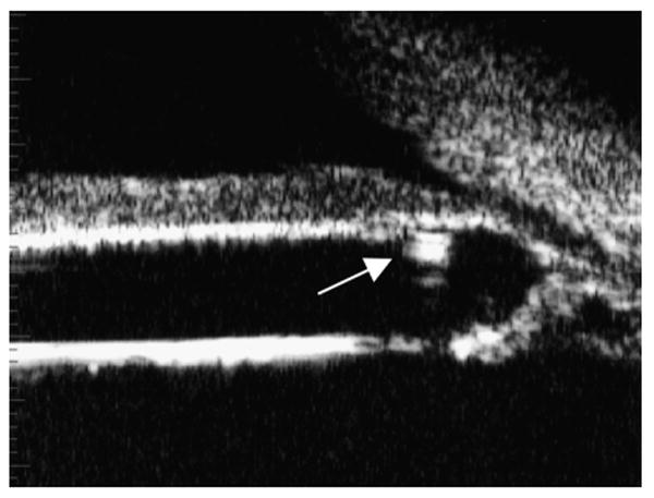
Zonular Injury. Radial scan shows the absence of the zonule normally extending from the ciliary process (arrow) to the lens surface (arrow). (Image courtesy of Charles J. Pavlin MD).

Descemet's membrane detachment. Radial scan shows irregularly thickened cornea with a smooth, highly reflective detachment of Descemet's membrane (arrow).
Similar articles
-
Role of B-scan ultrasonography in the localization of intraocular foreign bodies in the anterior segment: a report of three cases.BMC Ophthalmol. 2015 Aug 14;15:102. doi: 10.1186/s12886-015-0076-1. BMC Ophthalmol. 2015. PMID: 26268356 Free PMC article.
-
Case Report: Ultrasonography and Magnetic Resonance Imaging of Anterior Segment Dysgenesis in a Calf.Front Vet Sci. 2022 Apr 8;9:794255. doi: 10.3389/fvets.2022.794255. eCollection 2022. Front Vet Sci. 2022. PMID: 35464371 Free PMC article.
-
Evaluation of intra- and interobserver reliability and image reproducibility to assess usefulness of high-resolution ultrasonography for measurement of anterior segment structures of canine eyes.Am J Vet Res. 2005 Oct;66(10):1775-9. doi: 10.2460/ajvr.2005.66.1775. Am J Vet Res. 2005. PMID: 16273910
-
Ultrasound biomicroscopy of the anterior segment.J Am Optom Assoc. 1996 Aug;67(8):469-79. J Am Optom Assoc. 1996. PMID: 8888877 Review.
-
[Anterior segment optical coherence tomography in glaucoma].Klin Monbl Augenheilkd. 2008 Mar;225(3):194-9. doi: 10.1055/s-2008-1027241. Klin Monbl Augenheilkd. 2008. PMID: 18351532 Review. German.
Cited by
-
Ultrasound Biomicroscopy for the Detection and Characterization of Anterior Segment Cysticercosis.Clin Ophthalmol. 2024 Nov 25;18:3441-3448. doi: 10.2147/OPTH.S494556. eCollection 2024. Clin Ophthalmol. 2024. PMID: 39618987 Free PMC article.
References
-
- Kim T, Cohen EJ, Schnall BM, et al. Ultrasound biomicroscopy and histopathology of sclerocornea. Cornea. 1998;17(4):443–5. - PubMed
-
- Haddad AM, Greenfield DS, Stegman Z, et al. Peter's anomaly: diagnosis by ultrasound biomicroscopy. Ophthalmic Surg Lasers. 1997;28(4):311–2. - PubMed
-
- Chawla B, Agarwal A, Kashyap S, Tandon R. Diagnosis and management of corneal keloid. Clin Experiment Ophthalmol. 2007;35(9):855–7. - PubMed
Grants and funding
LinkOut - more resources
Full Text Sources