Review
. 2010 Jul;77(7):566-85.
doi: 10.1002/mrd.21175.

Regulation of the G2/M transition in rodent oocytes

Affiliations
Review

Regulation of the G2/M transition in rodent oocytes

Stephen M Downs. Mol Reprod Dev. 2010 Jul.

Abstract

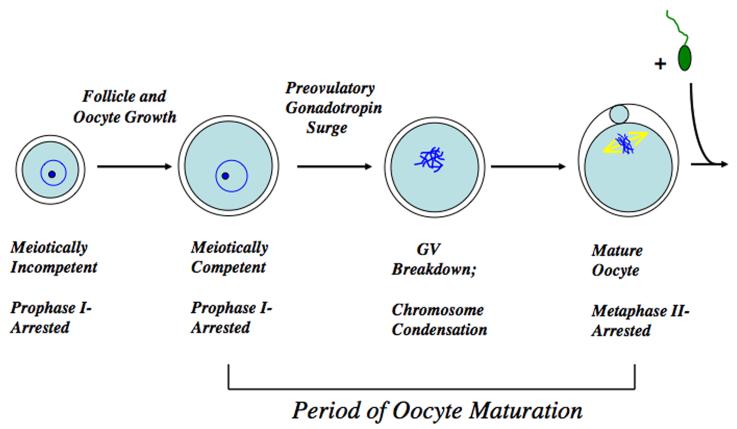
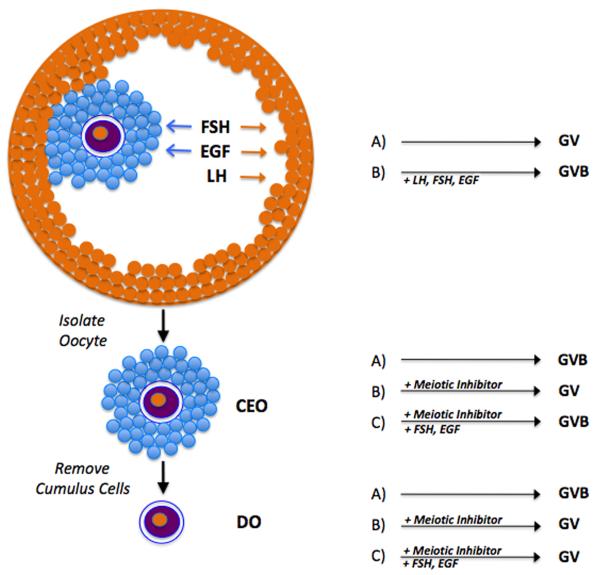
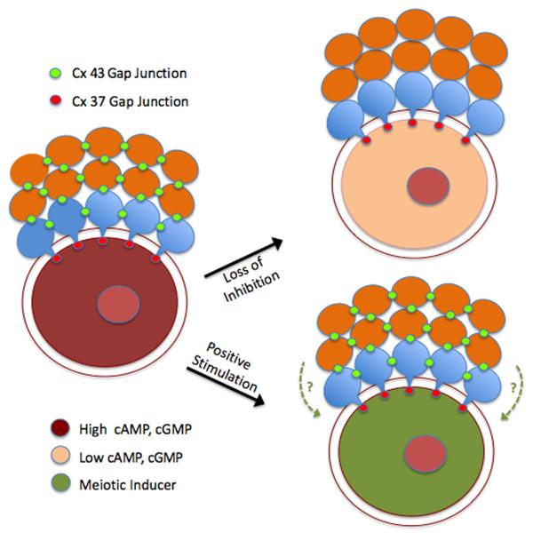
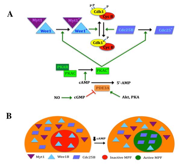
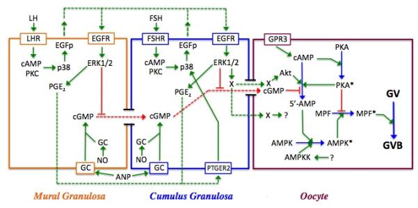
Regulation of maturation in meiotically competent mammalian oocytes is a complex process involving the carefully coordinated exchange of signals between the somatic and germ cell compartments of the ovarian follicle via paracrine and cell-cell coupling pathways. This review highlights recent advances in our understanding of how such signaling controls both meiotic arrest and gonadotropin-triggered meiotic resumption in competent oocytes and relates them to the historical context. Emphasis will be on rodent systems, where many of these new findings have taken place. A regulatory scheme is then proposed that integrates this information into an overall framework for meiotic regulation that demonstrates the complex interplay between different follicular compartments.


Figures

Figure 1
Figure 1
Overview of oocyte maturation. The oocyte remains meiotically incompetent and in prophase I arrest during the period of follicular and oocyte growth. At the time of meiotic resumption the nucleus, or germinal vesicle, breaks down as the chromosomes condense and meiosis proceeds until the metaphase II stage that immediately follows extrusion of the first polar body. The MII arrest is maintained in healthy oocytes until successful fertilization is achieved, whereupon the oocyte completes maturation, maternal and paternal chromatin is combined, and embryonic development is initiated.
Figure 2
Figure 2
In vitro model systems for studying oocyte maturation. The intact preovulatory follicle (top) maintains the oocyte in the germinal vesicle (GV) stage in the absence of hormonal stimulation. The addition of hormone (gonadotropins or EGF-like peptides) stimulates germinal vesicle breakdown (GVB) after binding to receptors on the follicle cells. Arrows show the cells responsive to each of the hormones. Removal of the cumulus-enclosed oocyte (CEO) results in spontaneous maturation in the absence of added inhibitor. If maintained in meiotic arrest with appropriate inhibitor, GVB can be induced by proper hormonal stimulation (FSH and EGF-like peptides, but not LH). Removal of cumulus cells produces a denuded oocyte (DO) that will also undergo spontaneous maturation, but is unresponsive to hormones if maintained in meiotic arrest.
Figure 3
Figure 3
Gap junction regulation of meiotic status. Homologous Cx 43 gap junctions couple mural granulosa cells (green) to one another and cumulus granulosa cells (blue) to one another, and heterologous gap junctions couple mural granulosa cells to cumulus granulosa cells. Heterologous Cx 37 gap junctions in the innermost layer of cumulus cells couple the somatic compartment of the follicle to the oocyte. Two different models exist for meiotic regulation. In the loss of inhibition model, loss of Cx 43 coupling terminates the flux of inhibitor (cAMP and cGMP) from mural to cumulus cells, thereby significantly reducing levels within the oocyte and leading to GVB. In the positive stimulation model, patent gap junctions mediate the flux of a positive stimulus into the oocyte from the surrounding follicle cells that can override elevated cyclic nucleotide levels, although contribution of a diffusible paracrine factor (dotted lines) originating from the granulosa cells cannot be discounted.
Figure 4
Figure 4
Regulation of MPF activity in the mouse oocyte. A, Control of MPF by PKA. MPF activity is regulated by the phosphorylation state of the Cdk1 subunit. PKA activity is dependent on cAMP levels. This cyclic nucleotide binds to the regulatory subunit (PKAR), driving dissociation from, and activation of, the catalytic subunit (PKAC; asterisk denotes activated state). When sufficient cyclin B is present, MPF is maintained in the inactive state by the action of Wee1 and Myt1 kinases that are positively regulated by PKA. Dephosphorylation of Cdk1 triggers activation of MPF and is principally driven by Cdc25B that is negatively regulated by PKA. This response occurs when PKA activity is lowered by a decrease in cAMP, brought about by the action of PDE3. Increased PDE3 activity results from a drop in cGMP levels or active stimulation by Akt or perhaps PKA. B, Effect of Myt1/Wee1/Cdc25B localization on MPF activity. In the competent oocyte, pre-MPF accumulates in the nucleus. Under meiosis-arresting conditions, Wee1B is present in the germinal vesicle, but Myt1 and Cdc25B are restricted to the cytoplasm, and this is under PKA control. When oocyte cAMP levels are reduced, Wee1B exits the nucleus and Cdc25B translocates into the nucleus. The loss of inhibitory Wee1B and the entry of stimulatory Cdc25B promotes dephosphorylation of pre-MPF in the germinal vesicle and its subsequent activation that will drive GVB.
Figure 5
Figure 5
AMPK in metabolism and oocyte maturation. A, general metabolic control. High levels of ATP suppress AMPK activity while elevated AMP has the opposite effect. Activation of AMPK (asterisk) suppresses anabolic pathways that consume ATP and stimulates catabolic pathways that generate ATP. B, proposed contribution of AMPK to meiotic resumption in the mouse oocyte. PKA can phosphorylate AMPK and maintain it in the inactive state. A number of different stimuli can activate AMPK, including stress, AMP produced by cAMP degradation, conversion of AICAR to the AMP analog, ZMP, and upstream kinases such as LKB1 and CaMKKβ. Active AMPK phosphorylates, and inactivates, mitochondria-associated acetyl CoA carboxylase II. This enzyme catalyzes the conversion of acetyl CoA to malonyl CoA, an important negative regulator of fatty acid transport into the mitochondrion, and, thus, fatty acid oxidation. Since fatty acid oxidation appears to be a vital component of meiotic induction in mouse oocytes, the positive action of AMPK on oocyte maturation is likely due, at least in part, to activation of fatty acid oxidation (see Downs et al, 2009),
Figure 6
Figure 6
Model for meiotic regulation in the mouse. Shown are the three cell types within the follicle--mural and cumulus granulosa cells and the oocyte—and the interactions between them. Red lines depict inhibitory processes; green lines are stimulatory; and dotted lines represent agents that diffuse between cells. Active enzymes within the oocyte are denoted by an asterisk. In the oocyte, PGR3-dependent cAMP production activates PKA, which negatively regulates MPF. cGMP produced within the granulosa cells by soluble or membrane bound guanylate cyclase (GC) is transmitted through gap junctions to the oocyte where it suppresses PDE3 activity and helps to maintain meiotic arrest by sustaining high cAMP levels and PKA activity. Soluble GC can be stimulated by nitric oxide (NO), while membrane-bound GC can be stimulated by atrial natriuretic peptide (ANP). LH stimulation of the mural granulosa cells initiates a signal transduction cascade that results in p38MAPK-dependent production of EGF-like peptides (EGFp) that act in autocrine fashion to stimulate the phosphorylation and activation of ERK1/2. ERK1/2 then drives the synthesis of prostaglandin E2 that diffuses to cumulus granulosa cells, binds to the prostaglandin receptor PTGER2 and triggers the synthesis of EGFp (see Shimada et al, 2006). FSH can mimic this action in cumulus cells. ERK1/2 terminates gap junction coupling between the two granulosa compartments and blocks cGMP transfer to the oocyte; it also generates a positive signal (X) for maturation that can be transmitted through gap junctions or possibly diffuses to the oocyte. PDE3 activity in the oocyte is increased via protein kinase B (Akt), the removal of cGMP inhibition, and perhaps also by a transient increase in oocyte cAMP and PKA activity that occurs following gonadotropin stimulation of the granulosa cells. The resulting decrease in cAMP levels leads to inactivation of PKA and elimination of its block to MPF activation. AMPK is activated in immature mouse oocytes in response to gonadotropin stimulation and helps drive MPF activation and GVB, most likely through indirect means. Its activation may result from AMP generated by PDE3 and/or perhaps by an upstream kinase (AMPKK), whose identity has not yet been identified.

Similar articles

Cited by

References

    1. Albertini DF, Combelles CMH, Benecci E, Carabatsos MJ. Cellular basis for paracrine regulation of ovarian follicle development. Reproduction. 2001;121:647–653. - PubMed
    1. Alessi DR, Cuenda A, Cohen P, Dudley DT, Saltiel AR. PD98059 is a specific inhibitor of activation of mitogen-activated protein kinase kinase in vitro and in vivo. J Biol Chem. 1995;270:27489–27494. - PubMed
    1. Araki K, Naito K, Haraguchi S, Suzuki R, Yokoyama M, Inoue M, Aizawa S, Toyoda Y, Sato E. Meiotic abnormalities of c-MOS knockout mouse oocytes: Activation after first meiosis or entrance into third meiotic metaphase. Biol Reprod. 1996;55:1315–1324. - PubMed
    1. Ashkenazi H, Cao X, Motola S, Popliker M, Conti M, Tsafriri A. Epidermal growth factor family members: mediators of the ovulatory response. Endocrinology. 2005;146:77–84. - PubMed
    1. Ben-Ami I, Freimann S, Armon L, Dantes A, Ron-El R, Amsterdam A. Novel function of ovarian growth factors: combined studies by DNA microarray, biochemical and physiological approaches. Mol Hum Reprod. 2006;12:413–419. - PubMed

LinkOut - more resources