. 2010 Jul;52(1):238-48.
doi: 10.1002/hep.23628.

Aquaporin-1 facilitates angiogenic invasion in the pathological neovasculature that accompanies cirrhosis

Affiliations

Aquaporin-1 facilitates angiogenic invasion in the pathological neovasculature that accompanies cirrhosis

Robert C Huebert et al. Hepatology. 2010 Jul.

Abstract

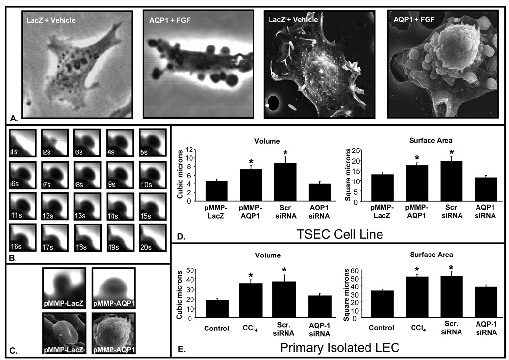
Increasing evidence suggests that hepatic fibrosis and pathological angiogenesis are interdependent processes that occur in parallel. Endothelial cell invasion is requisite for angiogenesis, and thus studies of the mechanisms governing liver endothelial cell (LEC) invasion during cirrhosis are of great importance. Emerging research implicates amoeboid-type motility and membrane blebbing as features that may facilitate invasion through matrix-rich microenvironments. Aquaporins (AQPs) are integral membrane water channels, recognized for their importance in epithelial secretion and absorption. However, recent studies also suggest links between water transport and cell motility or invasion. Therefore, the purpose of this study was to test the hypothesis that AQP-1 is involved in amoeboid motility and angiogenic invasion during cirrhosis. AQP-1 expression and localization was examined in normal and cirrhotic liver tissues derived from human and mouse. AQP-1 levels were modulated in LEC using retroviral overexpression or small interfering RNA (siRNA) knockdown and functional effects on invasion, membrane blebbing dynamics, and osmotic water permeability were assayed. Results demonstrate that AQP-1 is up-regulated in the small, angiogenic, neovasculature within the fibrotic septa of cirrhotic liver. AQP-1 overexpression promotes fibroblast growth factor (FGF)-induced dynamic membrane blebbing in LEC, which is sufficient to augment invasion through extracellular matrix. Additionally, AQP-1 localizes to plasma membrane blebs, where it increases osmotic water permeability and locally facilitates the rapid, trans-membrane flux of water.

Conclusion: AQP-1 enhances osmotic water permeability and FGF-induced dynamic membrane blebbing in LEC and thereby drives invasion and pathological angiogenesis during cirrhosis.


Figures

Figure 1
Figure 1. AQP-1 Expression is Increased in Cirrhotic Liver
A. cDNA from human liver tissue at various stages of NAFLD-induced cirrhosis amplified using quantitative RT-PCR (n=3; mean +/− SE). B-D. Representative immunoblots for AQP-1 or actin (50 µg/lane) in control or cirrhotic liver tissue from human or mouse and densitometric quantification (n=4 to 9; mean +/− SE). Panels represent: (B) multiple human NAFLD patients; (C) multiple human hepatitis C patients; and (D) CCL4 model. * P<0.05
Figure 2
Figure 2. AQP-1 is Localized to Endothelia within Fibrotic Septa
A. Control or cirrhotic human liver tissue stained with IHC for vWF (brown), counterstained with hematoxylin (blue), and quantified (n=5; mean +/− SE). * P<0.05. B. Control or cirrhotic human and mouse liver tissue stained with IF for AQP-1 (red) and nuclear TOTO-3 (blue). See Supplemental Figure 1 for AQP-1 IHC. C. IF co-staining on cirrhotic liver tissue for AQP-1 (red) and eNOS, PECAM, α-SMA, or CK-19 (green).
Figure 3
Figure 3. AQP-1 is Overexpressed in TSEC after Retroviral Transduction
A. TSEC transduced with retroviral AQP-1 or LacZ control and stained by IF for AQP-1 (red) and nuclear TOTO-3 (blue). B. Representative immunoblots (50 µg/lane) for AQP-1 or actin from TSEC treated with pMMP-AQP1 or LacZ control.
Figure 4
Figure 4. AQP-1 Enhances TSEC Invasion Capacity
A. TSEC transduced with retroviral AQP-1 or LacZ control subjected to chemotaxis assays in the presence or absence of FGF. Migrated cells are stained with DAPI (blue) and quantified (n=3; mean +/− SE). See supplemental Figure 2 for additional chemotaxis data. B. TSEC transduced with retroviral AQP-1 or LacZ controls were subjected to 3D collagen invasion assays in the presence or absence of FGF. Invading cells are stained with DAPI (blue) and quantified (n=3; mean +/− SE). * P<0.05 versus LacZ. # P<0.05 versus LacZ+FGF.
Figure 5
Figure 5. AQP-1 Enhances Dynamic Membrane Blebbing
A. TSEC transduced with retroviral AQP-1 or LacZ control, treated with FGF or vehicle, and imaged using phase-contrast, time-lapse, video microscopy (left panels) or scanning electron microscopy (right panels). See Supplemental Movie 1 and Supplemental Movie 2. B. Time course of a dynamic membrane bleb in TSEC at high magnification (1 second/frame). C. TSEC transduced with retroviral AQP-1 or LacZ control, treated with FGF and imaged using phase-contrast microscopy (upper panels) or scanning electron microscopy (lower panels). D. TSEC were transduced with retroviral AQP-1 or LacZ control and/or transfected with AQP-1 specific siRNA or scrambled siRNA, treated and imaged as above, and measured to calculate maximum bleb volume and surface area. E. Freshly isolated LEC from mice treated with CCl4 or vehicle were treated, imaged, and measured, as above. * P<0.05 versus control.
Figure 6
Figure 6. Dynamic Membrane Blebbing in TSEC is Non-Apoptotic
A. TSEC were transduced with retroviral LacZ, treated TNF-α or FGF, and subjected to Caspase 3,7 activity assays (n=6; mean+/−SE). B. TSEC transduced with retroviral AQP-1 were treated and assayed as above (n=6; mean+/−SE). * P<0.05 versus control. C-F. TSEC were imaged using phase-contrast, time-lapse, video microscopy during dynamic blebbing after withdrawal of FGF at times (C) O hr, (D) 1 hr, (E) 2 hr, and (F) 3 hr. See Supplemental Movie 3.
Figure 7
Figure 7. AQP-1 Localizes to Membrane Blebs and Facilitates Regional Water Flux
A TSEC overexpressing AQP-1 or LacZ control labeled with AQP-1 specific immunogold particles and imaged with scanning electron microscopy at 10,000X or 50,000X. B. TSEC overexpressing AQP-1 treated with FGF and stained by IF for AQP-1 (green) or both AQP-1 and Myosin II (red). C. TSEC overexpressing AQP-1 were pre-loaded with calcein AM, treated with FGF, and imaged during blebbing using phase-contrast or confocal microscopy. Pseudocolor enhancement shows increased fluorescence within blebs (representing localized dilution). Inset shows high magnification of a bleb. D-E. A 30 mOsm external osmotic gradient was applied to TSEC transduced with retroviral AQP-1 or LacZ, and/or transfected with AQP-1 specific siRNA or scrambled siRNA, and treated with FGF, imaged, and measured over time to calculate osmotic water permeability (D) and water flux (E).
Figure 8
Figure 8. Working model
Upper panel depicts FGF-stimulated, AQP-1 mediated water transport that enhances dynamic membrane blebbing and promotes amoeboid invasion through the cirrhotic microenvironment. Lower panel depicts a single bleb demonstrating a combination of hydraulic, mechanical, and osmotic forces driving dynamic membrane bleb expansion and retraction.

References

    1. Lim YS, Kim WR. The global impact of hepatic fibrosis and end-stage liver disease. Clin Liver Dis. 2008;12:733–746. vii. - PubMed
    1. Fernandez M, Semela D, Bruix J, Colle I, Pinzani M, Bosch J. Angiogenesis in liver disease. J Hepatol. 2009;50:604–620. - PubMed
    1. Tugues S, Fernandez-Varo G, Munoz-Luque J, Ros J, Arroyo V, Rodes J, Friedman SL, et al. Antiangiogenic treatment with sunitinib ameliorates inflammatory infiltrate, fibrosis, and portal pressure in cirrhotic rats. Hepatology. 2007;46:1919–1926. - PubMed
    1. Morales-Ruiz M, Jimenez W. Neovascularization, angiogenesis and vascular remodeling in portal hypertension. In: Sanyal A, Shah V, editors. Portal Hypertension: Pathobiology, Evaluation, and Treatment. Totowa, NJ: Humana Press; 2005. pp. 99–112.
    1. Fabris L, Cadamuro M, Libbrecht L, Raynaud P, Spirli C, Fiorotto R, Okolicsanyi L, et al. Epithelial expression of angiogenic growth factors modulate arterial vasculogenesis in human liver development. Hepatology. 2008;47:719–728. - PubMed

Publication types

MeSH terms