. 2011 Jun;32(6):947-61.
doi: 10.1002/hbm.21081. Epub 2010 Jun 24.

Correlations of striatal dopamine synthesis with default network deactivations during working memory in younger adults

Affiliations

Correlations of striatal dopamine synthesis with default network deactivations during working memory in younger adults

Meredith N Braskie et al. Hum Brain Mapp. 2011 Jun.

Abstract

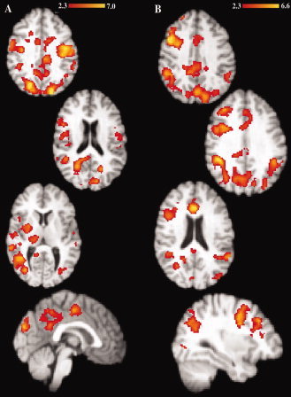
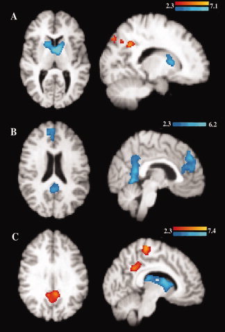
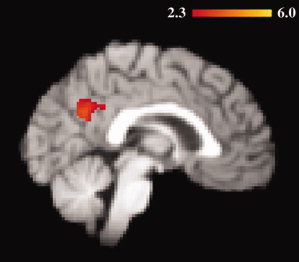
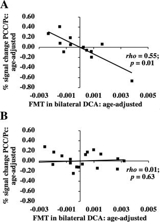
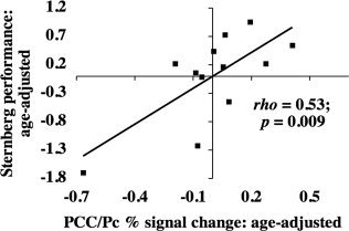
Age-related deficits have been demonstrated in working memory performance and in the dopamine system thought to support it. We performed positron emission tomography (PET) scans on 12 younger (mean 22.7 years) and 19 older (mean 65.8 years) adults using the radiotracer 6-[(18)F]-fluoro-L-m-tyrosine (FMT), which measures dopamine synthesis capacity. Subjects also underwent functional magnetic resonance imaging (fMRI) while performing a delayed recognition working memory task. We evaluated age-related fMRI activity differences and examined how they related to FMT signal variations in dorsal caudate within each age group. In posterior cingulate cortex and precuneus (PCC/Pc), older adults showed diminished fMRI deactivations during memory recognition compared with younger adults. Greater task-induced deactivation (in younger adults only) was associated both with higher FMT signal and with worse memory performance. Our results suggest that dopamine synthesis helps modulate default network activity in younger adults and that alterations to the dopamine system may contribute to age-related changes in working memory function.


Figures

Figure 1
Figure 1
Age‐related load‐dependent fMRI activity differences. Differences in fMRI activity were found between younger and older adults during the load‐dependent (A) cue phase and (B) delay phase when the activity was compared to fMRI signal during the baseline fixation. Functional MRI activity was not significantly different between age groups during the load‐dependent probe phase. Red to yellow scale thresholded Z score masks represent regions in which older adults showed significantly greater fMRI activity than younger adults. Significance was set at Z = 2.3, corrected for multiple comparisons using cluster thresholding to adjust the image‐wise significance to P < 0.05. [Color figure can be viewed in the online issue, which is available at wileyonlinelibrary.com.]
Figure 2
Figure 2
Age‐related load‐independent fMRI activity differences. Differences in fMRI activity were found between younger and older adults during the load‐independent (A) cue phase, (B) delay phase, and (C) probe phase when the activity was compared to fMRI signal during the baseline fixation. Red to yellow scale thresholded Z score masks represent regions in which older adults showed significantly greater fMRI activity than younger adults. Blue scale masks represent regions in which younger adults showed significantly greater fMRI activity. Significance was set at Z = 2.3, corrected for multiple comparisons using cluster thresholding to adjust the image‐wise significance to P < 0.05. [Color figure can be viewed in the online issue, which is available at wileyonlinelibrary.com.]
Figure 3
Figure 3
FMT modulates age‐related fMRI activity during the probe phase. The highlighted regions represent the intersection of two analyses: voxels in which the fMRI activity was significantly greater in older than in younger adults during the probe phase (independent of load), and also the relationship between FMT in bilateral DCA and fMRI signal was significantly different between age groups (P = 0.05 after cluster thresholding adjustment for multiple comparisons). The overlap of these contrasts suggests that the dopamine system may play a role in modulating fMRI activity in this region. [Color figure can be viewed in the online issue, which is available at wileyonlinelibrary.com.]
Figure 4
Figure 4
Age‐adjusted relationship between fMRI activity and FMT signal during the probe phase in (A) younger and (B) older adults. Residuals of fMRI in PCC/Pc (after age adjustment) were plotted against residuals of FMT signal in bilateral DCA (after age adjustment). The fMRI ROI was defined functionally as described in the Materials and Methods section. The ρ and P values shown on the graphs represent the individual contributions of FMT in bilateral DCA to describing fMRI activity after adjusting for age using a Spearman partial correlation as described in the Results section. When the outlying data point was removed in (A), the relationship was still significant (P = 0.01). Because these numbers have been age‐adjusted, the distribution of fMRI signal change in the negative versus positive range is different from the distribution of fMRI raw signal values reported in the Results section. Specifically, using raw fMRI signal values, 75% of younger adults, but only 26% of older adults showed any task‐induced deactivation in PCC/Pc during the probe phase.
Figure 5
Figure 5
Age‐adjusted relationship between fMRI activity and Sternberg task performance during the load‐independent probe phase in younger adults. Residuals of Sternberg task performance (after age adjustment) were plotted against residuals of fMRI percent signal change in PCC/Pc (after age adjustment). The fMRI ROI was defined functionally as described in the Materials and Methods section. The ρ and P values shown on the graphs represent the individual contributions of fMRI percent signal change in PCC/Pc during probe to describing Sternberg task performance after adjusting for age using a Spearman partial correlation as described in the Results section. When the outlying data point was removed from this analysis, the relationship still strongly trended toward significance (P [full model] = 0.069, P [partial contribution of fMRI % signal change] = 0.046).

References

    1. Alexander GE,DeLong MR,Strick PL ( 1986): Parallel organization of functionally segregated circuits linking basal ganglia and cortex. Annu Rev Neurosci 9: 357–381. - PubMed
    1. Apud JA,Mattay V,Chen J,Kolachana BS,Callicott JH,Rasetti R,Alce G,Iudicello JE,Akbar N,Egan MF,Goldberg TE,Weinberger DR ( 2007): Tolcapone improves cognition and cortical information processing in normal human subjects. Neuropsychopharmacology 32: 1011–1020. - PubMed
    1. Argyelan M,Carbon M,Ghilardi MF,Feigin A,Mattis P,Tang C,Dhawan V,Eidelberg D ( 2008): Dopaminergic suppression of brain deactivation responses during sequence learning. J Neurosci 28: 10687–10695. - PMC - PubMed
    1. Astafiev SV,Shulman GL,Stanley CM,Snyder AZ,Van Essen DC,Corbetta M ( 2003): Functional organization of human intraparietal and frontal cortex for attending, looking, and pointing. J Neurosci 23: 4689–4699. - PMC - PubMed
    1. Backman L,Ginovart N,Dixon RA,Wahlin TB,Wahlin A,Halldin C,Farde L ( 2000): Age‐related cognitive deficits mediated by changes in the striatal dopamine system. Am J Psychiatry 157: 635–637. - PubMed

Publication types