. 2010 Oct;217(4):300-11.
doi: 10.1111/j.1469-7580.2010.01259.x.

Investigating gradients of gene expression involved in early human cortical development

Affiliations

Investigating gradients of gene expression involved in early human cortical development

Bui K Ip et al. J Anat. 2010 Oct.

Abstract

The division of the neocortex into functional areas (the cortical map) differs little between individuals, although brain lesions in development can lead to substantial re-organization of regional identity. We are studying how the cortical map is established in the human brain as a first step towards understanding this plasticity. Previous work on rodent development has identified certain transcription factors (e.g. Pax6, Emx2) expressed in gradients across the neocortex that appear to control regional expression of cell adhesion molecules and organization of area-specific thalamocortical afferent projections. Although mechanisms may be shared, the human neocortex is composed of different and more complex local area identities. Using Affymetrix gene chips of human foetal brain tissue from 8 to 12.5 post-conceptional weeks [PCW, equivalent to Carnegie stage (CS) 23, to Foetal stage (F) 4], human material obtained from the MRC-Wellcome Trust Human Developmental Biology Resource (http://www.hdbr.org), we have identified a number of genes that exhibit gradients along the anterior-posterior axis of the neocortex. Gene probe sets that were found to be upregulated posteriorally compared to anteriorally, included EMX2, COUPTFI and FGF receptor 3, and those upregulated anteriorally included cell adhesion molecules such as cadherins and protocadherins, as well as potential motor cortex markers and frontal markers (e.g. CNTNAP2, PCDH17, ROBO1, and CTIP2). Confirmation of graded expression for a subset of these genes was carried out using real-time PCR. Furthermore, we have established a dissociation cell culture model utilizing tissue dissected from anteriorally or posteriorally derived developing human neocortex that exhibits similar gradients of expression of these genes for at least 72 h in culture.


Figures

Fig. 1
Fig. 1
Affymetrix Chip Analysis of genes differentially expressed across the anterior–posterior axis of the developing human neocortex. Condition tree (A), calculated by GeneSpring GX and showing levels of all genes indicates that clustering of samples occurs in an age-dependent manner. At each indicated age, two chips corresponding to levels of genes from anteriorally and posteriorally derived tissue are indicated. Scatter plot (B) shows expression levels of genes posteriorally (X-axis) and anteriorally (Y-axis), plotted against each other. Genes marked in white correspond to those found to be differentially regulated along the anterior–posterior axis by at least 1.75-fold. Colours correspond to an arbitrary ‘heatmap’, indicating expression levels.
Fig. 2
Fig. 2
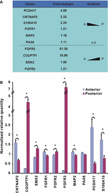
rtPCR confirmation of a subset of differentially regulated genes during early human neocortical development (8–12 PCW). Table indicating fold changes (A), and graphical representation (B) of NRQ of a subset of genes determined by rtPCR from RNA extracted from anterior and posterior regions of developing human neocortex aged between 8 and 12 PCW (n = 10). Genes exhibiting a high anterior to low posterior gradient include CNTNAP2, PCDH17, and S100A10. Genes exhibiting a high posterior, low anterior gradient include EMX2, FGFR3, and COUPTFI. No gradients were detected for PAX6. *P < 0.05, n.d., not detected; A, anterior, P, posterior.
Fig. 3
Fig. 3
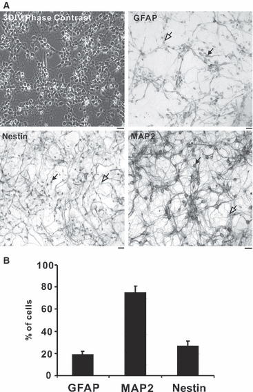
Characterization of human neuronal cortical cultures. Neuronal cell cultures were initiated from 11 PCW human neocortex and maintained for 3 DIV. Representative photographs in (A) depict cells under phase contrast, and stained with antibodies for GFAP, NESTIN and MAP2. Cell counts from random fields of vision (n = 4) indicate that MAP2-positive neurones account for most of the cells in culture (75.1 ± 6.1%), while GFAP- and Nestin-positive cells account for 19.2 ± 2.75% and 26.6 ± 4.8%, respectively. In each case, immuno-positive cells are indicated by filled arrowheads, and examples of negatively stained cells are indicated by empty arrowheads. Scale bar: 20 μm.
Fig. 4
Fig. 4
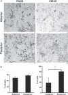
Anteriorally and posteriorally derived human cortical cultures exhibit differences in EMX2 but not in PAX6 expression. Immunohistochemistry was carried out for EMX2 and PAX6 on cultures derived from tissue dissected from the anterior and posterior poles of 11 PCW aged neocortex (A). Cell counts carried from random fields of vision (n = 4 per anterior and posterior) show a significant increase in numbers of cells expressing EMX2 (filled arrowheads) posteriorally than anteriorally. However, PAX6 (filled arrowheads) showed no differences between cultures. EMX2- or PAX6- negative cells are indicated by empty arrowheads. Scale bar: 20 μm. *P < 0.05.
Fig. 5
Fig. 5
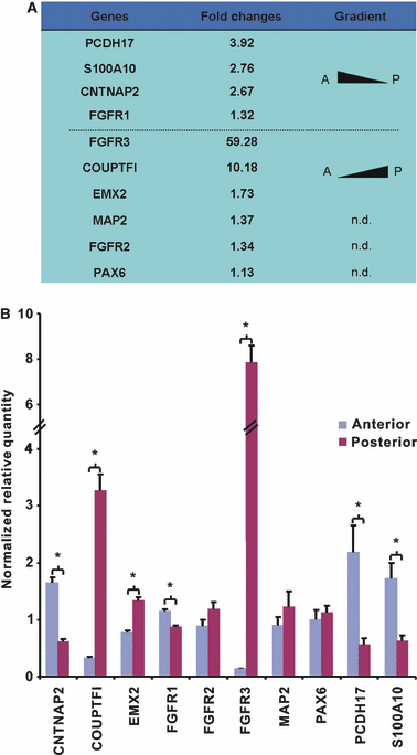
rtPCR confirmation for a subset of differentially regulated genes during early human neocortical development in cultures derived from 11 PCW human neocortex. Table indicating fold changes (A), and graphical representation (B) of NRQ of a subset of genes determined by rtPCR from RNA extracted from anteriorally and posteriorally derived cultures of human neocortex aged 11 PCW. CNTNAP2, PCDH17 and S100A10 exhibited a high anterior to low posterior gradient. EMX2, FGFR3 and COUPTFI exhibited a high posterior, low anterior gradient. No gradients were detected for PAX6, FGFR2 or MAP2. *P < 0.05, n.d., not detected; A, anterior, P, posterior.

References

    1. Abrahams BS, Tentler D, Perederiy JV, et al. Genome-wide analyses of human perisylvian cerebral cortical patterning. Proc Natl Acad Sci U S A. 2007;104:17849–17854. - PMC - PubMed
    1. Arlotta P, Molyneaux BJ, Chen J, et al. Neuronal subtype-specific genes that control corticospinal motor neuron development in vivo. Neuron. 2005;45:207–221. - PubMed
    1. Basu A, Graziadio S, Smith M, et al. Developmental plasticity connects visual cortex to motoneurons after stroke. Ann Neurol. 2010;67:132–136. - PubMed
    1. Bayatti N, Moss JA, Sun L, et al. A molecular neuroanatomical study of the developing human neocortex from 8 to 17 postconceptional weeks revealing the early differentiation of the subplate and subventricular zone. Cereb Cortex. 2008a;18:1536–1548. - PMC - PubMed
    1. Bayatti N, Sarma S, Shaw C, et al. Progressive loss of PAX6, TBR2, NEUROD and TBR1 mRNA gradients correlates with translocation of EMX2 to the cortical plate during human cortical development. Eur J Neurosci. 2008b;28:1449–1456. - PMC - PubMed

Publication types

MeSH terms

LinkOut - more resources